Kinetic basis for linking the first two enzymes of chlorophyll biosynthesis Mark Shepherd, Samantha McLean and C. Neil Hunter
Robert Hill Institute for Photosynthesis and Krebs Institute for Biomolecular Research, Department of Molecular Biology and Biotechnology, University of Sheffield, UK
Keywords chlorophyll; chelatase; methyltransferase; gun signalling
Correspondence M. Shepherd, Department of Biochemistry and Molecular Biology, A222 Life Sciences Building, Green Street, University of Georgia, Athens, GA 30602, USA Fax: +1 706 5427567 Tel: +1 706 5427252 E-mail: shepherd@secsg.uga.edu
Purified recombinant proteins from Synechocystis PCC6803 were used to show that the magnesium chelatase ChlH subunit stimulates magnesium protoporphyrin methyltransferase (ChlM) activity. Steady-state kinetics demonstrate that ChlH does not significantly alter the Km for the tetrapyr- role substrate. However, quenched-flow analysis reveals that ChlH dramat- ically accelerates the formation and breakdown of an intermediate in the catalytic cycle of ChlM. In light of the profound effect that ChlH has on the methyltransferase catalytic intermediate, the pre steady-state analysis in the current study suggests that ChlH is directly involved in the reaction chemistry. The kinetic coupling between the chelatase and methyltrans- ferase has important implications for regulation of chlorophyll biosynthesis and for the availability of magnesium protoporphyrin for plastid-to-nucleus signalling.
(Received 25 February 2005, revised 5 July 2005, accepted 19 July 2005)
doi:10.1111/j.1742-4658.2005.04873.x
enhances the magnesium chelatase reaction, and reduces the threshold Mg2+ concentration required for chelatase activity at low substrate concentrations, implying a possible role for this protein in substrate delivery.
The next step in chlorophyll biosynthesis, catalysed by magnesium protoporphyrin IX methyltransferase (ChlM in Synechocystis), involves the transfer of a methyl group from S-adenosyl-l-methionine (SAM) to the propionate group on ring C of magnesium proto- porphyrin IX (MgP) to form magnesium protopor- phyrin IX monomethylester (MgPME). Steady-state kinetic assays showed that the reaction proceeds via a random binding mechanism forming a ternary complex [13]. Stopped-flow fluorescence studies indicated that a relatively slow ((cid:1) 70 s)1) domain reorganization of ChlM alters the conformation of the MgD binding site and precedes rapid (> 600 s)1) substrate binding (Kd 3.36 lm) [14]. Rapid quenched-flow analysis showed that a catalytic intermediate is formed and
Magnesium chelatase lies at a branch point in tetrapyr- role biosynthesis where insertion of Mg2+ eventually results in the production of chlorophyll, or the insertion of Fe2+ produces heme. Magnesium chelatase is com- prised of three protein subunits, ChlI (38–42 kDa), ChlD (60–74 kDa) and ChlH (gun5, 140–150 kDa) (BchIDH in photosynthetic bacteria) [1–4]. ChlI is an AAA+ ATPase [5,6], contains a Mg2+ binding site [7], and forms a stable complex with ChlD [8]. The third subunit, ChlH, binds porphyrins [9,10] and presumably contains the active site for chelation. The steady-state kinetic characterization of magnesium chelatase quanti- fied the ATP hydrolysis required to complete a catalytic cycle and revealed a cooperativity with respect to Mg2+, which has important implications for regulation of chlo- rophyll biosynthesis [11]. The structure of Gun4, a pro- tein that binds to the tetrapyrrole substrate and product of the magnesium chelatase, has recently been solved [12]. Kinetic analysis revealed that Gun4 dramatically
Abbreviations Mg chelatase, magnesium chelatase; MgD, magnesium deuteroporphyrin IX; MgDME, Mg deuteroporphyrin IX monomethyl ester; MgP, magnesium protoporphyrin IX; MgPME, Mg protoporphyrin IX monomethyl ester; Mops, 4-morpholinepropanesulfonic acid; PIX, protoporphyrin IX; SAH, S-adenosyl-L-homocysteine; SAM, S-adenosyl-L-methionine; Synechocystis, Synechocystis PCC6803.
FEBS Journal 272 (2005) 4532–4539 ª 2005 FEBS
4532
M. Shepherd et al.
Enzyme interaction in chlorophyll biosynthesis
coincides with the
species
then depleted (rate constants of 11.9 ± 0.5 s)1 and 11.8 ± 0.5 s)1, respectively), and the decay of the intermediate evolution of magnesium deuteroporphyrin monomethylester (MgDME) product, which implies that MgDME is formed via the decay of this species [14].
The
tetrapyrrole
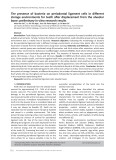
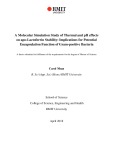
Fig. 1. Augmentation of the methyltransferase reaction by magnes- ium chelatase H subunit (ChlH). A plot of ChlM catalytic rate (lM min)1ÆlM ChlM)1 vs. ChlH concentration. Methyltransferase assays were performed in the presence of varying concentrations of ChlH protein. The reaction mixture contained 100 mM Tris pH 7.5, 100 mM glycerol, 0.2 lM ChlM, 20 lM MgD, 1 mM SAM and var- ious concentrations of ChlH. Error bars represent the standard errors when estimating the steady-state rate from timepoints in the stopped assay.
and the
ChlH, the porphyrin binding subunit of magnesium chelatase. MgD was used, instead of MgP, as the con- centration of this water-soluble analogue may be con- trolled more easily. Figure 1 depicts the increase in the catalytic rate of ChlM when the concentration of ChlH is increased, which implies that either a ChlH–MgD complex is acting as an activated substrate or that ChlH is directly accelerating the reaction chemistry. The plot of steady-state rate (vss) against ChlH concen- tration was fitted to a single rectangular hyperbola.
(MgP) and product substrate (MgPME) for the ChlM catalysed reaction have been implicated in plastid-to-nucleus signalling [15–20]. As MgP is also the product of the magnesium chelatase reaction, this emphasizes the importance of quantita- tive studies not only of the methyltransferase and chelatase, but also of the interaction between these enzymes. The coupling of the magnesium chelatase and MgP methyltransferase steps is not a new idea; in 1962, inhibition of methyltransferase activity by ethio- nine resulted in the accumulation of coproporphyrin (rather than MgP) by whole cells of Rhodobacter sph- aeroides, which suggested a degree of coupling between the magnesium chelation and methyltransferase steps. This coupling was proposed to take the form of a multienzyme complex for the conversion of proto- porphyrin to magnesium protoporphyrin IX mono- methylester (MgPME) [21]. Subsequently, it was shown that when Escherichia coli cell extracts containing the magnesium chelatase H subunit of R. capsulatus (BchH) corresponding methyltransferase (BchM) were mixed, stimulation of BchM activity was observed [22]. However, purified ChlH from Synecho- cystis was subsequently shown to have no effect on ChlM activity [23]. These are important observations, and the significance of these findings with respect to the current data is addressed in the Discussion.
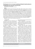
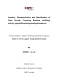
ChlM assays were performed as previously reported [13], and v vs. [MgD] curves were obtained in the pres- ence and absence of 4 lm ChlH; this concentration of ChlH gives almost maximal stimulation of methyltrans- ferase activity. 0.2 lm ChlM was assayed with 1 mm SAM and various concentrations of MgD. Figure 2 shows the rate of MgDME evolution (lmÆmin)1Ælm ChlM)1) vs. [MgD] in the presence and absence of 4 lm ChlH. Both data sets were fitted to single rectangular hyperbolae and apparent Km values were obtained. In the presence and absence of ChlH the apparent Km values were 17.3 ± 3.3 lm and 24.3 ± 7.5 lm, res- pectively.
In this paper we have used purified recombinant Synechocystis enzymes to demonstrate that ChlH has a dramatic stimulatory effect on ChlM catalysis. Quenched-flow experiments show that the magnesium chelatase H subunit markedly enhances ChlM catalysis by accelerating the formation and breakdown of the catalytic intermediate, providing a kinetic link between the first two reactions of chlorophyll biosynthesis, with the signalling molecule MgP as the common factor. Interactions between the methyltransferase and mag- nesium chelatase are likely to be crucial in determining the availability of MgP for both signalling [20] and biosynthetic roles in the chloroplast.
Results
The effect of ChlH on the lag phase prior to product formation
ChlH stimulates the methyltransferase reaction
ChlM (0.2 lm) was assayed in the presence of 50 lm MgD, 1 mm SAM and varying concentrations of
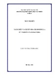
Figure 3A shows quenched-flow ChlM assays in the absence and presence of 0.75 lm ChlH. Figure 3B shows the evolution ⁄ decay of the catalytic intermediate
FEBS Journal 272 (2005) 4532–4539 ª 2005 FEBS
4533
M. Shepherd et al.
Enzyme interaction in chlorophyll biosynthesis
Fig. 2. Rate of ChlM catalysis vs. magnesium deuteroporphyrin IX (MgD) concentration in the presence and absence of 4 lM ChlH (Mg chelatase H subunit). Plots of ChlM catalytic rate (lMÆmin)1 per lM ChlM) vs. MgD concentration. The concentrations of ChlM and SAM were fixed at 0.2 lM, and 1 mM, respectively. Assays were performed in the presence (s) and absence (d) of 4 lM ChlH. MgD ¼ MgD ¼ 17.3 ± 3.3 lM in the presence of ChlH. K app K app 24.3 ± 7.5 lM in the absence of ChlH.
intermediate appears
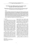
Fig. 3. Quenched-flow ⁄ HPLC analysis of product and intermediate evolution. All solutions contained 100 mM Tris pH 7.5, and 100 mM NaCl. Immediately after mixing, concentrations of ChlM, SAM and MgD were fixed at 0.5 lM, 1 mM and 30 lM, respectively. This was performed when ChlM and SAM were preincubated in the pres- ence (s) and absence (d) of 0.75 lM ChlH in syringe 1. Syringe 2 contained only MgD. (A) MgDME evolution was followed by integ- rating the peaks at 12.3 min on the HPLC chromatograms for each timepoint. (B) The evolution ⁄ depletion of the putative intermediate was followed by integrating the peaks at 12.7 min on the HPLC chromatograms for each timepoint. The data for the decay of inter- mediate in the presence of ChlH were fitted to a three-parameter exponential (k ¼ 31.7 ± 9.5 s)1). The evolution of intermediate in the absence of ChlH was characterized by a single exponential (k ¼ 24.3 ± 4.1 s)1). Units are in arbitrary fluorescence units (AU). (C) A typical HPLC chromatogram to show the elution of MgD, MgDME and the catalytic intermediate (Int).
in the absence and presence of 0.75 lm ChlH, and Fig. 3C depicts a typical chromatogram obtained dur- ing HPLC analysis of the quenched-flow samples. ChlM, SAM and ChlH (when present) were preincu- bated (in 100 mm Tris pH 7.5 ⁄ 100 mm NaCl) in syr- inge 1, and MgD was preincubated similarly in syringe 2. The quench solution used was the same as that in steady-state assays (acetone ⁄ H2O ⁄ 33% ammonia solu- tion, 80 : 20 : 1). In the absence of ChlH, the lag phase that precedes MgDME evolution is approximately 150 ms. The presence of ChlH reduces this lag phase the to approximately 50 ms, and the amplitude of burst phase is enhanced approximately fivefold. The evolution ⁄ depletion of the putative catalytic intermedi- ate was also monitored. Figure 3B demonstrates that the when ChlH is absent, the catalytic intermediate accumulates between 0 and 150 ms with a rate con- stant of 24.3 ± 4.1 s)1. When ChlH is present, the concentration of to decrease immediately, suggesting that its evolution occurs on a timescale more rapid than the dead time of the instru- ment (approximately 2 ms). Hence, this process must occur with a rate constant in excess of 500 s)1. The rate of intermediate decay (+ ChlH) was fitted to a single exponential with a rate constant of 31.7 ± 9.5 s)1. The rate constants for product accumulation (Fig. 3A) may be estimated from the rates of inter- mediate decay (Fig. 3B); rate constants for the accumu- lation of MgDME are 11.8 s)1 (–ChlH) and 31.7 s)1 (+ 2 lm ChlH). All these parameters are summarized in Table 1.
FEBS Journal 272 (2005) 4532–4539 ª 2005 FEBS
4534
M. Shepherd et al.
Enzyme interaction in chlorophyll biosynthesis
Table 1. Summary of kinetic parameters for Synechocystis ChlM, and the effects of the magnesium chelatase ChlH subunit (rate constants refer to Scheme 1).
Parameter
ChlM
ChlM + ChlH
MgD
K app Rate constant for formation of catalytic intermediate Rate constant for decay of intermediate Lag phase preceding MgDME product formation Magnitude of burst in product formation Rate constant for MgDME product formationa
24.3 ± 7.5 lM 24.3 ± 4.1 s)1 (k4) 11.8 s)1 s)1 (k5) [14] 50 ms Approx. 10 nM 11.8 s)1
17.3 ± 3.3 lM > 500 s)1 (k6) 31.7 ± 9.5 (k7) 150 ms Approx. 50 nM 31.7 s)1 [14]
a Rate constants for MgDME product formation have been estimated from the rates of intermediate decay.
Discussion
intermediate
The presence of ChlH clearly exerts a dramatic effect on the methylation of MgD catalysed by ChlM (Fig. 1). These data appear to conflict with previous work whereby purified ChlH was found to have no stimulatory effect on ChlM activity [23]. However, that study used a stopped assay where a single timepoint was taken after 30 min, which misses the much faster initial rate seen in the current study, the measurement of which is complete within 8 min. Three hypotheses present themselves: a ChlH–MgD complex is a pre- ferred substrate for the methyltransferase, ChlH binds to ChlM as an allosteric effector, or ChlH accelerates the reaction chemistry directly. The concentration dependence demonstrates that only a small excess of ChlH over ChlM is required for maximum rate enhancement (Fig. 1). The ChlH concentration at half the maximal rate is 1.2 ± 0.3 lm, which might repre- sent the binding constant (KD) for the binding of ChlH to ChlM.
When excess ChlH was present, the apparent K m
the K app
exponential decay phase of the intermediate occurs much earlier (Fig. 3B), and intermediate accumulation occurs within the 2 ms dead time of the instrument. This dramatic acceleration in formation of the interme- diate by ChlH, as well as reduction in its lifetime, is consistent with the concomitant decrease in lag phase of product evolution in Fig. 3A. The accumulation of intermediate in the absence of ChlH was fitted to an exponential with a rate constant of 24.3 ± 4.1 s)1, which compares to 11.9 s)1 with previous work [14]. The current value is a better estimate of intermediate accumulation, as the fit in Fig. 3B considers only the evolution of intermediate. The rate constant for the in the presence of ChlH decay of (31.7 ± 9.5 s)1) is three times as large as the value recorded in the absence of ChlH (11.8 s)1 [14]). These rate constants can be used to estimate the rates of MgDME accumulation in Fig. 3A, which suggests that ChlH elicits a threefold increase in the rate of product accumulation (Table 1). These data demonstrate that ChlH enhances both the accumulation and decay of this reaction intermediate, resulting in a reduction in the lag phase of product accumulation, and an increase in initial rate of product evolution. Furthermore, the presence of ChlH increases the magnitude of the burst phase approximately fivefold (Table 1), which implies that a greater concentration of enzyme is available to bind MgDME. As ChlM appears to bind the inter- mediate more transiently, this is likely to yield a higher available concentration of ChlM to bind other mole- cular species in the reaction.
MgD MgD) was 17.3 ± 3.3 lm (Fig. 2), whereas in the (K app MgD was 24.3 ± 7.5 lm absence of ChlH, (Fig. 2). The Kd for ChlM binding to free MgD, deter- mined by fluorimetric titration, is 2.4 lm [13]. There- fore, if a ChlH–MgD complex is indeed a preferred substrate for ChlM, the affinity of the methyltrans- ferase for such a complex does not appear to be greater than that of free MgD. Also, given that ChlH MgD, these observa- does not significantly alter the K m tions suggest an alternative role for ChlH in stimula- ting the methyltransferase reaction.
the
Figure 3 shows that ChlH reduces the lag phase of MgDME product evolution (Fig. 3A). This is consis- tent with the data in Fig. 3B, where the evolu- intermediate tion ⁄ depletion is putative of monitored. When ChlH is absent, the intermediate does not reach the exponential decay phase until at least 150 ms has elapsed, which coincides with the evo- lution of MgDME [14]. When ChlH is present, the
ChlH appears to enhance catalysis by accelerating the formation and decay of a catalytic intermediate. This is depicted in Scheme 1. One cannot yet pinpoint is the exact mode of action of ChlH, although it possible that ChlH may possess reactive sidechains involved in methyltransferase catalysis. Such roles may include the stabilization of the positive charge on the methyl carbon of SAM, or the enhancement of the negative charge on the propionate carboxyl groups of MgD. The rate constants quoted in Scheme 1 are all
FEBS Journal 272 (2005) 4532–4539 ª 2005 FEBS
4535
M. Shepherd et al.
Enzyme interaction in chlorophyll biosynthesis
Scheme 1. The ChlM reaction and the proposed involvement of the magnesium chelatase H subunit. The rate constants are described in Table 1. ChlM, ChlH and the catalytic intermediate are abbreviated as E, H, and Int, respectively.
faster than kcat. It has previously been proposed that product release is the slow step in the reaction [14]. This is consistent with the current work, as ChlH does not enhance kcat.
tetrapyrrole biosynthesis was proposed by Gibson et al. [26]. Our quantitative data reported here extend the influence of ChlH and show for the first time the way in which this protein exerts a strong effect on the next enzyme in the pathway, ChlM. Inspection of the ChlH titration in Fig. 1 leads to the conclusion that temporal variations in CHLH (ChlH in plants) concentration in vivo may greatly influence the cata- lytic rate of CHLM, the eukaryotic MgP methyl- transferase. This has important implications for the coupling between these steps and for the availability of MgP for signalling. Scheme 2 summarizes these conclusions in terms of variation the magnesium che- latase H subunit and its effect on the tetrapyrrole branchpoint and ChlM, but neglects the effect of Gun4. The fact that variations in ChlM activity are accompanied by altered ferrochelatase activity has been shown recently using transgenic approaches [30]. It would be necessary to establish the levels of these proteins in vivo in order to apply the enhancements measured in this study to a more physiologically rele- vant situation.
Experimental procedures
All pigments were purchased from Porphyrin Products (Logan, UT, USA). The remaining chemicals were pur- chased from Sigma-Aldrich unless otherwise specified.
Protein expression and purification
The plasmid pET9a-His6-ChlM [23] was transformed into E. coli BL21 (DE3) cells and the Synechocystis chlM gene was induced for 15 h at 20 (cid:1)C using 0.4 mm isopropyl
A recent study showed that MgP accumulation triggered the alleviation of repression of photosyn- thetic genes in Arabidopsis [20], and MgP is suggested to be a signal for one of the plastid to nucleus signal- ling pathways. However, the relative catalytic rates of magnesium chelatase and MgP methyltransferase may dictate that there is very little free MgP available for signalling. We have estimated that kcat for Mg chela- tion is 0.8 min)1 [24], whereas kcat for the subsequent methyltransferase step is estimated to be 3.4 min)1, and this is in the absence of ChlH [14]. This implies that in vivo, there may be little unbound MgP, especi- ally when ChlH is present in excess over that required for Mg chelation, since methyltransferase activity will be greatly stimulated. The discovery that another pro- tein, Gun4, can both stimulate Mg chelatase and bind MgP [12,25] adds another layer of complexity to both the regulation of chlorophyll biosynthesis, and the availability of MgP for signalling to the nucleus. We suggest that the relative amounts of both Gun4 and CHLH are crucial factors that regulate both flux down the early part of the chlorophyll biosynthetic pathway and the availability of the MgP signalling molecule. It is known that CHLH expression exhib- its diurnal fluctuations in Antirrhinum, Arabidopsis, barley and soybean [3,26–28] and that CHLH is regu- lated by a circadian clock [29]. A regulatory mecha- in magnesium chelatase nism whereby alterations H subunit levels affect partitioning between the mag- nesium (chlorophyll) and iron (haem) branches of
FEBS Journal 272 (2005) 4532–4539 ª 2005 FEBS
4536
M. Shepherd et al.
Enzyme interaction in chlorophyll biosynthesis
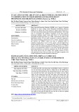
Scheme 2. Diagram of the branchpoint of tetrapyrrole biosynthesis, showing the parti- cipation of the Mg chelatase H subunit in both the chelatase and methyltransferase steps. The KD for the H ⁄ ID interaction was taken from Jensen et al. 1998 [24], and the KD for the association with ChlM is from Fig. 1. The possible effects of varying the concentration of the magnesium chelatase H subunit in the 0.2–0.5 lM range are also presented, although the effects of Gun4 or varying porphyrin concentrations are not included.
ChlM assays
buffer binding
thio-b-d-galactoside. The cells were harvested at 3000 g at 4 (cid:1)C and cells from 2 L of culture were resuspended in [20 mm citrate ⁄ KOH 20 mL chilled (pH 5.8), 500 mm NaCl, 500 mm glycerol, 5 mm imidazole]. The cells were disrupted by sonication for 6 · 30 s on ice, and the cell debris was removed at 39 000 g at 4 (cid:1)C. The supernatant was loaded at 2 mLÆmin)1 onto a 2.0 cm · 5.0 cm column packed with Chelating Sepharose Fast-Flow resin (Amersham Biosciences, Uppsala, Sweden) charged with 50 mm NiSO4 and pre-equilibrated with three column volumes of binding buffer. The column was washed with 10 column volumes of binding buffer and 6 column vol- umes of binding buffer containing 60 mm imidazole (wash buffer) to remove any loosely bound contaminants. The His-tagged ChlM was eluted with binding buffer containing 250 mm imidazole (elute buffer). A 50 mL column of P-6 desalting gel (Bio-Rad, Hercules, CA, USA) was equili- brated with 50 mm citrate ⁄ KOH (pH 5.8), 300 mm gly- cerol, 200 mm NaCl and used to remove imidazole from the buffer. A typical yield was 15 mg protein from a 2 L culture of E. coli.
Reactions were carried out at 30 (cid:1)C in 100 mm Tris pH 7.5, 100 mm glycerol, 0.2 lm ChlM, and MgD and SAM concentrations as indicated in the figure legends. The assay mixtures were incubated at 30 (cid:1)C in the absence of SAM for 5 min to allow for thermal equilibration. The SAM was added, and 20-lL aliquots were taken every 2 min over a period of 8 min and quenched in 400 lL stop solution (acetone ⁄ water ⁄ 33% ammonia solution, 80 : 20 : 1). These aliquots were centrifuged at 20 000 g for 5 min to pellet any aggregated protein. Pigments were sep- arated using reversed phase HPLC [13,14]. Between 10 and 70 lL of soluble phase, depending on the MgD concentra- tion, was loaded onto a Beckman ODS Ultrasphere column (150 · 4.6 mm; CA, USA). The pigments were separated by a 7-min linear gradient from 0% to 67% solvent B at 2 mLÆmin)1, and then the gradient was paused for a further 5 min for the porphyrins to elute (Solvent A ¼ 0.005% tri- ethylamine in water, solvent B ¼ acetonitrile). Eluted por- phyrins were detected with a Waters in-line fluorescence detector. Excitation and emission wavelengths were 394 ± 5 nm and 580 ± 5 nm, respectively.
Porphyrin stocks
The peaks that corresponded to MgD obtained in the elution profiles were integrated using Waters Millennium software. Known amounts of MgDME were analysed in the same way to produce a standard curve. The maximum rate during an assay was taken as the steady-state rate and occurred at the beginning of the reaction.
Quenched-flow measurements
FEBS Journal 272 (2005) 4532–4539 ª 2005 FEBS
4537
Pre-steady state time samples from ChlM-catalysed reac- tions were obtained using a Hi-Tech rapid quenched flow system. All solutions contained 100 mm Tris pH 7.5, and 100 mm NaCl. The reaction cell was maintained at a con- Porphyrin solutions were freshly prepared by dissolving a small amount of porphyrin in buffer. A more water-soluble analogue, magnesium deuteroporphyrin (MgD) was used instead of MgP. The presence of detergent in the assay buf- fer was no longer required. Porphyrin concentrations were determined in 0.1 m HCl using the e398 of 433 000 m)1Æcm)1 [31] after Mg2+ had been removed from the porphyrin by a 5-min incubation in 1 m acetic acid. SAM and S-adenosyl- l-homocysteine (SAH) stock solutions were prepared daily in 0.1 m HCl and 0.1 m NaOH, respectively. Their concen- trations were determined using the e256 of 15 200 m)1Æcm)1 in 1 m HCl for SAM and e260 of 16 000 m)1Æcm)1 at pH 7 for SAH [32].
M. Shepherd et al.
Enzyme interaction in chlorophyll biosynthesis
magnesium chelatase and formation of a heptameric AAA+ ring. Biochemistry 42, 6912–6920. 8 Jensen PE, Gibson LCD & Hunter CN (1999)
ATPase activity associated with the magnesium-proto- porphyrin IX chelatase enzyme of Synechocystis sp. PCC6803: evidence for ATP hydrolysis during Mg2+ insertion, and the MgATP–dependent interaction of the ChlI and ChlD subunits. Biochem J 339, 127–134.
stant temperature of 30 (cid:1)C by circulation of water from a thermostatically controlled water bath (Grant Instruments, Cambridge, UK). Reactions were quenched in stop solution (acetone ⁄ water ⁄ 33% ammonia solution, 80 : 20 : 1, v ⁄ v ⁄ v), and the concentrations of MgDME and intermediate were determined using reversed phase HPLC, as previously described [13,14]. The data obtained was analysed using nonlinear regression (sigmaplot 8.0), and apparent rate constants were obtained by fitting the plots to a single exponential [y ¼ y0 + (1-e–bx)].
Acknowledgements
We thank Mark Hoggins for assisting with the data analysis. This research was funded by the Biotechno- logy and Biological Sciences Research Council, UK.
9 Karger GA, Reid JD & Hunter CN (2001) Characteri- zation of the binding of deuteroporphyrin IX to the magnesium chelatase H subunit and spectroscopic prop- erties of the complex. Biochemistry 40, 9291–9299. 10 Willows RD & Beale SI (1998) Heterologous expression of the Rhodobacter capsulatus BchI-D, and -H genes that encode magnesium chelatase subunits and charac- terization of the reconstituted enzyme. J Biol Chem 273, 34206–34213. 11 Reid JD & Hunter CN (2004) Magnesium-dependent
References
1 Gibson LCD, Willows RD, Kannangara CG, von Wett- ATPase activity and cooperativity of magnesium chela- tase from Synechocystis sp. PCC6803. J Biol Chem 279, 26893–26899.
stein D & Hunter CN (1995) Magnesium-protopor- phyrin chelatase of Rhodobacter sphaeroides: reconstitiution of activity by combining the products of the bchH-I and -D genes expressed in Escherichia coli. Proc Natl Acad Sci USA 92, 1941–1944. 12 Davison PA, Schubert HL, Reid JD, Lorg CD, Heroux A, Hill CP & Hunter CN (2005) Structural and bio- chemical characterization of Gun4 suggests a mechan- ism for its role in chlorophyll biosynthesis. Biochemistry 44, 7603–7612. 2 Jensen PE, Gibson LCD, Henningsen KW & Hunter CN
13 Shepherd M, Reid JD & Hunter CN (2003) Purification and kinetic characterization of the magnesium proto- porphyrin IX methyltransferase from Synechocystis PCC6803. Biochem J 371, 351–360.
(1996) Expression of the chlI, chlD, and chlH genes from the cyanobacterium Synechocystis PCC6803 in Escherichia coli and demonstration that the three cognate proteins are required for magnesium-protoporphyrin chelatase activity. J Biol Chem 271, 16662–16667.
14 Shepherd M & Hunter CN (2004) Transient kinetics of the reaction catalysed by magnesium protoporphyrin IX methyltransferase. Biochem J 382, 1009–1013.
15 Johanningmeier U (1988) Possible control of transcript levels by chlorophyll precursors in Chlamydomonas. Eur J Biochem 177, 417–424. 3 Jensen PE, Willows RD, Petersen BL, Vothknecht UC, Stummann BM, Kannangara CG, von Wettstein D & Henningsen KW (1996) Structural genes for Mg-chela- tase subunits in barley: Xantha-f-g and -h. Mol Gen Genet 250, 383–394. 16 Kropat J, Oster U, Rudiger W & Beck CF (1997)
Chlorophyll precursors are signals of chloroplast origin involved in light induction of nuclear heat-shock genes. Proc Natl Acad Sci USA 94, 14168–14172. 17 Kropat J, Oster U, Rudiger W & Beck CF (2000)
4 Papenbrock J, Gra¨ fe S, Kruse E, Ha¨ nel F & Grimm B (1997) Mg-chelatase of tobacco: identification of a Chl D cDNA sequence encoding a third subunit, analysis of the interaction of the three subunits with the yeast two- hybrid system, and reconstitution of the enzyme activity by co-expression of recombinant CHLD, CHLH and CHLI. Plant J 12, 981–990. 5 Neuwald AF, Aravind L, Spouge JL & Koonin EV Chloroplast signalling in the light induction of nuclear HSP70 genes requires the accumulation of chlorophyll precursors and their accessibility to cytoplasm ⁄ nucleus. Plant J 24, 523–531.
(1999) AAA+: a class of chaperone-like ATPases asso- ciated with the assembly, operation, and disassembly of protein complexes. Genome Res 9, 27–43.
18 Susek RE, Ausubel FM & Chory J (1993) Signal trans- duction mutants of Arabidopsis uncouple nuclear CAB and RBCS gene expression from chloroplast develop- ment. Cell 74, 787–799.
6 Fodje MN, Hansson A, Hansson M, Olsen JG, Gough S, Willows RD & Al Karadaghi S (2001) Interplay between an AAA module and an integrin I domain may regulate the function of magnesium chelatase. J Mol Biol 311, 111–122.
FEBS Journal 272 (2005) 4532–4539 ª 2005 FEBS
4538
19 Mochizuki N, Brusslan JA, Larkin R, Nagatani A & Chory J (2001) Arabidopsis genomes uncoupled 5 (GUN5) mutant reveals the involvement of Mg-chela- tase H subunit in plastid-to-nucleus signal transduction. Proc Natl Acad Sci USA 98, 2053–2058. 7 Reid JD, Siebert CA, Bullough PA & Hunter CN (2003) The ATPase activity of the ChlI subunit of
M. Shepherd et al.
Enzyme interaction in chlorophyll biosynthesis
Mg chelatase subunit from Arabidopsis thaliana cv C24. Sequence and transcript analysis of the gene, import of the protein into chloroplasts, and in situ localization of the transcript and protein. Plant Physiol 111, 61–71. 27 Hudson A, Carpenter R, Doyle S & Coen ES (1993) 20 Strand A, Asami T, Alonso J, Ecker JR & Chory J (2003) Chloroplast to nucleus communication triggered by accu- mulation of Mg-protoporphyrin IX. Nature 421, 79–83. 21 Gorchein A (1972) Magnesium protoporphyrin chela- tase activity in Rhodopseudomonas spheroides. Studies with whole cells. Biochem J 127, 97–106. 22 Hinchigeri SB, Hundle B & Richards WR (1997) Olive: a key gene required for chlorophyll biosynthesis in Antirrhinum majus. EMBO J 12, 3711–3719. 28 Nakayama M, Masuda T, Bando T, Yamagata H,
Demonstration that the BchH protein of Rhodobacter capsulatus activates S-adenosyl-l-methionine: magne- sium protoporphyrin IX methyltransferase. FEBS Lett 407, 337–342. 23 Jensen PE, Gibson LCD, Shephard F, Smith V & Ohta H & Takamiya K (1998) Cloning and expression of the soybean chlH gene encoding a subunit of Mg-chelatase and localization of the Mg2+ concentra- tion-dependent ChlH protein within the chloroplast. Plant Cell Physiol 39, 275–284. 29 Harmer SL, Hogenesch JB, Straume M, Chang HS,
Han B, Zhu T, Wang X, Kreps JA & Kay SA (2000) Orchestrated transcription of key pathways in Arabidop- sis by the circadian clock. Science 290, 2110–2113. Hunter CN (1999) Introduction of a new branchpoint in tetrapyrrole biosynthesis in Escherichia coli by co-expression of genes encoding the chlorophyll- specific enzymes magnesium chelatase and magnesium protoporphyrin methyltransferase. FEBS Lett 455, 349–354.
30 Alawady AE & Grimm B (2005) Tobacco Mg protopor- phyrin IX methyltransferase is involved in inverse acti- vation of Mg porphyrin and protoheme synthesis. Plant J 41, 282–290. 31 Falk JE (1964) Porphyrins and Metalloporphyrins. Elsevier, London. 24 Jensen PE, Gibson LCD & Hunter CN (1998) Determi- nants of catalytic activity with the use of purified I, D and H subunits of the magnesium protoporphyrin IX chelatase from Synechocystis PCC6803. Biochem J 334, 335–344. 32 Dawson RMC, Elliot DC, Elliot WH & Jones KM 25 Larkin RM, Alonso JM, Ecker JR & Chory J (2003)
FEBS Journal 272 (2005) 4532–4539 ª 2005 FEBS
4539
(1986) Data for Biochemical Research, 3rd edn. Oxford University Press Inc., New York. GUN4, a regulator of chlorophyll synthesis and intra- cellular signaling. Science 299, 902–906. 26 Gibson LCD, Marrison JL, Leech RM, Jensen PE, Bassham DC, Gibson M & Hunter CN (1996) A putative