doi:10.1046/j.1432-1033.2003.03942.x
Eur. J. Biochem. 271, 418–428 (2004) (cid:1) FEBS 2003
New substrate analogues of human serotonin N-acetyltransferase produce insituspecific and potent inhibitors
Gilles Ferry1, Caroline Ubeaud1, Julien Mozo1, Christophe Pe´ an2, Philippe Hennig2, Marianne Rodriguez1, Catherine Scoul1, Anne Bonnaud1, Olivier Nosjean1, Jean-Pierre Galizzi1, Philippe Delagrange3, Pierre Renard3, Jean-Paul Volland2, Said Yous4, Daniel Lesieur4 and Jean A. Boutin1 1Pharmacologie Mole´culaire et Cellulaire, Institut de Recherches SERVIER, Croissy-sur-Seine; 2Physico-Chimie Analytique, Institut de Recherches SERVIER, Suresnes; 3Institut de Recherches Internationales SERVIER, Courbevoie; 4Faculte´ de Pharmacie, Lille, France
2
(N-acetyl[3H]PEA and coenzyme A-S[3H]acetyltryptamine, respectively). The biosynthesis of this bifunctional inhibitor derived from BAT was also followed by nuclear magnetic resonance during its catalytic production by the pure enzyme. In a similar manner we studied the production of another inhibitor generated from N-[2-(7-hydroxynaphth- 1-yl)ethyl]bromoacetamide. New derivatives were also screened for their capacity to inhibit a purified enzyme, in addition to enzyme overexpressed in a cellular model. Some of these compounds proved to be extremely potent, with IC50s of (cid:1) 30 nM. As these compounds, by definition, closely resemble the natural substrates of arylalkylamine N-acetyl- , we also show that they are potent ligands at the transferase melatonin receptors. Nevertheless, these inhibitors form a series of pharmacological tools that could be used to understand more closely the inhibition of pineal melatonin production in vivo.
Keywords: melatonin; serotonin N-acetyltransferase; cellu- lar model; inhibition; N-bromoacetyltryptamine.
Melatonin is synthesized by an enzymatic pathway, in which arylalkylamine (serotonin) N-acetyltransferase cata- lyzes the rate-limiting step. A previous study [Khalil, E.M., De Angelis, J., Ishii, M. & Cole, P.A. (1999) Proc. Natl Acad. Sci. USA 96, 12418–12423] reported the discovery of bromoacetyltryptamine (BAT), a new type of inhibitor of this enzyme. This compound is the precursor of a potent bifunctional inhibitor (analogue of the transition state), capable of interfering with both the substrate and the cosubstrate binding sites. This inhibitor is biosynthesized by the enzyme itself in the presence of free coenzyme A. In the present report, we describe the potency of new N-halo- genoacetyl derivatives leading to a strong in situ inhibition of serotonin N-acetyltransferase. The new concept behind the mechanism of action of these precursors was studied by following the biosynthesis of the inhibitor from tritiated- BAT in a living cell. The fate of tritiated-phenylethylamine (PEA), a natural substrate of the enzyme, in the presence or absence of [3H]BAT was also followed, leading to their incorporation into the reaction product or the inhibitor
Melatonin (5-methoxy-N-acetyltryptamine) is a neuro- hormone which is responsible for the transmission of photo- period circadian rhythm information to the periphery in vertebrates. It is synthesized in the pineal gland during the dark period [1]. Melatonin has been described as functioning in many physio-pathological processes including sleep
disorders, mild depression [2], obesity [3], cancer [4] and bone formation [5]. The synthesis of melatonin from serotonin is directed by a series of enzymes, of which arylalkylamine (serotonin) N-acetyltransferase (AANAT; EC 2.3.1.87) catalyses the penultimate and rate-limiting step. The human AANAT enzyme has been cloned [6] and purified, and the specificity of its substrates and cosubstrates have been described [7]. A large program of chemistry and screening was launched in order to discover potent inhibitors of this particular enzyme and to investigate the effects of the reduction (or total inhibition) of nocturnal melatonin production. We recently described a robust cellular model in which human AANAT (hAANAT) was overexpressed [8]. This cellular model completes the collection of tools required for the discovery of pharmacologically active inhibitors.
Five classes of hAANAT inhibitors have been reported in the literature to date: loose structural analogues of the indoleamine hormone [9,10], tetrapeptides issued from the deconvolution of combinatorial libraries [7], heterocyclic compounds from a large screen of natural plant extracts and synthetic molecules [11], bisubstrate analogues (i.e. Coen- zyme A derivatives) [12] and the N-bromoacetamido deriv- atives (precursors of potent inhibitors) [13]. Each of these
Correspondence to J. A. Boutin, Pharmacologie Mole´ culaire et Cellulaire, Institut de Recherches SERVIER, 125 Chemin de Ronde 78290 Croissy-sur-Seine, France. Fax: + 33 1 55 72 28 10, Tel.: + 33 1 55 72 27 48, E-mail: jean.boutin@fr.netgrs.com Abbreviations: BAT, bromoacetyltryptamine; CHO, Chinese hamster ovary cell line; CoASAT, coenzyme A-S-acetyltryptamine; CoA-SH, free coenzyme A; hAANAT, human arylalkylamine N-acetyltransferase; hMT1, human receptor to melatonin, isoform 1; hMT2, human receptor to melatonin, isoform 2; HNEBA, N-[2-(7-hydroxynaphth-1-yl)ethyl]bromoacetamide; PEA, phenyl- ethylamine; PKA, protein kinase A (cAMP-dependent). Enzyme: human arylalkylamine N-acetyltransferase (EC 2.3.1.87). (Received 11 August 2003, revised 14 November 2003, accepted 25 November 2003)
Mechanism of serotonin N-acetyltransferase inhibition (Eur. J. Biochem. 271) 419
(cid:1) FEBS 2003
inhibitor classes have their limitations, namely, the melato- nin-based derivatives can have strong affinities for one or the other (or both) melatonin receptors [10,14], with newly discovered chemical species having unknown pharmacolo- gical (particularly toxicological) properties that still remain to be explored, and the peptide inhibitors have poor cellular penetration properties as do the bisubstrate analogues [15]. The properties of the N-halogenoacetamido derivatives suggest that they might form a new class of inhibitors with the original mechanism(s) of action. The paucity of knowledge about these compounds necessitates more stud- ies on their mechanisms of production in addition to their specificity, particularly vis-a` -vis the melatonin receptors.
In the present
suspension was detergent Tween 85 (5% v/v). This sonicated eight for 1 min in ice, centrifuged times (20 000 g, 20 min) and the supernatant passed through an affinity column 40 mLÆh)1 (Glutathione-Sepharose, Pharmacia), that was previously equilibrated with buf- fer A (2· NaCl/Pi, pH 6.9 containing 10 mM dithiothre- itol). The column was washed (40 mL buffer A) and then eluted with 150 mL of buffer B (50 mM Tris/HCl, pH 8.0 containing 100 nM sodium citrate, 10 mM dithiothreitol and 10% (v/v) glycerol). The glutathione-S-transferase– AANAT was eluted sequentially with 40 mL of 10 mM glutathione in buffer B. Active fractions were pooled and stored frozen at )80 (cid:3)C until used. The protein concen- tration was determined by the Bradford assay [26] (Protassay, Bio-Rad, Ivry- sur-Seine, France) with bovine serum albumin as standard.
Assays for enzymatic and cellular AANAT
(Costar
6,7 6,7 8
report, we explore the inhibitory properties of new derivatives of the N-iodo- or N-bromo- acetamidoethylaryl derivatives on purified hAANAT, as well as in cells overexpressing this enzyme. We studied the mechanism of production of some of these inhibitors by nuclear magnetic resonance, and by using a radiolabelled inhibitor precursor. These experiments demonstrate the potency of the approach using N-halogenoacetamidoethyl derivatives of AANAT substrates to generate potent inhibitors of the enzyme activity. Indeed, our data suggest that they are acetyl-CoA binding, as well as substrate- binding site competitive inhibitors because they occupy the cosubstrate site. As these compounds are broad analogues of melatonin [16–25], we report their affinities for the MT1 and/or MT2 receptors.
Experimental procedures
Culture of AANAT-expressing cell line
3
4
9
Selection of the positive clone, CHO-hAANAT-8, was described in a previously published work [8]. Cells were seeded (1 · 106 cells per well, of a 96-well plate) and cultured for 72 h with 5% CO2:95% air (v/v) in a humidified incubator in 96-well plates containing Ham MF12 medium (GIBCO BRL, Invitrogen SARL, Cargy- Pontoise, France) supplemented with 10% (v/v) fetal bovine serum, 50 lgÆmL)1 penicillin/streptomycin, 2 mM L-gluta- mine, 5 mgÆmL)1 G418, all from Life Technologies.
Expression and production of hAANAT
5
coli
An enzymatic assay for AANAT was performed in a 100 lL final volume comprising 1 lg of enzyme, in a phosphate buffer (50 mM sodium phosphate, pH 6.8, 500 mM NaCl and 2 mM EDTA) with 1 mM acetyl-CoA and 4 mM phenylethylamine. Compounds were dispensed in black 96-well plates Issy-les-Moulineaux, , Dutscher, , at a starting concentration France) under a volume of 10 lL of 100 lM in pure dimethylsulfoxide . Cellular assays for AANAT were performed on subconfluent cells seeded in 96-well plates and incubated with 10 lL of the tritiated sub- strate [3H]phenylethylamine ([3H]PEA, Amersham Pharma- cia Biotech, 2.78 TBqÆmmol)1). In brief, for cell studies, 70 lL of incubation buffer (phosphate buffer detailed above) containing 0.5% Tween 85 were added to each well of the plates. Plates were shaken at room temperature for 30 min using an orbital shaker (Bioblock Scientific, Strasbourg, France). Each well was then sonicated once (setting 40/100, probe 0.2 cm diameter) for five seconds. For [3H]bromoace- tyltryptamine experiments ([3H]BAT, Amersham Pharmacia Biotech, 2.25 TBqÆmmol)1) , 10 lL of the compound was added along with cold PEA standard incubation, to enzyme or to cells in 96 wells plates. After incubation (30 min, 37 (cid:3)C), the reaction was stopped by the addition of 50 lL of 10% (v/ v) trichloroacetic acid in water. Thirty microliters of enzy- matic reaction, conditioned culture media or lysed cells were analyzed by reverse phase HPLC using a C4 ASTEC, 4.6 · 150 mm column (CIL Cluzeau, Ste-Foy-la-Grande, France). The column was developed with a linear gradient of 5–35 or 0–100% (v/v) acetonitrile in H2O, 0.1% (v/v) trifluoroacetic acid at a flow rate of 1 mLÆmin)1 for 15 or 30 min. The area under the peak of the reaction product was integrated by the software of the HPLC system (HP-CHEM- v06.01; Agilent Technologies, Waldbronn, STATION, Germany), by comparison of surfaces of the peak for N-acetylPEA in the presence and absence of compounds. At least two determinations were made for each experiment. Each inhibition factor was calculated using the mean values of those duplicates.
NMR studies
Expression, production and purification of hAANAT have been described by Ferry et al. [7]. In brief, the human arylalkylamine N-acetyltransferase cDNA coding region (kindly provided by D. C. Klein and S. L. Coon , NIH, Bethesda, USA) was inserted into the bacterial expression vector pGEX-4T (Pharmacia, Les Ulis, France), leading to the expression of a protein fused to glutathione-S-transferase. An Escherichia strain [BL21(DE3)pLysS] was transformed with the resulting plasmid. Expression of the construct was induced by adding isopropyl thio-b-D-galactoside (0.2 mM) at 24 (cid:3)C for 6 h. The cells were harvested by centrifugation (5000 g, 4 (cid:3)C, 10 min). All purification procedures were performed at 0–4 (cid:3)C. Approximately 10 g of a frozen bacteria pellet was thawed in 40 mL of 2· NaCl/Pi containing 10 mM dithiothreitol, a cocktail of protease inhibitors (Complete, Roche; 1 tablet per 50 mL) and the
NMR was used to follow the alkyltranferase activity of hAANAT. All kinetic studies followed by NMR spectro-
420 G. Ferry et al. (Eur. J. Biochem. 271)
(cid:1) FEBS 2003
other chemicals were of the highest purity available, from Sigma (Saint Louis, MO, USA).
Binding experiments at human MT1 and MT2 receptors
[38]. The CHO-K1 cell
scopy were performed on a Bruker Avance 500 MHz spectrometer (Bruker Instruments, Wissembourg, France) equipped with a triple axes gradient 5 mm BBI probe. The experiments were conducted in a medium containing the inhibitor (BAT or compound 1, 100 lM in 5% (v/v) d6- dimethylsulfoxide final volume), 1 mM acetyl-CoA and 4 mM serotonin (when required) in deuterated phosphate buffer (50 mM sodium phosphate pH 6.8, 500 mM NaCl, 2 mM EDTA) and 1 lg of enzyme. Monitoring of the enzymatic reaction was carried out by recording a series of 1D 1H NMR spectra (the temperature of the sample was continuously set to 300 K), using 16 K data points. The data were then Fourier-transformed, after zero filling to 32 K, using a line broadening exponential treatment (lb ¼ 2 Hz). All spectra were phased and the baseline was corrected before integration of the signals of interest. For the BAT studies, 128 transients were accumulated for each spectrum (157 s between each spectrum), whereas for the compound 1 studies, only 64 scans were accumulated (92 s interval time between each spectrum).
Chemicals
14
[10];
bromoacetamide}
[10];
compound 6
Binding experiments were carried out as described previously in Audinot et al. lines stably expressing either the hMT1 or hMT2 receptors were grown at confluence, harvested in phosphate buffer containing 2 mM EDTA and centrifuged at 1000 g for 5 min (4 (cid:3)C). The resulting pellet was suspended in 5 mM Tris/HCl, pH 7.4, containing 2 mM EDTA, and homogenized using a Kinem- atica (Lucerne, Switzerland) polytron. The homogenate was then centrifuged (20 000 g, 30 min, 4 (cid:3)C), and the final pellet was suspended in 75 mM Tris/HCl, pH 7.4, containing 2 mM EDTA and 12.5 mM MgCl2. The determination of protein content was performed according to Bradford [26] using the Bio-Rad kit (Bio-Rad SA, Ivry-sur-Seine, France). Aliquots of membrane preparations were stored at )80 (cid:3)C until use. 2-[125I]iodomelatonin binding assay experiments were con- ducted as follows: membranes were incubated for 2 h at 37 (cid:3)C in binding buffer (Tris/HCl 50 mM, pH 7.4, 5 mM MgCl2) in a final volume of 250 lL containing 2[125I]iodo- melatonin (25 or 200 pM for competition experiments in MT1-CHO and MT2-CHO cells, respectively, according to the slightly better affinity of this compound to MT1, as compared to MT2). Nonspecific binding was defined with 10 lM melatonin. The reaction was stopped by rapid filtration through GF/B unifilters (Whatmann, Maidstone, Kent, UK), followed by three successive washes with ice-cold binding buffer. Data were analyzed with PRISM software (GraphPad Software Inc., San Diego, CA, USA). For saturation assays, the density of binding sites (Bmax) and the dissociation constant of the radioligand (KD) values were calculated using Scatchard plot analysis. For displacement inhibition constants (Ki) were calculated experiments, according to the Cheng–Prussof equation: Ki ¼ IC50/ [1 + (L/KD)], where IC50 is the inhibitory concentration 50% and L is the concentration of 2[125I]iodomelatonin. For the correlation analysis of pKi values, the Pearson product- moment correlation coefficient was employed.
Results
and finally,
Synthesis of the various chemicals used in the present studies follows: N-bromoacetyltryptamine {N-[2-(1H- were as indol-3-yl)ethyl]bromoacetamide} (BAT) [27]; compounds 2 {N-[2-(7-methoxy-napht-1-yl)ethyl]bromoacetamide}, 3 {N-[2-(5-hydroxy-benzo[b]thiophen-3-yl)ethyl]bromoaceta- {N-[2-(5-ethyl-benzo[b]thiophen-3-yl)ethyl] mide}, bromoacetamide} and 15 {N-[2-(5-Bromo-benzo[b]thio- phen-3-yl)ethyl]bromoacetamide} compound 4 {N-[2-(5-fluoro-1H-indol-3-yl)ethyl]bromoacetamide} [29]; compound 9 {N-[2-(7-propoxy-napht-1-yl)ethyl]iodoacet- amide} [30]; compound 12 {N-[2-(5-chloro-benzo[b]thio- phen-3-yl)ethyl] compound 1 {N-[2-(7-hydroxy-napht-1-yl)ethyl]bromoacetamide} [31]; {N-[2-(2-benzyl-5-methoxy-benzofuran-3- compounds 5 yl)ethyl]iodoacetamide} and 18 {3-(2-(bromoacetylamino)- ethyl)-benzo[b]thiophen-5-yl]carboxylic acid methyl ester} {N-[2-(3-ethyl-7-methoxy-napht-1- [29]; yl)ethyl]iodoacetamide} [33]; compounds 10 {N-[2-(7-ethyl- 1,2,3,4-tetrahydronapht-1-yl)ethyl]iodooacetamide} and 17 {N-[2-(7-ethyl-napht-1-yl)ethyl]-bromoacetamide} [34]; compound 8 {N-[2-(7-methoxy-8-propenyl-napht-1-yl)ethyl]- iodoacetamide} [35]; compound 11 {N-[2-(7-methoxy-3- (3-trifluoromethylphenyl)-napht-1-yl) ethyl]iodoacetamide} {N-[2-(2-phenyl-ben- [36] compound 16 the zo[b]thiophen-3-yl)ethyl]bro-moacetamide} [37]. All
The hypothesis that compounds can be designed to produce bisubstrate analogue inhibitors of the enzyme formed in the catalytic site of its target, is the basis of the present work. Indeed, after the description of BAT
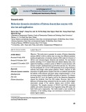
34 Fig. 1. Schematic representation of the two reactions catalyzed by AANAT: (A) acetylation and (B) alkylation. (A) Acetylation of the AANAT substrate, phenylethylamine, in the presence of its cosubstrate, acetyl-CoA. The products of this reaction are N-acetylphenyl- ethylamine and free CoA-SH. (B) Alkylation of N-bromoacetyltryptamine (BAT) using the CoA-SH released by the previous reaction, and leading to the biosynthesis of a potent bisubstrate inhibitor, CoA-S-acetyltryptamine (based largely on Khalil et al. [13]).
Mechanism of serotonin N-acetyltransferase inhibition (Eur. J. Biochem. 271) 421
(cid:1) FEBS 2003
expressing the enzyme. [3H]BAT was first incubated with hAANAT, cold cosubstrate and/or the substrate sero- tonin for 15 min and 1 h (Fig. 2), which gave rise to [3H]CoASAT. The formation of this inhibitor is not dependent on the chemical nature of the substrates. Indeed, the same compound production is recorded, whether PEA or tryptamine were used as substrate in the incubations (not shown). This tritiated same type of
[13], we searched our chemical library for compounds that have these features. We further studied the original in order to better understand its compound, BAT, complex mechanisms of production and action (Fig. 1). To perform these studies, [3H]BAT was used with the aim to visualize the tritiated coenzyme A–S-acetyltrypta- mine ([3H]CoASAT) that was formed by either the purified enzyme or in the conditioned media of cells
35 Fig. 2. In situ production of the CoA-S-acetyl- tryptamine inhibitor during hAANAT incuba- tions. Incubations contained the purified hAANAT, [3H]N-bromoacetyltryptamine, acetyl-CoA, and serotonin. Radioactivity (solid line) and UV (A280; dashed line) profiles after (A) 15 min incubation and (B) after 60 min incubation. Peak identifications are: a, acetyl-CoA (UV trace); b, serotonin (UV trace); c, N-acetylserotonin (UV trace); d, CoA-S-acetyl[3H]tryptamine (radioactivity trace); e, [3H]N-bromoacetyltryptamine (radioactivity trace).
Fig. 3. Inhibition of the cellular hAANAT activity by N-bromoacetyltryptamine and production of the bisubstrate inhibitor, CoA-S-acetyltryptamine. The histogram represents the amounts of the different products measured during an incubation containing the enzyme, the substrate ([3H]PEA), the cosubstrate (acetyl-CoA) with or without tritiated N-bromoacetyltryptamine over a period of 6 h. (A) represents the N-acetylation of [3H]phe- nylethylamine by hAANAT-expressing cells without (dark bars) or with (hatched bars) [3H]N-bromoacetyltryptamine. (B) represents the amount of CoA-S[3H]acetyltryptamine formed during the same experiment as in (A). Experiments were conducted as described in Experimental procedures. Analyses were carried out by HPLC, and the various tritiated species were separated easily, as indicated in Fig. 2. At least three separate experiments were conducted and the mean values are presented in the graphs. The units represent the direct output from the radioactivity detector, with identical settings in all experiments.
422 G. Ferry et al. (Eur. J. Biochem. 271)
(cid:1) FEBS 2003
compound has the same retention time as the synthetic product, CoASAT [7].
21
20
We incubated cells overexpressing hAANAT for 1, 3 and 6 h with [3H]PEA and with or without [3H]BAT (Fig. 3A). In the control experiments, the product of the reaction, [3H]N-acetylPEA, was present in the conditioned medium. In the presence of [3H]BAT, the reaction was slowed down significantly. Additionally, as a result of the HPLC separ- ation, we could measure the production of the bisubstrate inhibitor CoA–S[3H]acetyltryptamine and its presence in the conditioned medium (Fig. 3B), clearly showing that CoASAT had travelled freely out of the cell, despite its size and relative hydrophilicity. This result is in agreement with previously published data showing that BAT can react with present in small quantities of free Coenzyme A (CoA-SH) the reaction mixture to form CoASAT which, in turn, inhibits the hAANAT activity [12]. BAT incubation with the enzyme but without cosubstrate did not lead to CoASAT formation.
the influence of added serotonin,
Spectrum C, the last of the series (t ¼ 50 min, Fig. 4) shows that all the substrate molecules had been consumed and transformed in the final product during the 50 min incuba- tion. Moreover, a comparison between spectrum C (50 min incubation) and spectrum D (pure synthesized final product, CoASAT, Fig. 4) led to the nonambiguous identification of the reaction final product. An interesting point of using the NMR technique is that the evolution of the reaction (Fig. 4E) can be followed in real time without any further manipulation. Indeed, the integration values of the appro- priate proton (b¢) was recorded over time and plotted in Fig. 4F. This kinetic curve shows the progression of the reaction until the complete consumption of the substrate by the enzyme (after 50 min). The next step of our study was to look at another example of a potent AANAT inhibitor; compound 1 (Fig. 5 shows compound structures). As it has been demonstrated that BAT spontaneously formed its conjugate with CoA-SH [13], we wanted to ensure that HNEBA (compound 1), as a model of the other com- pounds of the series, was also able to form a CoA conjugate spontaneously (i.e. in the absence of enzyme). Indeed, it did, although in the presence of the enzyme, the speed at which the conjugate was formed was enhanced 10-fold. Further- the enzyme more, substrate, was studied as depicted in Figs 6 and 7. The presence (Fig. 6E–G) or absence (Fig. 6A–C) of the substrate serotonin did not influence the nature of the final product, as spectra C and G (showing the product of the
The kinetics of the formation of the bisubstrate inhibitors that arise from the incubation of either BAT (in the absence the substrate serotonin) or N-[2-(7-hydroxynaphth- of 1-yl)ethyl]bromoacetamide (HNEBA; compound 1) were studied by following the evolution of the NMR spectra of the reaction product over time. In spectrum B (Fig. 4), recorded 20 min after the initial spectrum A (Fig. 4), new signals were observed (labeled a¢ to e¢) that corresponded to the proton signals of the product of the enzymatic reaction.
Fig. 4. NMR spectroscopy analysis of the serotonin-N-acetyltransferase reaction in the presence of the inhibitor precursor, N-bromoacetyltryptamine, as a function of incubation time. The experiment was conducted as described in Experimental procedures. The enzymatic reaction, in deuterated buffer, comprised the cosubstrate, acetyl-CoA, the enzyme and the inhibitor precursor, N-bromoacetyltryptamine. (A) The spectrum was recorded at the beginning of the reaction (time ¼ 0 min); (B) the reaction after 20 min; (C) the reaction after 50 min – completion; (D) NMR spectrum of the synthetic product of the reaction, the bisubstrate inhibitor, CoA-S-acetyltryptamine; (E) schematic representation of the inhibitor precursor and of the final product, the bisubstrate inhibitor, CoA-S-acetyltryptamine and their proton attributions; (F) kinetic representation of the NMR spectroscopy analyses. The proton b¢ peak height was measured and plotted versus time, during the complete course of the experiment. (cid:1) 20 spectra, i.e. one spectrum every 150 s.
Mechanism of serotonin N-acetyltransferase inhibition (Eur. J. Biochem. 271) 423
(cid:1) FEBS 2003
22
Newly synthesized compounds presenting an N-halo- genoacetylethylamine moiety were tested as inhibitors of hAANAT, using a two-step strategy: initial screening at 10 lM, followed by full range concentration displacements for those that presented 40–50% inhibition at the initial concentration (Table 1). The structures of these compounds are presented in Fig. 5. Several of these compounds were potent inhibitors of hAANAT activity; their enzymatic IC50 ranged from above 10 lM to submicromolar concentra- tions, particularly, compounds 1, 3, 6, 12, 14 and 13 (Table 1). The best inhibitor under these experimental conditions was compound 12, with an IC50 of 180 nM.
23
protein 14-3-3,
A number of these compounds were then selected for testing in a cellular assay. Of the group, all were tested at 100 nM concentration and further tests were run only on those in which a significant inhibition (> 10%) could be measured. Only four compounds (compounds 1–4) had an IC50 below 1 lM. Compound 3 was particularly potent in this model, with an apparent IC50 of 30 nM. For a selected range of the actives, the IC50 was measured after treatment of the hAANAT-expressing cells with forskolin. Through the activation of protein kinase A by cAMP, hAANAT becomes phosphorylated and associates with the leading to a conformational chaperone change of AANAT [39,40]. Interestingly, this apparent change leads to significant variation of the inhibitory potency of some of the compounds. Table 2 summarizes the most important IC50 shifts between the two conditions (with and without forskolin). In all but one case, the general trend was that the phosphorylation of hAANAT by protein kinase A favors inhibition. Indeed, a lowering of their IC50 values is observed, at least four to one-hundred times even for the less active compounds, specifically the marginally active compounds 7 and 9. Only compound 3 showed an increase of its IC50 from 30 to 446 nM.
Finally, we tested the binding capacities of
these compounds to human recombinant MT1 and MT2 recep- tors. As shown in Table 3, the majority of the compounds were subnanomolar ligands at one or more receptors. For example, compound 2 had a Ki of 98 and 86 pM at MT1 and MT2 receptors, respectively, with compound 6 having a Ki of 30 pM and compound 16, 40 pM, both at MT2. For most it is apparent that their inhibitory of the compounds, potencies on AANAT are in concentration ranges signifi- cantly poorer than their Ki for MT1 and/or MT2.
Discussion
The present study describes new hAANAT inhibitor precursors that can be used to investigate the effect of lowering melatonin levels. New compounds, particularly enzyme inhibitors, but also receptor antagonists [38], might help greatly in deciphering the multiple protein targets by which melatonin exerts its many actions [41]. Indeed, although we succeeded in demonstrating the possibility of finding AANAT inhibitors by systematic screening [11], powerful inhibitors of this enzyme are still lacking. Previous studies have described a chemical, tryptamyl-CoA, which is derived from the transitional state of the enzyme [15,27]. Nevertheless, acetyl-CoA as well as other analogues of the Coenzyme A cannot cross the cell membrane [42] and are therefore unlikely to give rise to compounds that can be
reaction) were identical. In both systems, we observed the disappearance of the HNEBA signals, and the appearance of a second species. The attributions of NMR signals are as described on the chemical drawing (Fig. 6D). The appropriate proton signals (b¢; Fig. 6D) evolved according to time (Fig. 7). When the substrate was added, the production of the inhibitory species was accelerated, as more free CoA-SH molecules became available in the enzymatic mixture to enter the reaction and form the bisubstrate inhibitor according to the scheme (Fig. 1) proposed by Khalil et al. [13].
Fig. 5. Structures of the compounds studied. The compounds were separated into three clusters: (A) miscellaneous, (B) naphthalene-based and (C) benzothiophene-based compounds.
424 G. Ferry et al. (Eur. J. Biochem. 271)
(cid:1) FEBS 2003
used in the whole animal [12]. Subsequently, De Angelis et al. have been able to induce the enzyme to biosynthesize its own inhibitor by incubating the enzyme with the ad hoc precursor, namely, bromoacetyltryptamine (BAT) [43].
24
New chemical entities predisposed
to be inhibitor precursors should closely resemble the AANAT substrate in order to be recognized by the enzyme, but must bear a leaving atom that is susceptible to reacting inside the enzyme catalytic site with the thiol from Coenzyme A. The theory of this approach was demonstrated in part by Zheng & Cole [44]. In the present report, we further studied the mechanism by which the enzyme generates its own inhibitors from new pseudo-substrates. The melatonin receptor molecular pharmacology shows clearly that the indoleamine core of melatonin can be substituted easily by
selected bioisosteres, such as naphthalene, benzothiophene or benzofurane cores [36]. We showed that hAANAT shared a similar loose specificity [7]. We found potent inhibitor precursors from our chemical collection by systematic screening of the purified recombinant enzyme with selected chemicals, such as compounds 3 and 12, which both lead to enzyme inhibition with IC50s in the 100–200 nM range. The effect of the best compounds were then evaluated according to the amount of N-[3H]acetyl- PEA present in the conditioned media from the cells stably expressing hAANAT. The IC50s were measured for the most active compounds (Table 2). These compounds do not have the same inhibition capacities, however, more derivatives will be synthesized in order to establish structure-activity relationships, as has previously been
37 36 Fig. 6. NMR spectroscopy analyses of the serotonin-N-acetyltransferase reaction in the presence of the inhibitor precursor, N-[2-(7-hydroxynaphth-1- yl)ethyl]bromoacetamide. The experiment was conducted as described in Experimental procedures. The enzymatic reaction, in deuterated buffer, comprised the cosubstrate (acetyl-CoA), the enzyme and the inhibitor precursor (N-[2-(7-hydroxynaphth-1-yl)ethyl]bromoacetamide [com- pound 1]). (A) The spectrum was recorded at the beginning of the reaction (time ¼ 0 min); (B) the reaction after 15 min; (C) the reaction after 33 min; (D) schematic representation of the inhibitor precursor and of the final product, the bisubstrate inhibitor, N-[2-(7-hydroxynaphth-1-yl) . and proton attributions; (E–G) Spectra recorded in the presence of the substrate serotonin, at the same time courses as (A–C) ethyl]acetamide-CoA
Mechanism of serotonin N-acetyltransferase inhibition (Eur. J. Biochem. 271) 425
(cid:1) FEBS 2003
the biological
25
done for other types of inhibitors of this enzyme [45]. Briefly, tentative trends on structure-activity relationships can be drawn as follows: (a) the core of the molecule – planar, aromatic cores are acceptable, as the only compound with a nonplanar core (tetrahydronaphthalene, compound 10) is not recognized by the enzyme, (b) the a permanent feature in halogenoacetaminoethyl moiety is
all molecules – either a bromine or an iodine is suitable but no other variations, therefore, no conclusions can be drawn for this part and (c) the presence of a bulky and lipophilic substituent in the a (compounds 5 and 16) or b (com- pounds 6 and 11) position of the halogenoacetamidoethyl side chain preserves an inhibition activity on the purified enzyme but surprisingly this activity is totally eliminated when these compounds are tested in the cellular assay. Presently, we have no explanation for this loss of activity [4]. With regard to the substituent located at the same position as the hydroxyl group of serotonin, we could expect that it would play a significant role on the inhibition potency. We therefore tested the greatest number of pharmacomodulations at this position, but nevertheless it was very difficult to gain structure-activity relationships. The results cannot be correlated with the stereoelectronic or lipophilicity of the substituents and they differ strongly according to the nature of tests. For example, the best result with the purified enzyme is obtained with compound 12 (a chloro derivative) which is inactive in the cellular assay. The only exception concerns the hydroxyl substituent, with compounds 1 and 3 being among the most potent in each case. This is probably related to the fact that serotonin is the natural substrate of the enzyme and that its hydroxyl substituent is a recognition component. Obviously these trends are limited because of the restricted number of compounds reported in the present study. Some products present an enzymatic IC50 lower than 1 lM (compounds 1, 3, 6, 12, 13 and 14). It appears that most of the compounds are more
38 Fig. 7. Kinetic representation of the NMR spectroscopy analyses of the serotonin-N-acetyltransferase reaction in the presence of the inhibitor precursor (N-[2-(7-hydroxynaphth-1-yl)ethyl]bromoacetamide) and the substrate (serotonin). The proton b¢ peak height was measured and plotted versus time, during the complete course of the experiment (Fig. 6) in the presence (n) or in the absence of substrate (m) . (cid:1) 16 spectra, i.e. one spectrum every 200 s.
39
40
Table 1. Inhibition of pure and cellular human serotonin-N-acetyltransferase by halogenoderivatives. Experiments were conducted in three stages: the compounds were tested in vitro on the purified, recombinant enzyme at a fixed concentration (10 lM). For those presenting an inhibition better than 30%, full IC50s were measured. Similarly, for all the compounds presented herein, a test at 100 nM was conducted to observe their capacities to inhibit activity when supported by a CHO cell line stably expressing the enzyme. Only the compounds presenting an inhibition of at least 10% were further tested in a full range of concentrations to obtain IC50 values. All IC50 determinations were carried out at least three times (mean ± SE are presented) with the exception of the data obtained on the cellular experiment (at 100 nM), which were conducted twice. The variations for those experiments were often in the 10–20% range. Structures are shown in Fig. 5. ND, not determined.
Pure hAANAT hAANAT-expressing cells
Compounds Inhibition (% at 100 nM) Inhibition (% at 10 lM) IC50 (lM) IC50 (lM)
1.4 ± 1 2 ± 0 8.7 ± 2 0.6 ± 0.1 2.0 ± 0.5 >100 4.2 ± 0.2 >100
75 ± 5 71 ± 6 54 ± 4 89 ± 1 76 ± 1 32 ± 1 59 ± 1 33 ± 3 48 ± 3 58 ± 1 82 ± 2 89 87 83 ± 1 72 ± 2 85 ± 3 85 ± 2 58 80 ± 9 45 ± 8 0.18 ± 0.0 0.72 ± 0.0 0.18 ± 0.0 0.39 ± 0.2 0.71 ± 0.1 3.78 ± 0.1 1.0 ± 0.4 1.77 ± 0.7 5.6 ± 0.4 2.25 ± 0.0 14 13 4 3 13 0 20 7 5 4 15 51 5 4 5 9 2 10 12 1.3 ± 0.5 0.5 ± 0.0 ND ND 27 ± 3 ND 22 ± 2 ND ND ND 0.5 ± 0.01 0.03 ± 0.00 ND ND ND ND ND 0.71 ± 0.2 2.18 ± 0.5 Bromoacetyltryptamine Compound 2 Compound 5 Compound 6 Compound 7 Compound 8 Compound 9 Compound 10 Compound 11 Compound 12 Compound 1 Compound 3 Compound 13 Compound 14 Compound 15 Compound 16 Compound 17 Compound 4 Compound 18
426 G. Ferry et al. (Eur. J. Biochem. 271)
(cid:1) FEBS 2003
27
Table 2. Influence of the hAANAT phosphorylation state on the potencies of its inhibitors. CHO cells stably expressing the recombinant hAANAT were incubated with or without 10 lM forskolin, together with the substrate of the enzyme ([3H]phenylethylamine) and the candidate inhibitor compound at concentrations ranging from 1 nM to 1 mM. The conditioned media were analyzed by HPLC for their content of the reaction product (N-acetyl-[3H]-phenylethylamine). IC50s were calculated by nonlinear regression analysis. Data are pre- sented as mean ± SEM of at least three separate sets of experiments. Fsk, forskolin.
IC50 (lM)
Compounds Without Forskolin Forskolin (10 lM)
26
enzyme, with a diurnal rhythm, showed that it involves specific phosphorylation that either protects the protein from proteasome catabolism of AANAT or, on the contrary, directs the enzyme to it [39,40]. It is possible to obtain inhibitors with a greater affinity for the phosphor- of IC50 on ylated enzyme as shown by the measured values the cellular model, which decreased after forskolin treat- ment, i.e. after the activation of protein kinase A and the further phosphorylation of hAANAT [13]. A single chemical, compound 3, has greater affinity towards the unphosphorylated enzyme, a feature already observed [8]. From an enzymatic mechanical point of view, AANAT appears to be able to catalyse three different activities: (a) the acetylation of its ethylamine substrates such as PEA, tryptamine and serotonin, (b) the hydrolysis of acetyl-CoA and (c) the alkylation of N-bromoacetylamine derivatives. Indeed, while we provide the enzyme with N-bromoacetyl- tryptamine, one can observe that the enzymatic activity is efficiently inhibited, because it leads to a chemical species that is identical to genuine CoA-S-acetyltryptamine, a potent inhibitor of the acetylation reaction [7,27]. NMR analyses clearly show that this compound is produced during appropriate incubation reactions. Furthermore, this 28 product is stable in vitro for at least 6 h in cell conditioned medium (Fig. 3B). Despite the constant biosynthesis of the potent inhibitor, the production of the alkylated product remains, strongly suggesting that alkylation and acetylation occur at different catalytic sites of the enzyme as previously proposed [12]. Alternatively, as spontaneous (i.e. nonenzy- matical) formation of CoASAT occurs [12], depending only on the presence of free CoA, one can hypothesize that acetyl-CoA (the first compound that binds to AANAT [43]) is hydrolysed, thereby permitting the further spontaneous
potent in the cellular than in the cell-free assay. One the inhibitor is possible explanation for this is that produced in the cell in which it accumulates . Indeed, by comparing the radioactivity scales in Fig. 3A,B, one can see that almost 20· more bisubstrate analogue molecules than reaction product were produced. Our results also indicate that the inhibition potency of most compounds depends, at least in part, on the phosphorylation state of the enzyme. The description of the catabolism of the
1.3 ± 0.5 0.49 ± 0.02 0.50 ± 0.02 0.03 ± 0.004 0.71 ± 0.21 27.3 22.7 2.2 ± 0.5 0.20 ± 0.04 0.13 ± 0.054 0.42 ± 0.34 0.44 ± 0.21 0.21 ± 0.11 0.17 0.69 0.72 ± 0.5 Bromoacetyltryptamine Compound 1 Compound 2 Compound 3 Compound 4 Compound 7 Compound 9 Compound 18
Table 3. Binding affinities of the inhibitor precursors at hMT1 and hMT2 receptors expressed in CHO cell membranes. Concentration-response curves were analyzed by nonlinear regression. Data are expressed as mean Ki ± SE of at least three independent experiments. Structures of the compounds are given in Fig. 5. The functionality of each compound was determined using the GTPcS assay [38]. Ag, agonist; At, antagonist; pAg, partial agonist.
MT1 MT2
Compounds Functionality Functionality Ki ± SE (nM) Ki ± SE (nM)
13.5 ± 0.5 0.09 ± 0.02 8.7 ± 1.0 0.3 ± 0.01 1.3 ± 0.2 5.3 ± 0.7 1.3 ± 0.2 3.6 ± 0.3 32 ± 6 0.8 ± 0.05 36 ± 2 43 ± 1 21 ± 2 2.1 ± 0.1 1.2 ± 0.4 0.9 ± 0.1 0.59 ± 0.04 2.6 ± 0.4 0.8 ± 0.3 pAg Ag pAg Ag pAg At Ag pAg pAg Ag At At pAg pAg Ag Ag pAg Ag Ag 7.3 ± 0.7 0.08 ± 0.00 0.4 ± 0.05 0.03 ± 0.00 0.5 ± 0.03 0.8 ± 0.03 0.1 ± 0.02 0.7 ± 0.02 28 ± 2 0.15 ± 0.03 8.3 ± 0.7 3.6 ± 0.1 0.36 ± 0.02 1.0 ± 0.01 0.16 ± 0.02 0.04 ± 0.00 0.18 ± 0.03 0.48 ± 0.08 0.29 ± 0.02 Ag Ag At Ag Ag At Ag At pAg Ag pAg Ag Ag pAg Ag Ag Ag Ag Ag Bromoacetyltryptamine Compound 2 Compound 5 Compound 6 Compound 7 Compound 8 Compound 9 Compound 10 Compound 11 Compound 12 Compound 1 Compound 3 Compound 13 Compound 14 Compound 15 Compound 16 Compound 17 Compound 4 Compound 18
Mechanism of serotonin N-acetyltransferase inhibition (Eur. J. Biochem. 271) 427
(cid:1) FEBS 2003
alkylation by N-halogenoacetylethyl compounds, such as BAT (Fig. 4) or compound 1 (Fig. 6).
Renard, P., Canet, E., Fauchere, J.L. & Boutin, J.A. (2000) Sub- strate specificity and inhibition studies of human serotonin N-acetyltransferase. J. Biol. Chem. 275, 8794–8805.
8. Ferry, G., Mozo, J., Ubeaud, C., Berger, S., Bertrand, M., Try, A., Beauverger, P., Mesangeau, C., Delagrange, P. & Boutin, J.A. (2002) Characterization and regulation of a CHO cell line stably expressing human serotonin N-acetyltransferase (EC 2.3.1.87). Cell Mol. Life Sci. 59, 1395–1405.
that
subsequently inhibit
9. Shen, S., Bremont, B., Serraz, I., Andrieux, J., Poncet, A., Mathe- Allainmat, M., Chanut, E., Trouvin, J.H. & Langlois, M. (1996) Structure-activity relationships for substrates and inhibitors of pineal 5-hydroxytryptamine-N-acetyltransferase: preliminary stu- dies. Eur. J. Pharmacol. 307, 133–140.
10. Beaurain, N., Mesangeau, C., Chavatte, P., Ferry, G., Audinot, V., Boutin, J.A., Delagrange, P., Bennejean, C. & Yous, S. (2002) Design, synthesis and in vitro evaluation of novel derivatives as serotonin N-acetyltransferase inhibitors. J. Enzyme Inhib. Med. Chem. 17, 409–414.
11. Ferry, G. & Boutin, J.A. (2000) High-capacity screening of arylalkylamine N-acetyltransferase inhibitors using a high- performance liquid chromatography system. J. Biomol. Screen. 5, 361–368.
12. Kim, C.M. & Cole, P.A. (2001) Bisubstrate ketone analogues as serotonin N-acetyltransferase inhibitors. J. Med. Chem. 44, 2479–2485.
13. Khalil, E.M., De Angelis, J., Ishii, M. & Cole, P.A. (1999) Mechanism-based inhibition of the melatonin rhythm enzyme: pharmacologic exploitation of active site functional plasticity. Proc. Natl Acad. Sci. USA 96, 12418–12423.
30
The process by which inhibitor precursors are activated 29 should follow three successive steps: the cellular penetration of the substrate analogues, the alkyltransferase reaction generated by AANAT that leads to bisubstrate analogues at a different catalytic site than the acetyltranferase reaction [12], and the binding of those bisubstrate analogues to the catalytic site, leading to the inhibition of the enzyme activity. Using the present approach, it was possible to show that compounds could enter the cell and have adequate affinity for the enzyme to be alkylated by CoA-SH, in order to form bisubstrate analogues the AANAT catalytic activity. As stated earlier, because they have to be alkylated by AANAT, the substrate analogues must have a structure that closely resembles the natural substrates of AANAT despite the apparent plasticity of this catalytic site [12]. Amongst these analogues, a series of compounds was extremely potent in inhibiting AANAT, both in vitro and in situ. Despite these interesting properties, these analogues are potent ligands at both MT1 and MT2 melatonin receptors. This is a major drawback, because while these compounds inhibit the melatonin production, they have the potential to replace the compound whose synthesis we are trying to block. Future challenges, thus, will be to find precursor compounds with no affinity for the melatonin receptors, but that are capable of being recog- nized by the catalytic site of hAANAT, in order to be transformed into potent bisubstrate inhibitors. We provide evidence that the inhibition of hAANAT with new compounds, one of which is active in the 30 nM range, is feasible in a living system. Further studies are in progress in our laboratory concerning other compounds that are structurally more distant from the melatonin core than the ones presented herein.
14. Mesangeau, C., Yous, S., Chavatte, P., Ferry, G., Audinot, V., Boutin, J.A., Delagrange, P., Bennejean, C. & Lesieur, D. (2002) Design, synthesis and in vitro evaluation of novel benzo[b]thio- phene derivatives as serotonin N-acetyltransferase (AANAT) inhibitors. J. Enzyme Inhib. Med. Chem. 18, 119–126.
Acknowledgments
15. Wolf, E., De Angelis, J., Khalil, E.M., Cole, P.A. & Burley, S.K. (2002) X-ray crystallographic studies of serotonin N-acetyl- transferase catalysis and inhibition. J. Mol. Biol. 317, 215–224. 16. Depreux, P., Lesieur, D., Mansour, H.A., Morgan, P., Howell, H.E., Renard, P., Caignard, D.H., Pfeiffer, B., Delagrange, P. & Guardiola, B. (1994) Synthesis and structure-activity relationships of novel naphthalenic and bioisosteric related amidic derivatives as melatonin receptor ligands. J. Med. Chem. 37, 3231–3239. The authors are extremely grateful to Dr Roy Golsteyn, for his help with the manuscript.
References
17. Langlois, M., Bremont, B., Shen, S., Poncet, A., Andrieux, J., Sicsic, S., Serraz, I., Mathe-Allainmat, M., Renard, P. & Dela- grange, P. (1995) Design and synthesis of new naphthalenic deri- vatives as ligands for 2-[125I]iodomelatonin binding sites. J. Med. Chem. 38, 2050–2060.
1. Reiter, R.J. (1991) Pineal melatonin: cell biology of its synthesis and of its physiological interactions. Endocr. Rev. 12, 151–180. 2. Delagrange, P. & Guardiola-Lemaitre, B. (1997) Melatonin, its receptors, and relationships with biological rhythm disorders. Clin. Neuropharmacol. 20, 482–510. 3. Kordik, C.P. & Reitz, A.B. (1999) Pharmacological treatment of 18. Leclerc, V., Fourmaintraux, E., Depreux, P., Lesieur, D., Morgan, P., Howell, H.E., Renard, P., Caignard, D.H., Pfeiffer, B., Dela- grange, P., Guardiola-Lemaitre, B. & Andrieux, J. (1998) Synth- esis and structure-activity relationships of novel naphthalenic and bioisosteric related amidic derivatives as melatonin receptor ligands. Bioorg. Med. Chem. 6, 1875–1887. obesity: therapeutic strategies. J. Med. Chem. 42, 181–201.
4. Ram, P.T., Dai, J., Yuan, L., Dong, C., Kiefer, T.L., Lai, L. & Hill, S.M. (2002) Involvement of the mt1 melatonin receptor in human breast cancer. Cancer Lett. 179, 141–150.
19. Mathe-Allainmat, M., Gaudy, F., Sicsic, S., Dangy-Caye, A.L., Shen, S., Bremont, B., Benatalah, Z., Langlois, M., Renard, P. & Delagrange, P. (1996) Synthesis of 2-amido-2,3-dihydro-1H-phe- nalene derivatives as new conformationally restricted ligands for melatonin receptors. J. Med. Chem. 39, 3089–3095.
5. Ladizesky, M.G., Cutrera, R.A., Boggio, V., Somoza, J., Cent- rella, J.M., Mautalen, C. & Cardinali, D.P. (2001) Effect of mel- atonin on bone metabolism in ovariectomized rats. Life Sci. 70, 557–565.
20. Fourmaintraux, E., Depreux, P., Lesieur, D., Guardiola- Lemaitre, B., Bennejean, C., Delagrange, P. & Howell, H.E. (1998) Tetrahydronaphthalenic derivatives as new agonist and antagonist ligands for melatonin receptors. Bioorg. Med. Chem. 6, 9–13. 6. Coon, S.L., Mazuruk, K., Bernard, M., Roseboom, P.H., Klein, D.C. & Rodriguez, I.R. (1996) The human serotonin N-acetyl- transferase (EC 2.3.1.87) gene (AANAT): structure, chromosomal localization, and tissue expression. Genomics 34, 76–84.
21. Jellimann, C., Mathe-Allainmat, M., Andrieux, J., Kloubert, S., Boutin, J.A., Nicolas, J.P., Bennejean, C., Delagrange, P. & Langlois, M. (2000) Synthesis of phenalene and acenaphthene 7. Ferry, G., Loynel, A., Kucharczyk, N., Bertin, S., Rodriguez, M., Delagrange, P., Galizzi, J.P., Jacoby, E., Volland, J.P., Lesieur, D.,
428 G. Ferry et al. (Eur. J. Biochem. 271)
(cid:1) FEBS 2003
35. Lesieur, D., Depreux, P., Leclerc, V., Delagrange, P. & Renard, P. (1996) European Patent Application No. EP0745583. derivatives as new conformationally restricted ligands for mela- tonin receptors. J. Med. Chem. 43, 4051–4062.
36. Lefoulon, F., Demuynck, L., Lesieur, D., Depreux, P., Bennejean, C., Renard, P. & Delagrange, P. (1999) European Patent Appli- cation No. EP0919541.
22. Jellimann, C., Mathe-Allainmat, M., Andrieux, J., Renard, P., Delagrange, P. & Langlois, M. (1999) Melatonergic properties of the (+)- and (–)-enantiomers of N-(4-methoxy-2,3-dihydro- 1H-phenalen-2-yl) amide derivatives. J. Med. Chem. 42, 1100–1105. 37. Lesieur, D., Ruiz, N., Wallez, V., Boye, S., Bennejean, C., Renard, P. & Delagrange, P. (1999) European Patent Application No. EP0926145.
23. Charton, I., Mamai, A., Bennejean, C., Renard, P., Howell, E.H., Guardiola-Lemaitre, B., Delagrange, P., Morgan, P.J., Viaud, M.C. & Guillaumet, G. (2000) Substituted oxygenated hetero- cycles and thio-analogues: synthesis and biological evaluation as melatonin ligands. Bioorg. Med. Chem. 8, 105–114.
38. Audinot, V., Mailliet, F., Lahaye-Brasseur, C., Bonnaud, A., Le Gall, A., Amosse´ , C., Dromaint, S., Rodriguez, M., Nagel, N., Galizzi, J.P., Malpaux, B., Guillaumet, G., Lesieur, D., Lefoulon, F., Renard, P., Delagrange, P. & Boutin, J.A. (2003) New selective ligands of human cloned melatonin MT1 and MT2 receptors. Naunyn Schmiedebergs Arch. Pharmacol. 367, 553–561.
24. Pegurier, C., Curtet, S., Nicolas, J.P., Boutin, J.A., Delagrange, P., Renard, P. & Langlois, M. (2000) Synthesis of a small library of phenylalkylamide derivatives as melatoninergic ligands for human mt1 and MT2 receptors. Bioorg. Med. Chem. 8, 163–171.
39. Ganguly, S., Gastel, J.A., Weller, J.L., Schwartz, C., Jaffe, H., Namboodiri, M.A., Coon, S.L., Hickman, A.B., Rollag, M., Obsil, T., Beauverger, P., Ferry, G., Boutin, J.A. & Klein, D.C. (2001) Role of a pineal cAMP-operated arylalkylamine N-acetyl- transferase/14-3-3-binding switch in melatonin synthesis. Proc. Natl Acad. Sci. USA 98, 8083–8088. 25. Wallez, V., Durieux-Poissonnier, S., Chavatte, P., Boutin, J.A., Audinot, V., Nicolas, J.P., Bennejean, C., Delagrange, P., Renard, P. & Lesieur, D. (2002) Synthesis and structure-affinity-activity relationships of novel benzofuran derivatives as MT2 melatonin receptor selective ligands. J. Med. Chem. 45, 2788–2800.
40. Ganguly, S., Coon, S.L. & Klein, D.C. (2002) Control of mela- tonin synthesis in the mammalian pineal gland: the critical role of serotonin acetylation. Cell Tissue Res. 309, 127–137.
26. Bradford, M.M. (1976) A rapid and sensitive method for the quantitation of microgram quantities of protein utilizing the principle of protein-dye binding. Anal. Biochem. 72, 248–254. 27. Khalil, E.M. & Cole, P.A. (1998) A potent inhibitor of the mela- tonin rhythm enzyme. J. Am. Chem. Soc. 120, 6195–6196. 41. Delagrange, P., Atkinson, J., Boutin, J.A., Casteilla, L., Lesieur, D., Misslin, R., Pellissier, S., Pe´ nicaud, L. & Renard, P. (2003) Therapeutic perspectives for melatonin agonists and antagonists. J. Neuroendocrinol. 15, 442–448. 31 42. Robishaw, J.D. & Neely, J.R. (1985) Coenzyme A metabolism. 28. Reference withdrawn. 29. Lesieur Am. J. Physiol. 248, E1–E9. 32 , D., Klupsch, F., Guillaumet, G., Viaud, M.C., Langlois, M., Bennejean, C., Renard, P. & Delagrange, P. (1999) Interna- tional Patent Application No. WO9958496. 43. De Angelis, J., Gastel, J., Klein, D.C. & Cole, P.A. (1998) Kinetic the catalytic mechanism of serotonin N-acetyl- 33 30. Andrieux, J., Houssin, R., Yous, S., Renard, P., Lesieur, D. & European Patent Application No. analysis of transferase (EC 2.3.1.87). J. Biol. Chem. 273, 3045–3050. Guardiola-Lemaitre B. (1991) EP0447285.
relationship of
44. Zheng, W. & Cole, P.A. (2002) Serotonin N-acetyltransferase: mechanism and inhibition. Curr. Med. Chem. 9, 1187–1199. 45. Chavatte, P., Yous, S., Beaurain, N., Me´ sangeau, C., Ferry, G. & (2002) Three-dimensional quantitative structure- Lesieur, D. arylalkylamine N-acetyltransferase activity (AANAT) inhibitors: a comparative molecular field analysis. Quant. Struct.-Act. Relat. 20, 414–421. 31. Yous, S., Depreux, P., Adam, G., Caignard, D.H., Lesieur, D., Guardiola, B. & Renard, P. (1993) European Patent Application No. EP0562956. 32. Reference withdrawn. 33. Depreux, P., Lesieur, D., Renard, P., Delagrange, P., Ait-Man- sour, H., Lefoulon, F. & Adam, G. (1995) European Patent Application No. EP0662471.
34. Lesieur, D., Fourmaintraux, E., Depreux, P., Delagrange, P., Renard, P. & Guardiola-Lemaitre, B. (1996) European Patent Application No. EP0721938.