Two conserved regions within the tissue-type plasminogen activator gene promoter mediate regulation by brain-derived neurotrophic factor Philip B. Daniel1, Wolfram Lux2,*, Andre L. Samson1, Wolf-Dieter Schleuning2,(cid:2), Be’eri Niego1, Thomas W. Weiss1, Anna Tja¨ rnlund-Wolf3 and Robert L. Medcalf1
1 Monash University, Australian Centre for Blood Diseases, Melbourne, Australia 2 Research Laboratories at Schering AG, Berlin, Germany 3 Institute of Neuroscience and Physiology, Section for Clinical Neuroscience and Rehabilitation, Sahlgrenska Academy at Go¨ teborg
University, Sweden
Keywords brain-derived neurotrophic factor; cortical neurons; promoter; tissue-type plasminogen activator; transcription
Correspondence R. L. Medcalf, Monash University, Australian Centre for Blood Diseases, Level 6, Burnet Building, 89 Commercial Road, Melbourne 3004, Australia Fax: +61 39903 0228 Tel: +61 39903 0133 E-mail: Robert.medcalf@med.monash.edu.au
Present address *Schering Germany BU Oncology, Berlin, Germany (cid:2)KAIROSmed GmbH, Berlin, Germany
(Received 17 December 2006, revised 11 February 2007, accepted 7 March 2007)
Tissue-type plasminogen activator (t-PA) has recently been identified as a modulator of neuronal plasticity and can initiate conversion of the pro- form of brain-derived neurotrophic factor (BDNF) into its mature form. BDNF also increases t-PA gene expression implicating t-PA as a downstream effector of BDNF function. Here we demonstrate that BDNF- mediated induction of t-PA mRNA requires an increase in t-PA gene tran- scription. Reporter constructs harboring 9.5 kb of the human t-PA promoter conferred BDNF-responsiveness in transfected mouse primary cortical neurons. This regulation was recapitulated in HEK 293 cells coex- pressing the TrkB neurotrophin receptor. t-PA promoter-deletion analysis revealed the presence of two BDNF-responsive domains, one located between )3.07 and )2.5 kb and the other within the proximal promoter. The upstream region was shown to confer BDNF responsiveness in a TrkB-dependent manner when attached to a heterologous promoter. We also identify homologous regions within the murine and bovine t-PA gene promoters and demonstrate that the equivalent upstream murine sequence functions as a BDNF-responsive enhancer when inserted 5¢ of the human proximal t-PA promoter. Hence, BDNF-mediated induction of t-PA tran- scription relies on conserved modular promoter elements including a novel upstream BDNF-responsive domain and the proximal t-PA gene promoter.
doi:10.1111/j.1742-4658.2007.05777.x
activate locally produced plasminogen or act on other molecular targets in the CNS.
In addition to its well-studied biosynthesis in endothel- ial cells, tissue-type plasminogen activator (t-PA), a serine protease more commonly associated with intra- vascular fibrinolysis, is also produced and secreted by neuroendocrine cells including sympathetic neurons innervating the vasculature [1,2], chromaffin cells of the adrenal gland [3], and neurons of the CNS [4]. t-PA produced by neurons is unlikely to be released into the blood in significant amounts and may instead
In vivo, t-PA gene expression is rapidly induced by seizure [5,6], and in cerebellar Purkinje neurons during learning of a complex motor task [7]. t-PA expression is also increased in cultured mouse brain slices by high-frequency tetanic stimulation [5,8,9]. This type of stimulation can also induce changes in the efficiency of long-term potentiation (LTP) or long-term depression.
Abbreviations BDNF, brain-derived neurotrophic factor; BRE, BDNF-responsive enhancer; LTP, long-term potentiation; MHRE, multihormone responsive enhancer; NFAT, nuclear factor of activated T cells; t-PA, tissue-type plasminogen activator.
FEBS Journal 274 (2007) 2411–2423 ª 2007 The Authors Journal compilation ª 2007 FEBS
2411
P. B. Daniel et al.
Regulation of t-PA transcription by BDNF
Demonstrated deficiencies of LTP ⁄ long-term depres- sion in t-PA null mice [9–12] or in wild-type mice in the presence of a t-PA inhibitor [13] suggests that t-PA has an important role in the modulation of synaptic plasticity and consequently memory formation. This is corroborated by the reduced performance of t-PA null mice in several experimental models of learning [9,14–16].
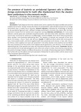
the induction was approximately fivefold for that both cell lines (data not shown). The BDNF-induced increase in t-PA mRNA at 4 h is blocked by actino- mycin D, suggesting that it is not an RNA stability effect, but rather due to an increase in the rate of transcription. By contrast, pretreatment with cycloh- eximide did not prevent this BDNF-mediated induc- tion, indicating that it is dependent upon changes in transcription factor activity rather than the synthesis of new transcription factors. These results are con- sistent with earlier observations in cultured mouse neurons [19].
Human t-PA promoter is regulated by BDNF in transfected mouse neurons
Neuronal expression of t-PA in the human brain is widespread [17,18]. Furthermore, neurotrophins acting via the TrkB receptor have been shown to increase t-PA mRNA levels in cultured neurons [19]. The TrkB-activating ligand, brain-derived neurotrophic fac- tor (BDNF), has also been identified as an essential modulator of synaptic plasticity [20,21]. BDNF-medi- ated increase in t-PA expression initiates a positive feedback loop, as t-PA, via plasmin formation, initi- ates the proteolytic processing of pro-BDNF into its active form [22].
lengths of
including
the multihormone
endogenous mouse
responsive
is can be
Studies on the transcriptional regulation of t-PA expression have revealed that the t-PA gene is regula- ted by a conserved core proximal promoter of (cid:2) 300 nucleotides [23], as well as by more distal enhancer elements, responsive enhancer (MHRE) between )7.1 and )8 kb [24]. Reg- ulatory regions within the t-PA promoter that convey BDNF-responsiveness are unknown. By transfecting primary mouse neurons with t-PA promoter–reporter gene constructs, we demonstrate that the human t-PA this promoter to BDNF and that response reconstituted in HEK 293 cells cotransfected with recombinant TrkB receptor. We have also defined t-PA promoter regions contributing to the BDNF response and provide evidence for the presence of multiple cooperating elements within 3 kb of the transcription start site, and for the functional conservation of an equivalent region in the mouse promoter.
Results
Having established a transcriptional component in the induction of t-PA biosynthesis by BDNF, we next examined BDNF activation of the human t-PA pro- moter in a primary neuronal cell system. To this end, primary cultures of mouse cortical neurons were tran- siently transfected with reporter constructs bearing different the human t-PA promoter (Fig. 2). BDNF treatment (20 ngÆmL)1) of cells trans- fected with constructs including 3 kb or more of pro- moter region (Fig. 3) produced sustained increased luciferase reporter gene expression between 2 and 2.5- fold, which is less than the estimated 7- to 9-fold induction reported for t-PA mRNA, as assessed by northern blotting [19]. None- theless, transfection experiments using truncations of the t-PA promoter showed a trend of decreasing BDNF responsiveness as the t-PA promoter sequence is shortened from )3069 to )238, with no BDNF induction of (pGL3Basic). the promoterless vector These results may also reflect the contribution of mul- tiple BNDF-responsive elements between )3069 and )238, as well as the proximal promoter region. The relatively small contribution from each region made further dissection of the specific component elements impractical.
Human t-PA gene expression is regulated by BDNF
The t-PA promoter responds to BDNF in a TrkB-dependent manner in HEK 293 cells
inhibitors of
the human t-PA transcript
Human neuroblastomal cell lines SY5Y and SK- N-SH were treated with BDNF for 4 or 8 h, with or transcription (actinomycin D) without or translation (cycloheximide). Specific mRNA levels were assessed using RNase protection assays. As shown in Fig. 1, is strongly induced after 4 h of BDNF treatment, but levels by 8 h. From quantitative returns to control it was estimated assessment of
three experiments
To further study the regulation of the human t-PA promoter by BDNF, we used a transient cotransfection system to reconstitute BDNF ⁄ TrkB signaling in human HEK 293 cells. As shown in Fig. 4, the 9.5 kb t-PA promoter conferred a 4.5-fold increase in reporter gene expression following BDNF treatment. Import- antly, this induction was critically dependent upon the cotransfection of TrkB, and did not occur with
FEBS Journal 274 (2007) 2411–2423 ª 2007 The Authors Journal compilation ª 2007 FEBS
2412
P. B. Daniel et al.
Regulation of t-PA transcription by BDNF
elements within the reporter vector or through post- transcriptional or post-translational effects of TrkB signaling.
An upstream enhancer contributes to the BDNF response in HEK 293 cells
Fig. 1. Induction of t-PA mRNA by BDNF in human neuroblastoma cells requires transcription but not translation. Human t-PA (tPA) and b-actin mRNA was detected in SY5Y and SK-N-SH cells by RNase protection assay following treatment of cells with BDNF for 4 or 8 h as indicated. Experiments were also performed in the pres- ence of 5 lgÆmL)1 actinomycin D or 5 lgÆmL)1 cycloheximide as indicated.
cotransfection of empty expression vector, thereby demonstrating successful reconstitution of the BDNF ⁄ TrkB signaling pathway in these cells. The promoter- less vector showed little or no response to BDNF, with or without TrkB, indicating that the effect was dependent on t-PA promoter elements, rather than
Analysis of the t-PA promoter-deletion series in the HEK 293-based system produced further evidence for the importance of cis-elements between )3069 and )238. As shown in Fig. 5A, BDNF induction is associ- ated with both the proximal promoter (up to )238; still conferring approximately two-fold induction) and the region between )3069 and )1566, with no signifi- cant contribution from the region upstream of )3069. That the p-3.07tpa construct retained BDNF-respon- siveness (in both transfected neurons and HEK 293 cells) also indicates that the NF-jB site at )3083 to )3074, which is absent from this construct, plays no role in the BDNF response. The pattern of response in HEK 293 cells differs from that observed in mouse cortical neurons, where deletion of the same region produced only a small, and not statistically significant, decrease in the BDNF response (Fig. 3). Nevertheless, the data indicate the presence of a potential enhancer element, possibly contributing only minor activity in
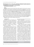
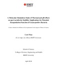
Fig. 2. The human t-PA promoter and derived reporter constructs. (A) The human t-PA promoter from )9.5 kb to the first intron with the relative positions of regula- tory elements and selected restriction enzyme sites indicated. (B) Schematic repre- sentation of the t-PA promoter 5¢ deletion series and an internal deletion mutant. The t-PA promoter variants were inserted into pGL3Basic that expresses the luciferase reporter gene. (C) Schematic representation of chimeric reporter constructs with upstream regions of the human t-PA promo- ter inserted into pGL3Promoter, which con- tains a minimal SV40-derived promoter and transcription start site (hatched box) fused to the luciferase reporter gene. The -3.07 ⁄ -2.93SV construct was prepared as a tandem construct, with two copies of the 135 bp region between )3069 and )2934 inserted in front of the minimal SV40 promoter.
FEBS Journal 274 (2007) 2411–2423 ª 2007 The Authors Journal compilation ª 2007 FEBS
2413
P. B. Daniel et al.
Regulation of t-PA transcription by BDNF
3.5
ns
ns
ns
*
*
3
2.5
2
n o i t c u d n
1.5
i
l
1
d o F
0.5
0
-9.5tpa
-3.2tpa
-3.07tpa
-1.5tpa
-238tpa
Basic
Construct
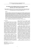
Fig. 3. The human t-PA promoter responds to BDNF stimulation in mouse cortical neurons. Mouse cortical neurons (days in vitro 10–12) were transfected with human t-PA reporter plasmids (-9.5tpa to -238tpa) or the promoterless pGL3Basic reporter plasmid (Basic). y-Axis values (fold induction) are the mean ratio of luciferase values for BDNF-treated wells to vehicle-treated wells after 8 h. ns, not sig- nificant. Asterisks indicate significant difference between indicated groups (P < 0.05; n ¼ 4 for all groups except for -3.07tpa, n ¼ 3).
Fig. 4. BDNF ⁄ TrkB regulation of the human t-PA promoter can be reconstituted in HEK 293 cells. HEK 293 cells were cotransfected with the reporter plasmids pGL3Basic or p-9.5tpa, along with expression plasmids pCIneo (neo) or pTrkB (TrkB). y-Axis values (fold induction) are the mean ratio of luciferase values for BDNF- treated wells to vehicle-treated wells after 8 h. ns, not significant. Asterisk indicates significant difference between indicated groups (P < 0.05, n ¼ 3).
mouse neurons, but with more important effects in other cell types or possibly species. To this end, the )3069 to )1566 region was selected for further analysis to define the minimal BDNF-responsive enhancer (BRE) in the context of HEK 293 cells.
Identification of multiple regions contributing to BDNF- Fig. 5. responsiveness in the human t-PA promoter. (A) HEK 293 cells were cotransfected with human t-PA promoter reporter plasmids representing a 5¢-deletion series starting at )9.5 kb and finishing at )238 bp, along with expression plasmid pTrkB. y-Axis values (fold induction) are the mean ratio of luciferase values for BDNF-treated wells to untreated wells after 8 h. ns, not significant. Asterisk indi- (B) HEK 293 cells cates significant difference (P < 0.05; n ¼ 3). were cotransfected with human t-PA promoter-driven reporter plas- mids representing 5¢ deletions and an internal deletion, along with expression plasmid pTrkB. y-Axis values (fold induction) are the mean ratio of luciferase values for BDNF-treated wells to untreated wells after 8 h. ns, not significant. Asterisk indicates significant dif- ference (P < 0.05; n ¼ 3). (C) HEK 293 cells were cotransfected with human t-PA promoter reporter plasmids representing a 5¢ dele- tion series starting at )3.05 kb and finishing at )1.5 kb, along with expression plasmid pTrkB. y-Axis values (fold induction) are the mean ratio of luciferase values for BDNF-treated wells to untreated wells after 8 h. ns, not significant. Asterisk indicates significant dif- ference (P < 0.05; n ¼ 3).
Using convenient PstI restriction sites, an internal deletion was generated that removed a 1.7 kb fragment between positions )2397 to )635. Co-transfection stud- ies revealed that this construct (pD-3.07tpa) exhibited
FEBS Journal 274 (2007) 2411–2423 ª 2007 The Authors Journal compilation ª 2007 FEBS
2414
P. B. Daniel et al.
Regulation of t-PA transcription by BDNF
BDNF-responsiveness similar to p-3.07tpa and this was significantly greater than the response produced from the p-1.5tpa construct (Fig. 5B). This further indicated the putative BRE resides between )3069 and that )2397. Further deletion experiments were performed on the region between )3069 and )2551. As shown in Fig. 5C, sequences between )3069 and )2963 contribu- ted to the response, but activity was also associated with sequence 3¢ of )2551. Given the results of Fig. 5B, any BDNF-responsive elements 5¢ of )2551 would be expected to reside between )2551 and )2397.
the results of Fig. 5C suggest that
Fig. 6. The human t-PA BDNF-responsive enhancer functions with a heterologous promoter. (A) HEK 293 cells were cotransfected with SV40 promoter reporter plasmids, some incorporating portions of the human t-PA promoter, along with expression plasmid pTrkB. y-Axis values (fold induction) are the mean ratio of luciferase values for BDNF-treated wells to untreated wells after 8 h. ns, not signifi- cant. Asterisk indicates significant difference (P < 0.05; n ¼ 3). (B) Primary neurons were transfected with SV40 reporter plasmid, or a human t-PA promoter ⁄ SV40 promoter construct. x-Axis values (fold induction) are the mean ratio of luciferase values for BDNF-treated wells to untreated wells after 8 h. Asterisk indicates significant dif- ference (P < 0.05; n ¼ 3).
To determine whether this upstream region of the t-PA promoter retained the capacity to independently confer BDNF-responsiveness, a 653 bp fragment con- taining the region between )3069 and )2416, and a 135 bp fragment containing only the region between )3069 and )2934 of the t-PA promoter were inserted separately into pGL3Promoter, a reporter construct containing a minimal SV40 promoter (Fig. 2C). Only the longer t-PA promoter fragment (p-3.07 ⁄ -2.42SV) was able to independently confer BDNF-mediated in- ducibility (Fig. 6A), even though the shorter fragment was tested as a tandem dimer. These results indicate the existence of at least two separate cis-elements ⁄ domains within the )3069 to )2416 region. The )3069 to )2963 region, although unable to independently confer BDNF responsiveness, may act cooperatively with the downstream region between )2551 to )2416. Conversely, the downstream region can confer some of the BDNF response without the presence of the upstream region. However, the relatively small contributions of both regions made further analysis problematic.
the presence of
We next decided to test independently the effective- ness of the putative BRE in the context of the heter- ologous reporter construct. As shown in Fig. 6B, the minimal SV40 promoter of pGL3Promoter was unre- the sponsive to BDNF however, 653 bp BRE in p-3.07 ⁄ -2.42SV conferred BDNF induction in primary cortical neurons, indicating func- tional significance for this t-PA promoter region.
The BRE is conserved across three mammalian species
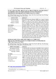
The vista plots presented in Fig. 7A identify substan- tial blocks of conserved sequence, which correspond to known regulatory elements. For instance, the human proximal promoter and first exon have a high degree of similarity with the orthologous mouse and bovine sequences (Fig. 7A, proximal area shaded in blue). In the area between )8 and the human promoter, )7.1 kb, previously identified as the MHRE [24], was also found to have similarity with both murine (between )4.8 and )4 kb) and bovine (between )5.2 and )4.5 kb) genomic DNA (area shaded in red). Sequence homology can arise from the presence of repeat elements, such as Alu repeats. vista software screening for such elements found no insertions of common repeat sequences within the MHRE, and only a short ((cid:2) 60 bp) fragment identified as part of a long interspersed nuclear element in the proximal promoter functionally (data not shown). This indicates that important regions within the t-PA promoter can exhi- bit significant cross-species sequence similarity.
No regulatory function has previously been demonstra- ted for the )3069 to )2416 region within the t-PA pro- moter. An important indicator for functional relevance would be sequence conservation between distantly rela- ted mammalian species [25]. To address this issue, we compared 9.0 kb of human, mouse and bovine t-PA genomic DNA, identified as t-PA promoter sequence by the UCSC genome browser, using lagan software.
FEBS Journal 274 (2007) 2411–2423 ª 2007 The Authors Journal compilation ª 2007 FEBS
2415
P. B. Daniel et al.
Regulation of t-PA transcription by BDNF
A
B
Fig. 7. Conserved regulatory regions in t-PA promoter from three species. (A) VISTA plots of comparisons between the human, mouse and bovine t-PA promoters. The position rel- ative to the start of transcription is given on the top axis in kb, and degree of similarity is indicated to the right. (a) Human promoter, mouse homology plotted; (b) mouse promo- ter, human homology plotted; (c) human promoter, bovine homology plotted; (d) bovine promoter, human homology plotted. Red domain, MHRE; blue domain, proximal promoter; yellow domain, BRE (also indica- ted with asterisks). (B) Human, bovine and murine t-PA promoter sequence from the conserved regions within the putative BRE. Stretches of sequence with high similarity in the human and bovine sequences detected by BLASTN tools are underlined. A canonical AP1 site present in the human and bovine species is also indicated in red. Regions of similarity in the mouse sequence are in smaller groups and are not indicated, except for a near-perfect consensus AP-1 site, which is indicated in red.
identifies
two small
still human and bovine t-PA promoters more conserved regions within 2 kb of the start of transcription.
start of
to the
Detection of repetitive DNA using vista software indicated the insertion of a short interspersed nuclear element between the upstream ()3 kb) and down- stream ()2.5 kb) segments of the BRE in the human t-PA promoter (data not shown). Indeed much of the expansion of the human t-PA promoter relative to those of the other species is a result of short inter- spersed nuclear element insertions.
(Fig. 7A,
together
closer
lower
In the area between )3.0 and )2.5 kb in the human t-PA promoter, coinciding with the experi- mentally defined BRE, segments were detected that are conserved among the three species (Fig. 7A, panels a and c, area marked with asterisk and highlighted in yellow). Within the mouse t-PA promoter, the corresponding regions are more closely linked and nearer transcription, between )1.30 and )1.55 kb. These regions are also conserved to a greater degree in the bovine genome, occurring between )3.5 and )3 kb (Fig. 7A, panel d, highlighted in yellow). As in the mouse, these two regions are two panels).
Downstream of
(Fig. 7B; 40 of 46 nucleotides
the BRE, a conserved region between )2.5 and )2.0 kb ()1.2 to )1.0 kb in the mouse t-PA promoter, and )3.0 to )2.5 kb in the bovine promoter) coincides with a promoter region reported to drive high-level t-PA expression in a mel- line [26], and the comparison between anoma cell
The similarity between mouse and human BRE appears to the eye as short fragments of identical sequence, which the blastn algorithm fails to detect (not shown). The higher level of conservation between bovine and human BRE appears to arise from two in the two extended motifs that are nearly identical the for promoters upstream and 37 of 41 nucleotides for the down- stream), and are detected using blastn.
FEBS Journal 274 (2007) 2411–2423 ª 2007 The Authors Journal compilation ª 2007 FEBS
2416
P. B. Daniel et al.
Regulation of t-PA transcription by BDNF
The mouse BRE confers BDNF-responsiveness to the human promoter
To confirm the bioinformatic analyses, we subcloned the putative BDNF-responsive region from the mouse t-PA promoter (nucleotides )1620 to )1261) into a vector already containing the proximal human t-PA promoter (p-238tpa) and tested the chimeric reporter construct in HEK 293 cells. In cotransfection experi- ments with pTrkB, pmBRE ⁄ -238tpa had a significantly increased response to BDNF stimulation compared with p-238tpa or p-1.5tpa (Fig. 8), proving that the region of the mouse t-PA promoter identified as partly homologous to the human t-PA promoter BRE can mediate activation by BDNF.
Discussion
We have transiently transfected primary cultures of mouse cortical neurons and also HEK 293 cells with t-PA promoter–reporter gene constructs to identify BDNF-responsive regions within the t-PA promoter. On the basis of promoter truncation and deletion analyses, we have defined at least two discrete domains within the t-PA promoter that contribute to the BDNF response. One of these maps to the proximal promoter, a region previously implicated in the response of the t-PA promoter to other effectors [29]. The second domain is located within a 653 bp region between )3069 and )2416, and contributes significantly to the BDNF-mediated regulation of human t-PA promoter in both primary cortical neurons and HEK 293 cells expressing TrkB. This second domain is comprised of two distinct contributing regions. The more 5¢ region located within a 135 bp fragment, is unable to confer BDNF-responsiveness independently, but may coope- rate with a downstream region that resides between positions )2551 and )2416. The entire 653 bp upstream domain is partly conserved among human, mouse and bovine t-PA gene promoters.
BDNF released from glutamatergic synapses is capable of signaling pre- and post-synaptically through the TrkB receptor, and is known to modulate LTP in part via modulating the expression of downstream target genes [27], including t-PA. t-PA itself is now recog- nized as a modulator of neuronal transmission and synaptic plasticity [28], and a model has been proposed that links t-PA as a downstream effector of BDNF [20]. In this study, we demonstrate that BDNF induces a transient induction of endogenous t-PA expression. This occurs within 4 h of treatment, includes a tran- scriptional component and is independent of on-going protein synthesis.
We note that the magnitude and duration of the response of the endogenous t-PA gene to BDNF was different to the response of the transfected t-PA pro- moter-driven luciferase gene. Whereas levels of t-PA mRNA in mouse neurons and human neuroblastomal cell lines reach a peak after 3–4 h of BDNF stimula- tion, and decline thereafter, luciferase activity from t-PA promoter reporter constructs continues to accu- mulate during the 8 h stimulation, both in transfected mouse neurons and HEK 293 cells. It is important to note that luciferase reporter constructs respond only to inducible transcription factors acting upon included cis-elements. Gene expression can also be governed by post-transcriptional mechanisms via elements within the 3¢-UTR. Because the reporter gene does not include the 3¢-UTR of the t-PA transcript, the normal post-transcriptional regulatory processes that influence the decay rate of t-PA mRNA are absent. This may explain, at least in part, why there are differences in the regulation of the endogenous t-PA gene and the transfected reporter gene to BDNF. Also, we cannot exclude the possibility that the differences in respon- siveness of the t-PA promoter to BDNF between the cell lines and the primary neurons are due to differ- ences in levels of total or phosphorylated TrkB.
Fig. 8. The mouse t-PA BDNF-responsive enhancer functions the human proximal t-PA promoter. HEK 293 cells were transfected with human t-PA promoter reporter plasmids as indicated together with plasmid pTrkB. y-Axis values (fold induction) are the mean ratio of luciferase values for BDNF-treated wells to untreated wells after 8 h. ns, not significant. Asterisk indicates significant difference (P < 0.05; n ¼ 4).
The relatively small response of the t-PA promoter to BDNF made it problematic to define the specific cis-acting element responsible for the induction. Also, as our data indicate, it is likely that the BDNF-medi- ated induction is a cooperative response of more than the one element(s)
located in different regions of
FEBS Journal 274 (2007) 2411–2423 ª 2007 The Authors Journal compilation ª 2007 FEBS
2417
P. B. Daniel et al.
Regulation of t-PA transcription by BDNF
was particularly strong in the hippocampus for both the endogenous t-PA mRNA [39] and the transgene. Transgenic mice incorporating 4 kb of mouse t-PA promoter with the same reporter gene also exhibit some CNS expression, primarily in the hippocampus [40], further suggesting conservation of functional ele- ments defining tissue-specific expression. A transgenic line with a shorter human t-PA promoter fragment (1.4 kb) had substantially curtailed CNS expression, suggesting that as yet undefined elements between )3 and )1.4 kb are essential for widespread expression in the mouse brain [38]. The BRE we have identified also resides within this region, but whether it is involved in constitutive neuronal t-PA expression remains an open question.
some responsiveness
t-PA promoters. However,
three human cell
site for
promoter. We initially considered the recently identi- fied functional jB site located at )3083 to )3074 in the human t-PA promoter [30] as a candidate BDNF- responsive element since NF-jB is considered to be a downstream target of TrkB activation [31]. However, our promoter-deletion analysis excludes a major role for this element given the sustained BDNF-responsive- ness of the p-3.07tpa construct in both primary neu- rons and HEK 293 cells. Other transcription factors reportedly activated by TrkB include CREB [32,33], AP1 [32], STAT5 [34], and NFATc4 [35]. The upstream and downstream segments of the human, mouse and bovine BRE were analyzed with transcrip- tion factor binding-site prediction software tess to identify likely consensus sites. Emphasis was placed on signal activated transcription factors common to all three species. One attractive candidate for rapidly inducible transcription factors is AP1, for which a con- sensus response element (5¢-TGACTCA-3¢) is found in both human ()2430 to )2424) and bovine ()3066 to )3060) this site is only partly conserved in the mouse promoter (5¢-TGACA CA-3¢; located )1304 to )1298). The region surround- ing this AP-1 site has been analyzed by in vivo foot- print analysis, and protected residues were found in lines and around this (HepG2, SK-N-SH and SNB-19 cells) [30], suggesting it may be an important regulatory element.
Our findings also show that the proximal promoter to BDNF. The t-PA retains proximal promoter is controlled, at least in part, by a CRE-like element present between )227 and )220 and this element has been shown to provide a binding site for the transcription factor CREB [29]. Because for CREB is also a recognized downstream target BDNF, it is reasonable to suggest that the stimulatory effect of BDNF on the proximal promoter may involve CREB. The relationship between cAMP and BDNF, two factors implicated in the formation of LTP, involves some cross-talk. It has been suggested that in cerebellar neurons BDNF can stimulate cAMP accumulation [41]. In hippocampal neurons, however, this was not observed [42] and instead cAMP was shown to be permissive for BDNF-induced translation of synaptic proteins. Finally, phosphorylation of TrkB in response to BDNF was found to be enhanced by cAMP but reduced by inhibitory cAMP analogs [43]. It would be interesting to observe the combined effects of cAMP and BDNF on our reporter con- structs and to determine the contribution of the BRE in this response.
localization,
Also of interest is a 5¢ nucleotide motif (5¢-TTT CC-3¢) in all three promoters in positions within the downstream BRE segment (human: )2565 to )2561; mouse: )1443 to )1439; bovine: )3162 to )3158). tess identifies this as the core of a CCAAT ⁄ enhancer-bind- ing protein beta site (C ⁄ EBPb) in human and bovine promoter, or as a c-Ets-2 site in the mouse promoter. A separate potential C ⁄ EBPb site was also identified in the mouse promoter (5¢-CTGGGAA-3¢; )1429 to )1422). The C ⁄ EBP family of transcription factors is diverse and signal activated [36], but to date has not been linked to neurotrophin responses. The 5¢-TTTCC-3¢ motif is also a common component of the binding site for nuclear factor of activated T cells (NFAT) family members [37]. These are Rel-related transcription factors activated by calcineurin-regulated nuclear least one member and at (NFATc4) has been shown to be activated as a conse- quence of TrkB signaling [35].
Results from studies with transgenic mice harboring the human t-PA promoter driving a lacZ reporter gene suggest some conservation of functional elements defi- ning neuronal expression. Extensive CNS expression of the transgene was observed in animals harboring 9.5 and 3 kb of human t-PA promoter [38]. Expression
Our study of the human t-PA promoter and its potential for mediating BDNF-inducible expression corroborates the recently described effect of neurotro- phins on mouse t-PA mRNA levels in neuronal cul- tures [19]. This earlier study also indicated that BDNF was unable to increase expression of t-PA inhibitors, including neuroserpin and protease nexin-1, but did increase levels of expression of plasminogen activator inhibitor type 2 (PAI-2) which is a relatively poor inhibitor of t-PA. The overall effect of BDNF was shown to be a net increase in proteolytic activity, as determined by fibrin zymography. Hence it is likely that BDNF induction of t-PA would result in a net increase in plasminogen activation, at least in regions containing t-PA and TrkB-positive neurons. This
FEBS Journal 274 (2007) 2411–2423 ª 2007 The Authors Journal compilation ª 2007 FEBS
2418
P. B. Daniel et al.
Regulation of t-PA transcription by BDNF
Neuroblastoma cultures and ribonuclease protection assays
increase in t-PA activity, and presumably in plasmin formation, may have a bearing on the mechanisms by which BDNF modulates LTP because t-PA has been postulated as a mediator of many of the long-term effects underlying L-LTP and memory formation [11,13,44].
A 172 bp human t-PA probe for ribonuclease protection assays was amplified from a t-PA cDNA clone using the 5¢-GCTCTAGAGCGCCCAGATAGTCCCCAG primers ATC-3¢ and 5¢-CGGGATCCCGTGTCTGAACGATGGC CGCATG-3¢, and cloned into Bluescript II SK vector. Ra- diolabeled antisense RNA was generated from BamHI- digested vector with the MAXIscript (Ambion, Austin, TX) in vitro transcription system. A 127 bp b-actin control probe was similarly generated from pTRI-b-actin-human plasmid (Ambion).
The enhancer activity we describe in this study may also have wider significance than neurotrophin recep- tor-mediated regulation, and may be important for the effects of other tyrosine kinase receptors known to upregulate t-PA mRNA in endothelial cells. This includes receptors for vascular endothelial growth fac- (bFGF, tor and basic fibroblastic growth factor FGF2) [45]. Whether the BRE we have identified in this study is involved in the response of the t-PA gene to other tyrosine kinase receptors awaits further study. In considering the significance of mutations within the t-PA promoter, it is noteworthy that t-PA null mice survive to maturity and breed effectively, with only moderate effects on vascular fibrinolysis [46]. The effects on behavior are more noticeable, with impacts on learning and memory [9,14–16] and stress-induced anxiety [47]. Whether these are the consequence of reduced fibrinolysis within the CNS [48,49] or from other mechanisms [28], remains to be elucidated. The t-PA levels on human behavior are not effects of known because no homozygous inactivating mutation of human t-PA has been reported to date.
von Mikroorganismen
Reporter and expression plasmids
The results we observed studying human neuroblas- lines (Fig. 1) are very similar to those tomal cell observed for primary mouse cortical neurons, while our in vitro reporter gene systems reveal a less pro- nounced but still significant degree of stimulation by BDNF. We conclude that rather than being controlled by a single transcription factor or cis-acting element, the t-PA promoter response to BDNF is highly modu- lar in nature, with multiple elements contributing in concert to the observed regulation.
The SK-N-SH cell line was obtained from the American Type Culture Collection (Manassas, VA) and cultured in DMEM with Glutamax I, pyruvate and 10% fetal bovine serum. SH-SY5Y cells were obtained from the Deutsche Sammlung und Zellkulturen (DSMZ; Braunschweig, Germany) and were cultured in modified Eagle’s medium:F12 with nonessential amino acids and 10% fetal bovine serum. Cells used were semiconfluent, nonsynchronized and undifferentiated. They were stimula- ted with BDNF for 4 or 8 h, or for 4 h following 30 min pretreatment with either cycloheximide or actinomycin D (both 5 lgÆmL)1, Sigma-Aldrich). Ribonuclease protection assays were performed with the Direct Protect kit (Ambi- on), and protected fragments were resolved with PAGE. Dried gels were visualized with a PhosphorImager SI (Amersham Pharmacia Biotech, Molecular Dynamics Division, Piscataway, NJ).
The p-9.5tpa luciferase reporter plasmid was created in pGL3Basic by insertion of a 9.8 kb XhoI fragment from a ptPA9578CAT plasmid [24] modified to include an XhoI site at the 5¢-end of the human t-PA promoter sequence. It includes over 9.5 kb of promoter sequence, the two tran- scription start sites and first exon (183 or 68 nucleotides depending on start site) and 131 nucleotides of first intron sequence inserted into the pGL3 Basic reporter vector (Promega, Madison, WI).
Experimental procedures
BDNF
Inc. Reporter plasmids p-3.2tpa and p-1.5tpa were subcloned from p-9.5tpa as HindIII and BamHI fragments, respect- ively, inserted into the HindIII or BglII sites of pGL3 Basic and checked for correct orientation. The p-3.07tpa reporter plasmid was derived from p-3.2tpa by NheI ⁄ XbaI digestion to excise the 5¢-end of the promoter including a functional NF-jB site [30], followed by religation. The internal dele- tion construct (pD-3.07tpa) was produced from p-3.07tpa by complete digestion with PstI and religation. This dele- tion removed 1761 bp sequence between )2397 and )635.
FEBS Journal 274 (2007) 2411–2423 ª 2007 The Authors Journal compilation ª 2007 FEBS
2419
also at a final Other constructs were derived from p-3.2tpa or p-9.5tpa by PCR using the reverse primer GL3R1 (5¢-CTT CCAGCGGATAGAATGG-3¢; residues 142–124 [reverse BDNF used in these experiments was obtained from three sources. For ribonuclease protection assays (Fig. 1), BDNF was obtained from Sigma-Aldrich (St Louis, MO), and was used at a final concentration of 5 ngÆmL)1. For the experi- ments in Figs 3–6, BDNF was obtained from R&D Sys- tems (Minneapolis, MN), and used at a final concentration of 20 ngÆmL)1. Experiments in Fig. 8 were performed with BDNF from Chemicon International (Temecula, CA), concentration of 20 ngÆmL)1.
P. B. Daniel et al.
Regulation of t-PA transcription by BDNF
strand] within pGL3Basic) and specific forward primers (p-238tPA: 5¢-CTGCGATTCAATGACATCACGGCTGT GAAT-3¢; p-2.96tpa: 5¢-AGTTTAGACACAGAATGTCC-3¢; p-2.75tPA: 5¢-TGTAATCTCAGCTACTCAGG-3¢; p-2.55tPA: 5¢-GACATCATTTCTTACCCAGC-3¢). Reactions were 50 lL total volume with 2 ng of template with 2.5 mm each of dNTPs and 0.2 units of Vent polymerase (New England Biolabs, Ips- wich, MA). Reactions were denatured for 2 min at 94 (cid:2)C and amplified over 25 cycles of 94 (cid:2)C for 5 s, 54 (cid:2)C for 10 s and 72 (cid:2)C for 2–4 min, depending on product size. A final extension of 2 min at 72 (cid:2)C was also employed. Products were cut with HindIII and ligated into pGL3Basic using SmaI and HindIII sites.
the coding sequence retaining the (AA11),
The product was used as a template for a further amplifica- tion with Vent polymerase under the same conditions for 16 cycles. PCR product was purified and digested with EcoRI and XbaI, then ligated into the corresponding sites of pCIneo (Promega; GenBank accession number U47120). Two clones capable of mediating BDNF upregulation of p-9.5tpa transcription were identified by functional assay and sequenced. The clone used in experiments presented here contained a single amino acid difference (AA306 Val to Leu) compared with the mouse TrkB protein (GenBank accession number X17647), while the other functional TrkB clone has two amino acid changes (AA46 Thr to Ile; AA376 Thr to Ala). Both clones are also truncated at the 5¢-end, with translation starting at the second methionine of signal sequence cleavage site. In comparative experiments, both clones mediated BDNF responsiveness, indicating that these amino acid changes did not compromise function.
Neuronal culture and transfections and luciferase assay
All oligonucleotides were obtained from Proligo Australia (Lismore, Australia). Sequencing was performed by AgGe- nomics (Bundoora, Australia). All plasmid constructs exclu- ding pmBRE ⁄ -238tpa and pTrkB are described in Fig. 2.
Heterologous reporter constructs were created by PCR amplification of fragments from p-3.2tpa using forward pri- mer GL3F1 (5¢-CCAGTGCAAGTGCAGGTG-3¢, identical to residues 4779–4796 in pGL3Basic) and reverse primers for 5¢-CTAGAATGCATGAGTCATGGAGGTGGCG-3¢ p-3.07 ⁄ -2.42SV and 5¢-CCCGCTAGCCCAGAAGACGGA CATTCTG-3¢ for p-3.07 ⁄ -2.93SV, with PCR and condi- tions as described above. The t-PA promoter fragment for p-3.07 ⁄ -2.42SV was digested with XbaI and cloned into the NheI and SmaI sites of pGL3Promoter. For p-3.07 ⁄ -2.93Prom, the t-PA promoter fragment was digested with both NheI and XbaI and ligated into the NheI site of pGL3Prom. Correctly oriented clones (5¢ to 3¢ relative to the luciferase gene) were selected by PCR. The clone of p-3.07 ⁄ -2.93SV used in the experiments in Fig. 6A had two identical 135 bp tandem repeats of the t-PA promoter fragment.
transfection was
Primary cortical neurons were prepared from trypsin- dissociated E15 mouse embryo cortices, plated on poly (d-lysine)-coated 24-well plates. Cultures were maintained in neurobasal media with 2.5% B27 supplement (Invitro- gen) in a humidified 5% CO2, 8% O2 incubator. Cultures were utilized for transfection between 10 and 12 days in vitro. Eight hours prior to transfection media was replaced with antibiotic-free neurobasal media +2% B27, and accomplished with Lipofecta- mine 2000 (Invitrogen), used at 2 lL per well with 1 lg of plasmid DNA combined in 100 lL of neurobasal media. Cultures were treated with agonist 18–20 h later in fresh neurobasal media +2% B27, and harvested after 8 h of treatment.
The chimeric reporter construct pmBRE ⁄ -238tpa contain- ing the putative mouse BRE upstream of the proximal human t-PA promoter was created as follows. A PCR product was amplified from mouse genomic DNA with the primers mBREF (5¢-AAATCTAGATGGCATGCATGA GGAGTCC-3¢) and mBRER (5¢-AAAGCTAGCAAGG CTAGCTTCTGCAATGG-3¢), producing a 360 bp product from )1620 to )1261. Amplification was carried out with Taq DNA polymerase (Invitrogen, Carlsbad, CA) for 35 cycles of 94 (cid:2)C for 5 s, 55 (cid:2)C for 10 s, 72 (cid:2)C for 40 s, fol- lowed by reamplification of the purified product with Vent polymerase (20 cycles of 94 (cid:2)C for 5 s, 58 (cid:2)C for 10 s). PCR product was digested with NheI and XbaI and ligated into the NheI site of p-238tpa. Correctly oriented clones were identified by PCR and the mBRE insert then verified by sequencing.
Cultures were treated with agonist 18–20 h later in fresh neurobasal media +2% B27, and harvested after 8 h of treatment in 200 lL per well of Cell Lysis Buffer (Promega). luciferase assays were performed in a Wallac Victor-21420 multilabel plate reader (Perkin–Elmer, Norwalk, NJ) by injecting 20 lL of lysate with 20 lL luciferase assay reagent (Promega) and determining emitted light (counts per sec- ond) for a period of 1 s.
FEBS Journal 274 (2007) 2411–2423 ª 2007 The Authors Journal compilation ª 2007 FEBS
2420
All results are presented as ‘fold-induction’. This was cal- culated from the average of the luciferase value obtained from BDNF-treated wells divided by the average luciferase value obtained from the control wells. Control wells were either the same construct treated with either vehicle (0.1% BSA) or left untreated, as indicated in figure legends. The expression vector for TrkB (pTrkB) was constructed using a PCR amplified TrkB coding sequence from mouse cDNA using primers TRKBF (5¢-AAAGAATTCCCTGG CTGAAGTGGCATGGACC-3¢) and TRKBR (5¢-AAA TCTAGATGGGCAGAAGGAGGACCCTAGC-3¢). Vent polymerase was used to amplify TrkB from mouse brain cDNA. PCR conditions were an initial denaturation of 94 (cid:2)C for 2 min followed by 35 cycles of 94 (cid:2)C for 10 s and 72 (cid:2)C for 4 min, with a final extension of 72 (cid:2)C for 2 min.
P. B. Daniel et al.
Regulation of t-PA transcription by BDNF
References
Statistically significant
Transfection of HEK 293 cells
Results shown are the average of at least three independent experiments. The bar graphs show results as mean fold induction ± SD. differences (P < 0.05) were identified using a 2-tailed t test. Nonsignif- icant comparisons are indicated as ‘ns’. 1 O’Rourke J, Jiang X, Hao Z, Cone RE & Hand AR (2005) Distribution of sympathetic tissue plasminogen activator (tPA) to a distant microvasculature. J Neurosci Res 79, 727–733.
2 Hao Z, Guo C, Jiang X, Krueger S, Pietri T, Dufour S, Cone RE & O’Rourke J (2006) New transgenic evidence for a system of sympathetic axons able to express tissue plasminogen activator (t-PA) within arterial ⁄ arteriolar walls. Blood 108, 200–202. 3 Parmer RJ, Mahata M, Mahata S, Sebald MT,
O’Connor DT & Miles LA (1997) Tissue plasminogen activator (t-PA) is targeted to the regulated secretory pathway. Catecholamine storage vesicles as a reservoir for the rapid release of t-PA. J Biol Chem 272, 1976– 1982.
4 Verrall S & Seeds NW (1988) Tissue plasminogen acti- vator binding to mouse cerebellar granule neurons. J Neurosci Res 21, 420–425.
5 Qian Z, Gilbert ME, Colicos MA, Kandel ER & Kuhl D (1993) Tissue-plasminogen activator is induced as an immediate-early gene during seizure, kindling and long- term potentiation. Nature 361, 453–457.
HEK 293 cells were transfected in 24- or 48-well plates. For 24-well plates, 200 ngÆwell)1 of reporter plasmid and 1 ngÆwell)1 of expression plasmid using 0.6 lLÆwell)1 of Fugene 6 reagent (Roche Diagnostics, Indianapolis, IN) combined in 20 lL well)1 serum-free DMEM were added to wells containing 2 · 105 HEK 293 cells plated 6–8 h pre- viously in 0.5 mL of DMEM ⁄ 10% iron-fortified calf serum with antibiotics. Stimulations were performed the following day in ambient media by adding agonist in 10 lL of serum- free media per well. Cells were harvested 8 h after simula- tion in cell lysis buffer (95 lLÆwell)1; Promega) and frozen prior to assay for luciferase activity (above). Reagent vol- umes and quantities were reduced by half for experiments performed in 48 well plates. Experiments were performed and analyzed as described above for neuronal cultures, except that treatments were performed in triplicate. Results shown are the average of either three or four separate experiments as indicated in the figure legends.
Bioinformatic analysis
6 Popa-Wagner A, Fischer B, Platt D, Schmoll H & Kess- ler C (2000) Delayed and blunted induction of mRNA for tissue plasminogen activator in the brain of old rats following pentylenetetrazole-induced seizure activity. J Gerontol A Biol Sci Med Sci 55, B242–B248.
7 Seeds NW, Williams BL & Bickford PC (1995) Tissue plasminogen activator induction in Purkinje neurons after cerebellar motor learning. Science 270, 1992–1994.
Genomic sequences were obtained from UCSC genome browser [50] (http://genome.ucsc.edu/index.html). The fol- lowing assemblies were used: the May 2004 human refer- ence sequence (NCBI Build 35); the March 2005 Bos taurus draft assembly (Btau_2.0); the February 2006 mls musculus genome data (Build 36). Comparisons of
vista suite software
8 Napolitano M, Marfia GA, Vacca A, Centonze D, Bellavia D, Di Marcotullio L, Frati L, Bernardi G, Gulino A & Calabresi P (1999) Modulation of gene expression following long-term synaptic depression in the striatum. Brain Res Mol Brain Res 72, 89–96. 9 Calabresi P, Napolitano M, Centonze D, Marfia GA, Gubellini P, Teule MA, Berretta N, Bernardi G, Frati L, Tolu M et al. (2000) Tissue plasminogen activator controls multiple forms of synaptic plasticity and mem- ory. Eur J Neurosci 12, 1002–1012.
t-PA promoter DNA from different species were carried out using lagan software [51] from the (http://genome.lbl.gov/vista/ index.shtml). Conserved regions were analyzed for potential transcription factor binding sites using the tess program [52] (http://www.cbil.upenn.edu/cgi-bin/tess/tess). Nucleo- tide alignment of sequences was performed with the blastn program through the National Centre for Biotechnology (http://www.ncbi.nlm.nih.gov/blast/ Information website bl2seq/wblast2.cgi).
Acknowledgements
supported with grants
10 Frey U, Muller M & Kuhl D (1996) A different form of long-lasting potentiation revealed in tissue plasminogen activator mutant mice. J Neurosci 16, 2057–2063. 11 Huang YY, Bach ME, Lipp HP, Zhuo M, Wolfer DP, Hawkins RD, Schoonjans L, Kandel ER, Godfraind JM, Mulligan R et al. (1996) Mice lacking the gene encoding tissue-type plasminogen activator show a selective interference with late-phase long-term potentia- tion in both Schaffer collateral and mossy fiber path- ways. Proc Natl Acad Sci USA 93, 8699–8704. 12 Centonze D, Napolitano M, Saulle E, Gubellini P,
This project was to RLM obtained from the National Health and Medical Research Council of Australia (Canberra, Australia). The authors also would like to acknowledge technical assistance from Jonathan Luk, Prue Hogg and Marina Demyanenko.
FEBS Journal 274 (2007) 2411–2423 ª 2007 The Authors Journal compilation ª 2007 FEBS
2421
Picconi B, Martorana A, Pisani A, Gulino A, Bernardi G & Calabresi P (2002) Tissue plasminogen activator is
P. B. Daniel et al.
Regulation of t-PA transcription by BDNF
required for corticostriatal long-term potentiation. Eur J Neurosci 16, 713–721. far upstream from the human tissue-type plasminogen activator gene. J Biol Chem 272, 663–671. 13 Baranes D, Lederfein D, Huang YY, Chen M, Bailey 25 Dubchak I, Brudno M, Loots GG, Pachter L, Mayor
C, Rubin EM & Frazer KA (2000) Active conservation of noncoding sequences revealed by three-way species comparisons. Genome Res 10, 1304–1306. CH & Kandel ER (1998) Tissue plasminogen activator contributes to the late phase of LTP and to synaptic growth in the hippocampal mossy fiber pathway. Neu- ron 21, 813–825. 14 Ripley TL, Horwood JM & Stephens DN (2001)
26 Fujiwara J, Kimura T, Ayusawa D & Oishi M (1994) A novel regulatory sequence affecting the constitutive expression of tissue plasminogen activator (tPA) gene in human melanoma (Bowes) cells. J Biol Chem 269, 18558–18562. Evidence for impairment of behavioural inhibition in performance of operant tasks in tPA– ⁄ – mice. Behav Brain Res 125, 215–227.
27 Bramham CR & Messaoudi E (2005) BDNF function in adult synaptic plasticity: the synaptic consolidation hypothesis. Prog Neurobiol 76, 99–125. 28 Samson AL & Medcalf RL (2006) Tissue-type plas- 15 Pawlak R, Nagai N, Urano T, Napiorkowska-Pawlak D, Ihara H, Takada Y, Collen D & Takada A (2002) Rapid, specific and active site-catalyzed effect of tissue- plasminogen activator on hippocampus-dependent learn- ing in mice. Neuroscience 113, 995–1001.
minogen activator: a multifaceted modulator of neuro- transmission and synaptic plasticity. Neuron 50, 673– 678. 29 Costa M & Medcalf RL (1996) Differential binding of 16 Seeds NW, Basham ME & Ferguson JE (2003) Absence of tissue plasminogen activator gene or activity impairs mouse cerebellar motor learning. J Neurosci 23, 7368– 7375. 17 Scarisbrick IA, Isackson PJ, Ciric B, Windebank AJ &
Rodriguez M (2001) MSP, a trypsin-like serine protease, is abundantly expressed in the human nervous system. J Comp Neurol 431, 347–361. 18 Teesalu T, Kulla A, Simisker A, Siren V, Lawrence DA, cAMP-responsive-element (CRE)-binding protein-1 and activating transcription factor-2 to a CRE-like element in the human tissue-type plasminogen activator (t-PA) gene promoter correlates with opposite regulation of t-PA by phorbol ester in HT-1080 and HeLa cells. Eur J Biochem 237, 532–538. 30 Lux W, Klobeck HG, Daniel PB, Costa M, Medcalf
Asser T & Vaheri A (2004) Tissue plasminogen activator and neuroserpin are widely expressed in the human central nervous system. Thromb Haemostat 92, 358–368. 19 Fiumelli H, Jabaudon D, Magistretti PJ & Martin JL RL & Schleuning WD (2005) In vivo and in vitro analy- sis of the human tissue-type plasminogen activator gene promoter in neuroblastomal cell lines: evidence for a functional upstream kappaB element. J Thromb Haemo- stat 3, 1009–1017. 31 Jiang X, Zhu D, Okagaki P, Lipsky R, Wu X, (1999) BDNF stimulates expression, activity and release of tissue-type plasminogen activator in mouse cortical neurons. Eur J Neurosci 11, 1639–1646.
20 Pang PT & Lu B (2004) Regulation of late-phase LTP and long-term memory in normal and aging hippocam- pus: role of secreted proteins tPA and BDNF. Ageing Res Rev 3, 407–430. Banaudha K, Mearow K, Strauss KI & Marini AM (2003) N-Methyl-d-aspartate and TrkB receptor activa- tion in cerebellar granule cells: an in vitro model of pre- conditioning to stimulate intrinsic survival pathways in neurons. Ann NY Acad Sci 993, 134–145 [discussion 159–160]. 21 Barco A, Patterson S, Alarcon JM, Gromova P, 32 Gaiddon C, Loeffler JP & Larmet Y (1996) Brain-
Mata-Roig M, Morozov A & Kandel ER (2005) Gene expression profiling of facilitated L-LTP in VP16-CREB mice reveals that BDNF is critical for the maintenance of LTP and its synaptic capture. Neuron 48, 123–137. 22 Pang PT, Teng HK, Zaitsev E, Woo NT, Sakata K, derived neurotrophic factor stimulates AP-1 and cyclic AMP-responsive element dependent transcriptional activity in central nervous system neurons. J Neurochem 66, 2279–2286.
Zhen S, Teng KK, Yung WH, Hempstead BL & Lu B (2004) Cleavage of proBDNF by tPA ⁄ plasmin is essen- tial for long-term hippocampal plasticity. Science 306, 487–491. 33 Finkbeiner S, Tavazoie SF, Maloratsky A, Jacobs KM, Harris KM & Greenberg ME (1997) CREB: a major mediator of neuronal neurotrophin responses. Neuron 19, 1031–1047.
34 Klein M, Hempstead BL & Teng KK (2005) Activation of STAT5-dependent transcription by the neurotrophin receptor Trk. J Neurobiol 63, 159–171.
23 Feng P, Ohlsson M & Ny T (1990) The structure of the TATA-less rat tissue-type plasminogen activator gene. Species-specific sequence divergences in the promoter predict differences in regulation of gene expression. J Biol Chem 265, 2022–2027.
FEBS Journal 274 (2007) 2411–2423 ª 2007 The Authors Journal compilation ª 2007 FEBS
2422
24 Bulens F, Merchiers P, Ibanez-Tallon I, De Vriese A, Nelles L, Claessens F, Belayew A & Collen D (1997) Identification of a multihormone responsive enhancer 35 Groth RD & Mermelstein PG (2003) Brain-derived neu- rotrophic factor activation of NFAT (nuclear factor of activated T-cells)-dependent transcription: a role for the transcription factor NFATc4 in neurotrophin-mediated gene expression. J Neurosci 23, 8125–8134.
P. B. Daniel et al.
Regulation of t-PA transcription by BDNF
45 Pepper MS, Rosnoblet C, Di Sanza C & Kruithof EK
36 Kalvakolanu DV & Roy SK (2005) CCAAT ⁄ enhancer binding proteins and interferon signaling pathways. J Interferon Cytokine Res 25, 757–769. 37 Badran BM, Wolinsky SM, Burny A & Willard-Gallo
(2001) Synergistic induction of t-PA by vascular endothelial growth factor and basic fibroblast growth factor and localization of t-PA to Weibel–Palade bodies in bovine microvascular endothelial cells. Thromb Hae- mostat 86, 702–709.
KE (2002) Identification of three NFAT binding motifs in the 5¢-upstream region of the human CD3gamma gene that differentially bind NFATc1, NFATc2, and NF-kappa B p50. J Biol Chem 277, 47136–47148.
46 Carmeliet P, Schoonjans L, Kieckens L, Ream B, Degen J, Bronson R, De Vos R, van den Oord JJ, Collen D & Mulligan RC (1994) Physiological consequences of loss of plasminogen activator gene function in mice. Nature 368, 419–424. 47 Pawlak R, Magarinos AM, Melchor J, McEwen B & 38 Yu H, Schleuning WD, Michl M, Liberatore G, Tan SS & Medcalf RL (2001) Control elements between )9.5 and )3.0 kb in the human tissue-type plasminogen acti- vator gene promoter direct spatial and inducible expres- sion to the murine brain. Eur J Neurosci 14, 799–808. 39 Sappino AP, Madani R, Huarte J, Belin D, Kiss JZ, Strickland S (2003) Tissue plasminogen activator in the amygdala is critical for stress-induced anxiety-like beha- vior. Nat Neurosci 6, 168–174. 48 Akassoglou K, Kombrinck KW, Degen JL &
Strickland S (2000) Tissue plasminogen activator- mediated fibrinolysis protects against axonal degenera- tion and demyelination after sciatic nerve injury. J Cell Biol 149, 1157–1166. Wohlwend A & Vassalli JD (1993) Extracellular proteo- lysis in the adult murine brain. J Clin Invest 92, 679–685. 40 Carroll PM, Tsirka SE, Richards WG, Frohman MA & Strickland S (1994) The mouse tissue plasminogen acti- vator gene 5¢ flanking region directs appropriate expres- sion in development and a seizure-enhanced response in the CNS. Development 120, 3173–3183.
49 East E, Baker D, Pryce G, Lijnen HR, Cuzner ML & Gveric D (2005) A role for the plasminogen activator system in inflammation and neurodegeneration in the central nervous system during experimental allergic encephalomyelitis. Am J Pathol 167, 545–554.
50 Kent WJ, Sugnet CW, Furey TS, Roskin KM, Pringle TH, Zahler AM & Haussler D (2002) The human gen- ome browser at UCSC. Genome Res 12, 996–1006.
FEBS Journal 274 (2007) 2411–2423 ª 2007 The Authors Journal compilation ª 2007 FEBS
2423
51 Brudno M, Do CB, Cooper GM, Kim MF, Davydov E, Green ED, Sidow A & Batzoglou S (2003) LAGAN and multi-LAGAN: efficient tools for large-scale multiple alignment of genomic DNA. Genome Res 13, 721–731. 52 Schug J & Overton GC (1997) TESS: Transcription 41 Gao Y, Nikulina E, Mellado W & Filbin MT (2003) Neurotrophins elevate cAMP to reach a threshold required to overcome inhibition by MAG through extracellular signal-regulated kinase-dependent inhibi- tion of phosphodiesterase. J Neurosci 23, 11770–11777. 42 Tartaglia NJ, Tyler WJ, Neale E, Pozzo-Miller L & Lu B (2001) Protein synthesis-dependent and -independent regulation of hippocampal synapses by brain-derived neurotrophic factor. J Biol Chem 276, 37585–37593. 43 Ji Y, Pang PT, Feng L & Lu B (2005) Cyclic AMP con- trols BDNF-induced TrkB phosphorylation and dendri- tic spine formation in mature hippocampal neurons. Nat Neurosci 8, 164–172. 44 Kandel ER (2001) The molecular biology of memory Element Search Software on the WWW. Technical report, CBIL-TR-1997-1001-v0.0. storage: a dialogue between genes and synapses. Science 294, 1030–1038.