J. Vet. Sci. (2005),(cid:1)6(1), 81–82
(cid:1) (cid:2) (cid:3) (cid:4) (cid:5) (cid:6) (cid:7) (cid:8) (cid:2) (cid:9) (cid:10)(cid:11)(cid:12)(cid:11)(cid:13)(cid:14)(cid:15)(cid:16)(cid:13)(cid:17)(cid:8) (cid:18)(cid:19)(cid:14)(cid:11)(cid:15)(cid:19)(cid:11)
Short Communication
Cystic endometrial hyperplasia and endometritis in a dog following prolonged treatment of medroxyprogesterone acetate
Kyung-Suk Kim1, Okjin Kim2,3,*
1NY Animal Hospital, Anyang 431-065, Korea 2Department of Laboratory Animal Sciences, College of Medicine, Seoul National University, Seoul 110-799, Korea 3Center for Animal Resource Development, Seoul National University, Seoul 110-799, Korea
parturition at 1 year-old age and had been treated with MPA, a derivative of progesterone, till 5 year-old age for contraception from other local animal clinic. The bitch had been fed a commercial pellet diet. Blood analysis was indicated slight decrease of total protein and slight increase of WBC. Urine analysis revealed mild protein loss. The possibility associated with canine distemper, canine parvovirus infection and heartworm was ruled out from the results of rapid test kits (SD, Korea). Abdominal sonography and radiography was demonstrated abnormal enlargement of left horn of uterus, and ovariohysterectomy was performed. The bitch made a complete recovery following an ovariohysterectomy.
An 8-year-old female Yorkshire Terrier was presented for investigation of reduced appetite, and occasional vomiting. She has been treated with medroxyprogesterone acetate (MPA) from past 3 year-old age for contraception. Abdominal sonography showed abnormal enlargement of uterus, and ovariohysterectomy was performed. Main gross findings of uterus were enlarged lesions in two areas of the left horn, which had thickened wall and yellowish sticky material in the lumen. Histopathologically, cystic endometrial hyperplasia (CEH) and endometritis were present in the thickened area. In this case, CEH and endometritis may be attributed to prolonged treatment of MPA. It was concluded that further study is needed to clarify the association of MPA treatment with age, its pathogenesis and abnormal uterine changes in dogs.
Keywords: dog, cystic endometrial hyperplasia, endometritis, medroxyprogesterone
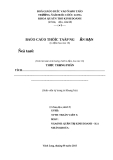
Gross examination of the surgical operated uterus revealed enlarged lesions in two areas of the left horn, which had thickened wall and yellowish sticky material in the lumen (Fig. 1). The trimmed tissues were fixed in 10% neutral buffered formalin, and paraffin embedded. Four µm sections were made and stained with hematoxylin and eosin for
Cystic endometrial hyperplasia (CEH) and pyometra in the bitch are dioestral syndromes, supposed to be caused by hormonal disturbances and changes in endometrial steroid hormone receptor levels [3]. Histologically, the endometria show cystic dilated glands and, if bacteria succeed in invading the uterus, pyometra may develop in the following metoestrus [2]. The occurrence of CEH has been reported in connection with the administration of progesterone-related contraceptives in cats and zoo animals [5]. In this report, we described a case of CEH with endometritis in a bitch that had been treated with medroxyprogesterone acetate (MPA), progesterone derivative, for 3 years.
An 8-year-old female Yorkshire Terrier was presented to NY Animal Hospital for investigation of reduced appetite, and occasional vomiting. The dog had not delivered since
*Corresponding author Tel: 82-2-740-8077; Fax: 82-2-763-5206 E-mail: kimoj@snu.ac.kr
Fig. 1. Uterus taken from surgical operation. (a) It revealed enlarged lesions in two areas of the left horn. The cut surface of enlarged horn had thickened wall (b) and yellowish sticky materials in the lumen (c).
82
Kyung-Suk Kim, Okjin Kim
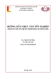
Fig. 2. Cystic endometrial hyperplasia of uterus, characterized by dilated cystic glands. H & E stain, ×100.
study is needed to clarify the association of MPA treatment with age, its pathogenesis and abnormal uterine changes for small animal practice.
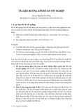
focal Fig. 3. Endometrial hyperplasia accompanied by calcification of uterus. Scattered inflammatory cells were observed in the endometrial stroma and mucinous material was protruded from endometrial surface. H & E stain, ×200.
Acknowledgments
The author greatly appreciates Jin-Uk Lee and Seung-Hee
Kim for excellent technical assistance.
References
in
histopathological examination. Histopathologically, cystic endometrial hyperplasia, endometritis and endometrial calcification were present in the thickened area. The most prominent endometrial lesion was CEH, characterized by dilated cystic glands, glandular proliferation in adenomatous clusters, or hyperplasia of the surface endometrium resulting in irregular folds or polypoid projections into the lumen (Fig. 2). Cystic glands were variable in size and were lined by densely packed epithelium that was usually compressed by retained secretory material. Lesions of severe hyperplasia were often accompanied by focal calcification (Fig. 3). Scattered the inflammatory cells were observed endometrial stroma and mucinous material was protruded from endometrial surface.
1. Chu P, Salamonsen LA, Lee CS, Wright PJ. Matrix metalloproteinases (MMPs) in the endometrium of bitches. Reproduction 2002, 123, 467-477. 2. Cockcroft PD. Focal cystic endometrial hyperplasia in a bitch. J Small Anim Pract 1995, 36, 77-78.
3. Leitner M, Aurich JE, Galabova G, Aurich C, Walter I. Lectin binding patterns in normal canine endometrium and in bitches with pyometra and cystic endometrial hyperplasia. Histol Histopathol 2003, 18, 787-795. 4. Middleton DJ. Megestrol acetate and the cat. Vet Annu 1986, 26, 341-347.
5. Munson L, Gardner IA, Mason RJ, Chassy LM, Seal US. Endometrial hyperplasia and mineralization in zoo felids treated with melengestrol acetate contraceptives. Vet Pathol 2002, 39, 419-427.
6. Noakes DE, Dhaliwal GK, England GC. Cystic endometrial hyperplasia/pyometra in dogs: a review of the causes and pathogenesis. J Reprod Fertil Suppl 2001, 57, 395-406.
Significant changes in the structure of the canine endometrium are associated with degeneration of luminal epithelium, cystic endometrial hyperplasia, pyometra and adenocarcinoma [1,6]. The basic mechanisms associated with these changes are poorly understood. Dogs given the higher dose levels of megestrol acetate, a progesterone derivative, were marked mammary stimulation, hyperplastic and neoplastic changes in the mammary glands, and clinical and pathologic changes typical pathologic changes of diabetes mellitus [8]. Side effects of exogenous progesterones were focused on induction of mammary tumors. Although the proliferative effects of progesterones counteracts estrogen and protects against endometrial hyperplasia [7], it has been reported that progesterone and its derivatives may increase CEH risk in uterus [5]. Prolonged exposure to megestrol acetate has been associated with endometrial hyperplasia and pyometra in domestic cats and progestin contraceptives may have similar effects on zoo felids [4,5]. MPA is a derivative of progesterone and used commonly both in contraception and hormone replacement therapy [7]. In this case, CEH and endometritis may be attributed to prolonged treatment of MPA. It was concluded that further
7. Pazol K, Wilson ME, Wallen K. Medroxyprogesterone acetate antagonizes the effects of estrogen treatment on social and sexual behavior in female Macaques. J Clin Endocrinol Metab 2004, 89, 2998-3006.
8. Weikel JH Jr, Nelson LW. Problems in evaluating chronicol toxicity of contraceptive steroids in dogs. J Toxicol Environ Health 1977, 3, 167-177.