J. Vet. Sci. (2007),
8(1), 27–38
J O U R N A L O F Veterinary Science
Estrogen receptor independent neurotoxic mechanism of bisphenol A, an environmental estrogen
Yoot Mo Lee1, Min Jae Seong1, Jae Woong Lee1, Yong Kyung Lee1, Tae Myoung Kim2, Sang-Yoon Nam2, Dae Joong Kim2, Young Won Yun2 , Tae Seong Kim3, Soon Young Han3, Jin Tae Hong1,*
1College of Pharmacy and CBITRC, and 2College of Veterinary Medicine and Research Institute of Veterinary Medicine, Chungbuk National University, Cheongju 361-763, Korea 3National Institute of Toxicological Research, Korea Food and Drug Administration, Seoul 122-704, Korea
Introduction
Bisphenol A
(BPA), a ubiquitous environmental contaminant, has been shown to cause developmental toxicity and carcinogenic effects. BPA may have physiological activity through estrogen receptor (ER) -α and -β, which are expressed in the central nervous system. We previously found that exposure of BPA to immature mice resulted in behavioral alternation, suggesting that overexposure of BPA could be neurotoxic. In this study, we further investigated the molecular neurotoxic mechanisms of BPA. BPA increased vulnerability (decrease of cell viability and differentiation, and increase of apoptotic cell death) of undifferentiated PC12 cells and cortical neuronal cells isolated from gestation 18 day rat embryos in a concentration-dependent manner (more than 50 µM). The ER antagonists, ICI 182,780, and tamoxifen, did not block these effects. The cell vulnerability against BPA was not significantly different in the PC12 cells overexpressing ER-α and ER-β compared with PC12 cells expressing vector alone. In addition, there was no difference observed between BPA and 17-β estradiol, a well-known agonist of ER receptor in the induction of neurotoxic responses. Further study of the mechanism showed that BPA significantly activated extracellular signal-regulated kinase (ERK) but inhibited anti-apoptotic nuclear factor kappa B (NF-κB) activation. In addition, ERK-specific inhibitor, PD 98,059, reversed BPA-induced cell death and restored NF-κB activity. This study demonstrated that exposure to BPA can cause neuronal cell death which may eventually be related with behavioral alternation in vivo. However, this neurotoxic effect may not be directly mediated through an ER receptor, as an ERK/NF-κB pathway may be more closely involved in BPA-induced neuronal toxicity.
Environmental estrogen-like chemicals have been increasingly recognized as potentially hazardous factors for human health. Some examples of this diverse group of compounds include industrial chemicals (e.g., bisphenol A, alkyl phenols, phthalates, polychlorinated biphenyls) and pesticides (e.g., DDT, methoxychlor, dieldrin, toxaphene, endosulfan). These estrogen-like chemicals are becoming ubiquitous in the environment, and humans are being exposed to these chemicals through the food chain [11]. Bisphenol A (BPA), an environmental estrogen, has been used as a compound in food and drink cans [5], as well as being a component of plastic in dental fillings. BPA has been reported to have not only estrogenic activity in the E- screen test and luciferase activity assay [31], but it also has several other biological toxicities.
Key words: bisphenol A, estrogen receptor, extracellular sig- nal regulated kinase, neurotoxicity
Eye, nose, throat, and skin irritation and a number of cases of skin sensitization and photosensitization have been reported in man in relation to BPA toxicity. BPA also possessed immunotoxicity and reduced the non-specific host defense system [39]. In addition, recent reports demonstrated that maternal exposure to BPA affected reproductive function [41]. BPA administration during the entire period of pregnancy in rats was reported to produce pregnancy failure, pre- and post-implantation loss, fetal developmental delay, and severe maternal toxicity [18]. BPA decreased the frequency of blastocyst development in vitro [40]. However, little is known regarding the effects of BPA on the neurons, even though BPA causes embryo and developmental toxicity [18,40]. We previously found that exposure of immature mice (3-week-old) to BPA for 3 weeks resulted in neurobehavioral alteration [36].
*Corresponding author Tel: +82-43-261-2813; Fax: +82-43-268-2732 E-mail: jinthong@chungbuk.ac.kr
The toxic effects of BPA have been proposed to be mediated through binding to estrogen receptor (ER)-α or -β [42]. For example, BPA reduced hepatic metallothionein synthesis and increased damage to the liver after Cd injection, and these effects occurred via an ER-mediated
28
Yoot Mo Lee et al.
complete medium to the desired concentrations immediately prior to use. PD 98,059, SB 203,580, SP 600,125, Tamoxifen, and ICI 182,780 pre-treatments were performed 30 min before the addition of BPA. The final concentration of DMSO was less than 0.2%.
Cell culture
mechanism [38]. BPA-induced increases in uterine wet weight and in luminal epithelial height in the ovariectomized B6C3F1 mouse are mediated by ERs [30]. The ligand binding domains of ER-α and ER-β are very similar in their tertiary architecture, and many compounds bind ER-α and ER-β with similar affinities [20] or with similar potencies in activation of estrogen responsive element-mediated receptor gene expression [3]. However, there is a difference in the distributions of ER-α and ER-β [19]. The uterus, breast, pituitary, bone, and cardiovascular tissue are known to be ER-α target organs [8], whereas the ventral prostate, ovarian granulosa cells [26], and gonadotropin-releasing hormone- containing neurons in the brain [12], sympathetic ganglia [44], and immune system [37] are targets of ER-β. In addition, differential biological responses have been reported to have estrogenic compound-induced toxic effects depending on whether those chemical agents act through ER-α or ER-β [23]. However, the neurotoxic mechanism of BPA, and the relevance of its neurotoxicity to ER have not yet been studied.
PC12 cells that have differentiation capacity were maintained on tissue culture plastic in Dulbecco’s modified Eagle’s medium (DMEM) and Ham’s F-12 nutrient (Invitrogen, USA) supplemented with 10% heat-inactivated horse serum, 5% fetal bovine serum, 100 µg/ml penicillin, and 100 µg/ml streptomycin at 37oC in a 5% CO atmosphere. PC12 cells 2 overexpressing ER as well as a control and PC12 cells expressing vector alone were routinely maintained in the above conditions to compare their viabilities. To induce differentiation of PC12 cells, nerve growth factor (NGF) (50 ng/ml) was added in DMEM supplied with only 1% heat- inactivated horse serum as described elsewhere [16]. Neuronal cells were prepared from E18 rat cortex (Sprague-Dawley rat brains) trypsinized (trypsin/EDTA) for 15 min at 37oC and dissociated using a fire-polished Pasteur pipette. The resulting cell suspension was added to poly-L-lysine-coated dishes containing in neurobasal media supplemented with B 27 serum (Invitrogen, USA). The dishes were incubated at for the designated amounts of time. 37oC in 5% CO 2
Transfection of PC12 cells
Activation of the mitogen activation protein (MAP) kinase family is known to be related to cellular toxic events and numerous physiological processes such as neuronal cell death and differentiation [32]. Transcription factor, nuclear factor kappa B (NF-κB), is linked to neurite formation, as well as survival and death of neuronal cells [9]. Extracellular signal-regulated kinase (ERK) has an important temporal regulator in the form of NF-κB activation and NF-κB- dependent gene expression [16]. NF-κB also down regulates c-Jun N-terminal kinase (JNK) activation, which promotes cell death [34]. These signals have been implicated in the neurotoxic mechanisms of estrogenic environmental neurotoxic materials, unless they do not act through ER. Our previous study demonstrated that interference of differentiation of neuronal cells may be a critical factor in neuronal cell survival, and differential activation of the MAP kinase family and transcription factors are involved in survival processes [17], ochratoxin-induced neurotoxicity [27], TNF- α-induced cortical neuronal cell death [39], and Zn-induced interference of PC12 cell differentiation [35]. Therefore, in the present study, we investigated whether BPA causes PC12 cells and neuronal cell death in a dose-dependent manner, and further investigated whether the neurotoxic effects may be mediated through ER or may be related by other signals.
Materials and Methods
Chemicals
ICI 182,780 (Tocris, USA), Tamoxifen, PD 98,059, SB 203,580, SP 600,125, BPA, and 17-β estradiol (Sigma- Aldrich, USA) were dissolved in dimethyl sulfoxide (DMSO; Sigma-Aldrich, USA). These chemicals were dissolved with Human ER-β cDNA was produced from human testis mRNA (BD Bioscience Clontech, USA) by PCR amplification, and it was then ligated into the PGEM-T easy vector to yield a hERβ cDNA fragment (1,672 bp) containing Not I and Spe I restriction sites. Neuron specific enolase (NSE)-hERβ cDNA (6,264 bp) was obtained by ligation of the above- mentioned hERβ cDNA with NSE promoter that was cloned in PCMVβ with Not I and Spe I digestion. Human ERα cDNA (pCMV5-hERα) or NSE-hERβ cDNA was transiently transfected into PC12 cells. PC12 cells were seeded onto 96-well tissue culture plates and grown in normal growth medium. The cells were incubated until they reached approximately 50-60% confluence. We prepared solution A (200 ng ER-α or β plasmid in 100 µl serum-free DMEM) and solution B (5 µl Lipofectamine Plus reagents (Life Technology, USA) in 95 µl serum-free DMEM per well). We combined the two solutions, which were mixed and incubated at room temperature for 20 min, and then overlaid the diluted complex solution onto the cells washed once with serum-free DMEM. The medium was exchanged with normal growth medium at 4 h after transfection. The transiently ER-α and -β transfected cells were referred to as PC12/ER-α and PC12/ER-β cells, respectively. The cells stimulated in the transient transfection conditions with only vector were named PC12/neo cells.
Estrogen receptor independent neurotoxic mechanism of bisphenol A, an environmental estrogen
29
WST-1 assay
After deleting the medium and then adding 90 µl PBS, 10 µl of WST-1 reagent (Roche, Germany) was added to each well (96-well plates). The cells were then incubated with the at 37oC. Following WST-1 reagent for 3 h in 5% CO 2 incubation, the absorbance at 450 nm was determined using a microplate reader (Megellan Software; Tecan, Austria). Cell viability was expressed as the percentage of absorbance obtained in the treated wells relative to that in the untreated control wells.
DAPI staining
buffer containing 20 mM HEPES, 20% glycerol, 420 mM , 0.5 mM dithiothreitol, NaCl, 0.2 mM EDTA, 1.5 mM MgCl 2 and 0.2 mM phenylmethylsulfonyl fluoride. After centrifugation, the supernatant contained the nuclear protein. The protein level was determined by a microplate modification of the Bradford method [4]. Then, 10 µg of nuclear protein was incubated in a total volume of 10 µl of binding buffer , 0.5 mM EDTA, 50 mM (10 mM Tris, pH 7.5, 1 mM MgCl 2 NaCl, 0.5 mM dithiothreitol, 0.05 µg/µl poly(dl-dC) · poly(dl-dC), 4% glycerol) at 4oC for 15 min, followed by another 20 min incubation with 100 µCi of γ-32P ATP-labeled oligonucleotide containing NF-κB binding sites. The DNA protein binding complex was run a 6% nondenatured polyacrylamide gel at 150 V for 2 h. Gels were dried and autographed using Kodak MR film at −80oC overnight. The relative density of the DNA binding bands was evaluated.
Statistics
The data were expressed as mean ± SE. In cases of significant variation, the individual values were compared by t-test in all experiments. Cells were cultured on 8-chamber slides. After treatment with compound for 24 h, the cells were washed with PBS and fixed with 4% paraformaldehyde at room temperature for 30 min. After fixation, dishes were washed with PBS and incubated at room temperature with 1 drop of 4,6- diamidino-2-phenylindole (DAPI) solution (Vector, USA) for 30 min in the dark. After DAPI staining, apoptotic cells were determined according to the morphological changes through fluorescence microscope (Leica Microsystems AG, Germany).
Results
Western blotting
BPA inhibits neural extension and induces neuronal cell death
Cells were homogenized with lysis buffer: 50 mM Tris, pH 8.0, 150 mM NaCl, 0.02% sodium azide, 0.2% SDS, 1 mM phenylmethylsulfonyl fluoride, 10 µg/ml aprotinin, 1% igepal CA 630 (Sigma-Aldrich, USA), 10 mM NaF, 0.5 mM EDTA, 0.1 mM EGTA, and 0.5% sodium deoxycholate, and were then centrifuged. Equal amounts of proteins were separated on a SDS/12% polyacrylamide gel and then transferred to a nitrocellulose membrane. Blots were blocked for 2 h with 5% (w/v) nonfat dried milk in Tris- buffered saline (10 mM Tris, pH 8.0, and 150 mM NaCl) solution containing 0.05% Tween-20. The membrane was then incubated for 3 h with specific primary antibodies, followed by incubation for 1 h with secondary antibodies. Rabbit polyclonal antibodies against ERK, p38, JNK, and mouse monoclonal antibodies against phosphorylated forms of MAP kinase (1 : 500) (Santa Cruz Biotechnology, USA) were used. Immunoreactive proteins were detected with the enhanced chemiluminescence Western blotting detection system. The relative density of the protein bands was evaluated.
Nuclear extract and gel mobility shift assay
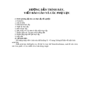
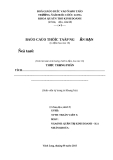
In this study, we examined the cytotoxic effects of BPA assessed by the WST-1 method. PC12 cells were cultured for 12, 24, 48, and 72 h in the condition of non-treated medium or with several doses of BPA-treated medium. The viability of cortical neuronal cells was determined after 72 h treatments with several concentrations of BPA. As seen in Fig. 1A and 2A, concentrations below 50 µM slightly increased cell viability, but at concentrations higher than 100 µM, BPA reduced the viability significantly in both types of cells. Untreated PC12 cells as well as cortical neurons contained round nuclei with homogeneous chromatin, whereas cells exposed to more than 100 µM BPA for 24 h showed a reduction in nuclear size and chromatin condensation, as well as nuclear fragmentation, all of which are typical features of apoptosis (Fig. 1B & 2B). These findings suggest that BPA can induce cell death through apoptosis. Considerable extension of neurites was observed in PC12 cells and cortical neurons after treatment with NGF (50 ng/ ml) for 5 days (Fig. 1C & 2C). BPA at low concentrations (0-10 µM in PC12 and cortical neuronal cells) could increase neurite extension, but high concentrations (more than about 50 µM) reduced neurite extension.
Effects of estrogen inhibitors against BPA-induced cell death
The gel mobility shift assay was performed using a slight modification of a method described previously [17]. In brief, the cultured cells were washed three times with ice-cold phosphate-buffered saline, pH 7.6, and pelleted. The pellets were resuspended in 400 µl of nonradioactive buffer , 10 mM KCl, containing 10 mM HEPES, 1.5 mM MgCl 2 0.5 mM dithiothreitol, and 0.2 mM phenylmetylsulfonyl fluoride, and were then centrifuged to remove everything except the nuclei. The pellets were resuspended in a second To investigate whether damage to cells induced by BPA was mediated by ER, cells were pretreated with ER antagonists (tamoxifen, ICI 182,780) 30 min prior to
30
Yoot Mo Lee et al.
Fig. 1. Effect of BPA on PC12 cell viability. (A) Cell viability of PC12 cells after 12, 24, 48, and 72 h treatments with/without several concentrations of BPA. Living cells were represented as a percentage of the total population of living and dead cells. (B) Photomicrographs of DAPI-stained PC12 cells treated for 24 h with several concentrations of BPA. Cells were stained with DAPI to visualize nuclear morphology. The percentage of apoptosis which presents a reduction in nuclear size, chromatin condensation, and nuclear fragmentation from two experiments was entered into a graph. (C) Photomicrographs of PC12 cells treated with the indicated materials for 5 days, respectively. PC12 cells were cultivated for 1 day in the condition of non-treated DMEM supplemented with Ham’s F12 nutrient, 1% horse serum, 50 ng/ml NGF, 100 units/ml penicillin, and 100 µg/ml streptomycin, and were then cultured with/ without several concentrations of BPA added to the medium. Control (Con), those cultured with/without BPA and NGF. *Significant difference from control (p < 0.05).
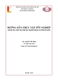
ER-β cDNA after culturing with several concentrations of BPA. The toxic responses among the cells did not differ (Fig. 5B) even though PC12 cells expressing ER-α and ER- β showed 3- to 4-fold increases in ER expression (Fig. 5A).
No difference between BPA and 17-β estradiol in the induction of neurotoxic responses
treatment with BPA, and we examined PC12 and cortical neuronal cell survival using the WST-1 assay or neurite extension. ER antagonists did not abolish BPA-induced toxicity in PC12 cells, as well as cortical neuronal cells (Fig. 3A & 4A). In addition, ER antagonists also did not have significant effects on BPA-induced neurite shrinkage of the 2 types of cells (Fig. 3B & 4B). These findings seem to reflect that ER may not be related with neuronal cell damage induced by BPA.
Cell vulnerability against BPA in PC12 cells overexpressing ER or expressing vector alone
To further confirm the involvement of ER-α or ER-β in BPA-induced neuronal cell damage, we compared the viability of PC12 using PC12 cells expressing vector alone (PC12/neo cells) and PC12 cells transfected with ER-α or We also compared the susceptibility of PC12 and cortical neuronal cells against BPA or 17-β estradiol using the WST- 1 assay. The cell viability was determined after 72 h incubation with these chemicals. The 17-β estradiol-induced viability was similar to that induced by BPA in both cell types (Fig. 5C). This result may indicate that BPA-induced cell damages are exerted at lower levels through the classical ER- mediated intracellular receptor signal transduction system, and suggests the possibility that one or more additional
Estrogen receptor independent neurotoxic mechanism of bisphenol A, an environmental estrogen
31
Fig. 2. Effect of BPA on cortical neuronal cell viability. (A) Cell viability of neuronal cells after 72 h treatments with/without various concentrations of BPA. Living cells were represented as a percentage of the total population of living and dead cells. (B) Photomicrographs of DAPI-stained neuronal cells treated for 24 h with various concentrations of BPA. Cells were stained with DAPI to visualize nuclear morphology. The percentage of apoptosis which presents a reduction in nuclear size, chromatin condensation, and nuclear fragmentation from two experiments was entered into a graph. (C) Photomicrographs of neuronal cells treated with BPA for 5 days, respectively. Neuronal cells were cultivated for 1 day in the condition of non-treated neurobasal medium supplemented with B 27 serum, and then cultured with/without several concentrations of BPA added to the medium. Control (Con), those cultured with/without BPA. *Significant difference from control (p < 0.05).
signal-mediated mechanisms are more significant in BPA- induced neurotoxicity.
Activation of MAP kinase signals in BPA-induced neurotoxicity
the MAP kinase inhibitors (PD 98,059, SB 203,580, and SP 600,125) assessed by the WST-1 method. Among them, only PD 98,059 (an ERK inhibitor) reversed BPA-induced toxicity of PC12 cells and neuronal cells. The other MAP kinase inhibitors did not reverse against BPA toxicities. However, it seems that the proper dose of PD 98,059 needed to protect BPA-induced neuronal damage varied. That is to say, 20 µM PD 98,059 showed the highest protective effect on PC12 cells, but a much higher dose (50-100 µM) of PD 98,059 was required to protect against BPA-induced cortical neuronal cell death (Fig. 7).
BPA-induced neurotoxic effect is related with a decrease of NF-κB activity
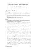
MAP kinase activation was determined by examining the induction of p38, JNK, and ERK, as well as their phosphorylation (p-p38, p-JNK, and p-ERK). After 2 h exposure to BPA, protein was extracted from PC12 cells and cortical neuronal cells, and was then subjected to Western analysis using each antibody that recognizes the MAP kinase family (p38, JNK, and ERK) and their phosphorylations. BPA increased the expression of p38, JNK, ERK and their phosphorylations, but only activation of ERK was clearly increased in a dose-dependent manner (Fig. 6). To study protective effects of MAP kinase inhibitions on BPA toxicities, we examined the cytotoxic effects of BPA with or without We also examined the activation of transcription factor NF-κB since it is known to act as a cell survival factor. The highest activations of NF-κB were seen in BPA (10 µM)-
32
Yoot Mo Lee et al.
Fig. 3. BPA-induced PC12 cell death was not mainly mediated by estrogen receptor. PC12 cells were treated with ER antagonist 30 min prior to treatment with BPA. (A) Viability of PC12 cell cultures was determined after treatment with compounds for 3 days. (B) Photomicrographs of PC12 cells cultured with BPA with/without ER antagonists added to DMEM supplemented with Ham’s F12 nutrient, 5% fetal bovine serum, 10% horse serum, 50 ng/ml NGF, 100 units/ml penicillin, and 100 µg/ml streptomycin for 5 days. *Significant difference from control (p < 0.05).
Fig. 4. BPA-induced neuronal cell death was not mainly mediated by estrogen receptor. Neurocortical (neuronal) cells were treated with ER antagonist 30 min prior to treatment with BPA. (A) Viability of neuronal cell cultures was determined after treatment with compounds for 3 days. (B) Photomicrographs of neuronal cell cultures cultured with BPA with/without ER antagonists added to neurobasal media supplemented with B 27 serum for 5 days. *Significant difference from control (p < 0.05).
Estrogen receptor independent neurotoxic mechanism of bisphenol A, an environmental estrogen
33
Fig. 5. BPA toxicity in PC12, PC12/neo, PC12/ER-α, or β cells. (A) Expression of ERs in established PC12 cells expressing ER-α and β. (B) Comparison of four cell types according to a viability assay performed 72 h after BPA treatments. The four cell types were cultured with/without several concentrations of BPA. (C) Comparison between BPA and 17-β estradiol on cell viability according to concentration using the WST-1 method. Viability was assayed in PC12 cells and neuronal cells treated with different concentrations of BPA and 17-β estradiol for 72 h. *Significant difference from control (p < 0.05).
Fig. 6. Effect of BPA concentrations for activations of the MAP kinase family. Cells were treated with BPA, and after 2 h exposure to BPA, activation of MAP kinase family was determined by Western blotting. Similar patterns of expression were found from three individual experiments.
treated cells for 90 min. To determine the effects of BPA for NF-κB activations, PC12 and cortical neuron cells were treated with 0, 10, 50, 100, and 200 µM BPA for 90 min. The highest activations of NF-κB were seen in PC12 and cortical neuron cells treated with 10 µM BPA (Fig. 8A & B), which corresponded to the promoting effect of BPA on cell survival. However, decreases of NF-κB occurred in toxic concentrations of BPA (50-200 µM) as compared with
34
Yoot Mo Lee et al.
Fig. 7. Protective effects of ERK inhibitor on BPA toxicity. PC12 and neuronal cells were pretreated with MAP kinase inhibitors (PD 98,059, SB 203,580, and SP 600,125) followed by BPA (300 µM) treatment. After 72 h of exposure, we examined PC12 and neuronal cell survival using the WST-1 assay. Living cells were represented as a percentage of the total population of living and dead cells. *Significant difference from control (p < 0.05). #Significant difference from only BPA-treated group (p < 0.05).
However, the susceptibility of ER-overexpressing PC12 cells was similar to that of PC12 cells overexpressing vector alone. Consistent with the dose-dependent increase of susceptibility, our experiments showed that BPA increased activation of ERK. However, the NF-κB activation level was reduced. Moreover, ERK inhibited the reversal of BPA- induced neuronal cell death and NF-κB. These results show that BPA causes neurotoxic events, and also indicated that activation of the ERK pathway accompanied with decreases of NF-κB rather than the ER pathway seems to be related to BPA-induced neurotoxicity.
10 µM BPA-induced NF-κB activity. Next, we tested the effects of tamoxifen, ICI 182,780, and PD 98,059 for BPA- dependent NF-κB inactivation. PC12 and neuronal cells were pretreated with ER antagonists for 30 min and then stimulated with BPA for 90 min. As shown in Fig. 8C and D, a decrease of NF-κB activation induced by a toxic dose of BPA (100 µM) obviously was not reversed by ER antagonists in PC12 and neuronal cells. To further study the relevance of the ERK pathway in BPA-induced inactivation of NF-κB to cause a neurotoxic response, both cells were pretreated with ERK inhibitor PD 98,059 at 30 min prior to treatment with BPA, and NF-κB activity was then determined after 90 min of treatment with BPA. As expectated and in accordance with cell viability, PD 98,059 reversed BPA (100 µM)-induced NF-κB inactivation in both types of cell (Fig. 8E & F).
Discussion
The present study showed that BPA reduced cell survival of PC12 and cortical neuronal cells dose-dependent manner, and this reduction was not reversed by ER antagonists. The dose causing neuronal cell death in this study (more than 50 µM) is comparable to the dose inducing cell death in rat Sertoli cells [14]. However, a low dose of BPA (less than 50 µM) may aid in the neurite extension of cells, which resulted in increased cell viability. It was reported that low concentrations of BPA exert neuroprotective effects against glutamate and amyloid beta toxicities [12]. The low dose- induced cell protective effect was partially prevented in the presence of ER antagonists (data not shown), which suggests that the protective effect may be mediated by ER. However, the neurotoxic response induced by a high dose was not mediated by ER. Therefore, these data suggest that BPA has a dose-differential effect on cell survival, and a concentration of about 50 µM may be a cut-off dose to cause either protective or toxic effects on neuronal cells. Although it has been reported that BPA toxicity may be mediated by ER in other cells [43], it may not have had much influence in neuronal cell death. This notion was supported by the present data showing that ER antagonists did not significantly affect BPA-induced neurotoxicities. In addition, the toxic effect of 17-β estradiol was similar to that of BPA in PC12 cells and neuronal cells, although it was reported that the estrogenic activity of BPA is one-third to one-quarter that of estradiol [2]. Moreover, there were no significant differences in the susceptibility to BPA between the PC12 cells overexpressed with ER-α or ER-β cDNA (ER-α or β/PC12) and vector alone transfected in PC12 cells. In connection with our findings, it was reported that BPA induced marked malformations and specific apoptosis of central nervous system cells during early development of X. laevis embryos, and that these BPA effects appeared to be due to non- estrogenic activities on developmental processes [28]. Neuronal apoptosis resulting from high doses of the isoflavone genistein, known to be an estrogenic compound, also is not blocked by an ER antagonist, ICI 182,780. In their study, increased intracellular calcium and p42/44 MAP kinase were suggested as ER-independent toxic mechanisms [23]. It was also noteworthy that estrogenic response Sertoli cells incubated with nonestrogenic compounds such as 2',3',4',5'- tetrachloro-4-biphenylol and 3,3',4,4'-tetrachlorobiphenyl (estrogenic contaminants), but not with 10−7 M 17-β estradiol, showed morphological changes [33]. Moreover, a recent study showed that BPA strongly bound to ER-related receptor
Estrogen receptor independent neurotoxic mechanism of bisphenol A, an environmental estrogen
35
Fig. 8. Effect of BPA, ER antagonists, and ERK inhibitor on NF-κB activation. PC12 cells (A) and cortical neuronal cells (B) were treated with 0, 10, 50, 100, and 200 µM (BPA) for 90 min with/without pretreatment of different doses of ER antagonists (tamoxifen and ICI 182,780) (C & D) or ERK inhibitor (E & F) 30 min prior to treatment with BPA. Nuclear extracts were prepared and assayed for NF-κB by EMSA from two individual experiments performed in duplicate.
compared to ER-α and ER-β, which may interrupt direct binding of BPA to ER-α and ER-β [42]. Taken together, that BPA causes these results clearly demonstrated neurotoxicities, but BPA-induced neurotoxic effects may not be directly mediated by ER.
(ROS), and its inhibition rescues cells from ROS-induced death [35]. MPTP, a neurotoxin used widely in Parkinson’s disease experimental paradigms, induces activation of ERK, while its inhibition rescues the affected cells [10]. ERK inhibition also reduces toxicity following cerebral ischemia [1]. The elements leading to ERK activation include , intracellular signal molecules such as thromboxane A 2 intracellular calcium, angiotensin II, insulin, and ROS [24,35]. BPA-induced ERK activation may be affected by these elements in various ways. Among them, alteration of intracellular calcium concentration could be a significant contributor to the activation of ERK, thereby inhibiting cell survival. In fact, we previously found that BPA significantly lowered intracellular calcium concentrations of PC12 cells and cortical neuron cells in a dose-dependent manner [22]. In connection with the direct calcium lowering effect of BPA, estradiol inhibited ATP-induced intracellular calcium concentration in dorsal root ganglia neurons, which may modulate nociceptive signaling in the peripheral nervous system [6]. It was also reported that angiotensin-induced -coupled Ras/ERK activation is dominantly regulated by G q Ca2+/calmodulin signaling in cardiac fibroblast cell death [25]. Therefore, it seems that the activation of ERK by depletion of intracellular calcium may be linked to the neurotoxicity of BPA. Activation of members of the MAP kinase family has been known to mediate death of several cell types, including neuronal cells [32]. Our previous data have also shown that the MAP kinase family is activated depending on the cell type and nature of stimuli in the neuronal cell survival [17,27]. Therefore, we investigated the alteration of MAP kinase signals to determine whether these signals may be related with BPA-induced neurotoxic effects. BPA activated all three types in the MAP kinase family, but only activation of ERK was clearly dose-dependent. Moreover, in the presence of ERK inhibitor, a BPA-induced neurotoxic effect was prevented, but that of other inhibitors was not, which suggested that ERK pathway activation could be involved in the neurotoxic effect of BPA. Activation of the ERK pathway as a neurotoxic mechanism has been reported on extensively. Stimulations of intracellular calcium signaling and CREB phosphorylation in mid-brain neuron death during development are associated with neurotrophin signaling by activation of MAP kinase [21]. ERK is also activated in neuronal cell death by reactive oxygen species
36
Yoot Mo Lee et al.
that a In addition, our results demonstrated
References
1. Alessandrini A, Namura S, Moskowitz MA, Bonventre JV. MEK1 protein kinase inhibition protects against damage resulting from focal cerebral ischemia. Proc Natl Acad Sci USA 1999, 96, 12866-12869.
2. Aloisi AM, Della Seta D, Rendo C, Ceccarelli I, Scaramuzzino A, Farabollini F. Exposure to the estrogenic pollutant bisphenol A affects pain behavior induced by subcutaneous formalin injection in male and female rats. Brain Res 2002, 937, 1-7.
3. Barkhem T, Carlsson B, Nilsson Y, Enmark E, Gustafsson J, Nilsson S. Differential response of estrogen receptor α and estrogen receptor β to partial estrogen agonist/ antagonists. Mol Pharmacol 1998, 54, 105-112.
4. Bradford MM. A rapid and sensitive method for the quantitation of microgram quantities of protein utilizing the principle of protein-dye binding. Anal Biochem 1976, 72, 248-254.
5. Brotons JA, Olea-Serrano MF, Villalobos M, Pedraza V, Olea N. Xenoestrogens released from lacquer coating in food cans. Environ Health Perspect 1995, 103, 608-612.
6. Chaban VV, Mayer EA, Ennes HS, Micevych PE. Estradiol inhibits atp-induced intracellular calcium concentration increase in dorsal root ganglia neurons. Neuroscience 2003, 118, 941- 948.
7. Coleman KM, Dutertre M, El-Gharbawy A, Rowan BG, Weigel NL, Smith CL. Mechanistic differences in the activation of estrogen receptor-α (ERα)-and ERβ-dependent gene expression by cAMP signaling pathway(s). J Biol Chem 2003, 278, 12834-12845.
low concentration of BPA resulted in the development of a significant increase of the transcription factor, NF-κB, which is thought to protect cell survival. However, higher doses of BPA decreased NF-κB activity. Some evidence has accumulated to indicate that the activation of NF-κB might rescue cells from oxidative stress-induced neuronal cell death, suggesting that NF-κB activation may be an important signal to prevent cellular degeneration [29]. Based on our experiments, the transcription factors stimulated by BPA (low dose) may lead to support weak neurite outgrowths and maintainance of survival or proliferation, but high doses of BPA resulted in decreasing the effect of NF-κB, which may not be able to protect cells against BPA-induced cell death. However, the BPA-induced decrease of NF-κB activation was not reversed by ER antagonists, which again suggests that the ER pathway is not involved in BPA- induced neurotoxicity or NF-κB inactivation. In contrast, ERK inhibitor reversed a BPA-induced down-regulation of NF-κB and neuronal cell death, suggesting the involvement of the ERK pathway in the BPA-induced neurotoxic effects. These data suggests that down-activation of NF-κB may be another significant contributing factor in the BPA-induced neurotoxic mechanism.
8. Couse JF, Korach KS. Estrogen receptor null mice: what have we learned and where will they lead us? Endocr Rev 1999, 20, 358-417.
(about 40 nM) [15]. Therefore,
9. Foehr ED, Bohuslav J, Chen LF, DeNoronha C, Geleziunas R, Lin X, O’Mahony A, Greene WC. The NF- κB-inducing kinase induces PC12 cell differentiation and prevents apoptosis. J Biol Chem 2000, 275, 34021-34024. 10. Gomez-Santos C, Ferrer I, Reiriz J, Vinals F, Barrachina M, Ambrosio S. MPP+ increases α-synuclein expression and ERK/MAP-kinase phosphorylation in human neuroblastoma SH-SY5Y cells. Brain Res 2002, 935, 32-39.
range in
11. Guillette LJ Jr, Gross TS, Gross DA, Rooney AA, Percival HF. Gonadal steroidogenesis in vitro from juvenile alligators obtained from contaminated or control lakes. Environ Health Perspect 1995, 103 (Suppl), 31-36.
In conclusion, BPA has a dose-dependent effect on neuronal toxicity, but the toxic effect may not be mediated by ER. The excess ERK stimulation accompanied by NF- κB inactivation is linked to BPA-induced neurotoxicity. It was reported that the normal serum BPA concentration in women is approximately 1-2 ng/ml (about 5-10 nM), and the amniotic fluid concentration of BPA is an approximately the 8.5 ± 0.2 ng/ml concentration of BPA used in the present study was much higher (about 100-2,000 higher level) than the normal environmental exposure level. However, it is thought that the concentration of BPA in the blood could reach in the micro-molar the case of high-dose oral supplementation or intravenous therapy. In addition, BPA may increase physiological effects in combination with natural hormones. Thus, even though it does not waken the public interest at present, the potential for neurotoxicity of BPA needs to be considered in future studies.
Acknowledgments
12. Gursoy E, Cardounel A, Kalimi M. The environmental estrogenic compound bisphenol A exerts estrogenic effects on mouse hippocampal (HT-22) cells: neuroprotection against glutamate and amyloid beta protein toxicity. Neurochem Int 2001, 38, 181-186.
13. Hrabovszky E, Steinhauser A, Barabas K, Shughrue PJ, Petersen SL, Merchenthaler I, Liposits Z. Estrogen receptor-β immunoreactivity in luteinizing hormone-releasing hormone neurons of the rat brain. Endocrinology 2001, 142,
This work was supported by a Korea Food and Drug Administration Research Grant, and by the Regional Research Centers Program of the Ministry of Education & Human Resources Development, Korea. We thank Dr. Carolyn L. Smith (Department of Molecular and Cellullar Biology, Baylor College of Medicine, Houston, Texas, USA) who provided human ERα cDNA (pCMV5-hERα).
Estrogen receptor independent neurotoxic mechanism of bisphenol A, an environmental estrogen
37
3261-3264.
14. Iida H, Maehara K, Doiguchi M, Mori T, Yamada F. Bisphenol A-induced apoptosis of cultured rat Sertoli cells. Reprod Toxicol 2003, 17, 457-464.
NG, Yun YW, Kim DJ, Ha TY, Song YS, Lee YM, Oh K, Hong JT. Inhibitory effects of ochratoxin A on nerve growth factor-induced neurite extension through downregulation of p38 MAP kinase and AP-1 activation in cultured pheo- chromocytoma cells. J Toxicol Environ Health A 2004, 67, 357-371.
15. Ikezuki Y, Tsutsumi O, Takai Y, Kamei Y, Taketani Y. Determination of bisphenol A concentrations in human biological fluids reveals significant early prenatal exposure. Hum Reprod 2002, 17, 2839-2841.
28. Oka T, Adati N, Shinkai T, Sakuma K, Nishimura T, Kurose K. Bisphenol A induces apoptosis in central neural cells during early development of Xenopus laevis. Biochem Biophys Res Commun 2003, 312, 877-882.
16. Jiang B, Xu S, Hou X, Pimentel DR, Brecher P, Cohen RA. Temporal control of NF-κB activation by ERK differentially regulates interleukin-1β-induced gene expression. J Biol Chem 2004, 279, 1323-1329.
29. O’Neill LAJ, Kaltschmidt C. NF-κB: a crucial transcription factor for glial and neuronal cell function. Trends Neurosci 1997, 20, 252-258.
17. Jung KM, Park KS, Oh JH, Jung SY, Yang KH, Song YS, Son DJ, Park YH, Yun YP, Lee MK, Oh KW, Hong JT. Activation of p38 mitogen-activated protein kinase and activator protein-1 during the promotion of neurite extension . Mol of PC-12 cells by 15-deoxy-∆12,14-prostaglandin J 2 Pharmacol 2003, 63, 607-616.
30. Papaconstantinou AD, Umbreit TH, Fisher BR, Goering PL, Lappas NT, Brown KM. Bisphenol A-induced increase in uterine weight and alterations in uterine morphology in ovariectomized B6C3F1 mice: role of the estrogen receptor. Toxicol Sci 2000, 56, 332-329.
18. Kim JC, Shin HC, Cha SW, Koh WS, Chung MK, Han SS. Evaluation of developmental toxicity in rats exposed to the environmental estrogen bisphenol A during pregnancy. Life Sci 2001, 69, 2611-2625.
31. Paris F, Balaguer P, Térouanne B, Servant N, Lacoste C, Cravedi JP, Nicolas JC, Sultan C. Phenylphenols, biphenols, bisphenol-A and 4-tert-octylphenol exhibit α and β estrogen activities and antiandrogen activity in reporter cell lines. Mol Cell Endocrinol 2002, 193, 43-49.
32. Purves T, Middlemas A, Agthong S, Jude EB, Boulton AJ, Fernyhough P, Tomlinson DR. A role for mitogen- the etiology of diabetic in activated protein kinases neuropathy. FASEB J 2001, 15, 2508-2514.
33. Raychoudhury SS, Flowers AF, Millette CF, Finlay MF. Toxic effects of polychlorinated biphenyls on cultured rat Sertoli cells. J Androl 2000, 21, 964-973.
34. Sakon S, Xue X, Takekawa M, Sasazuki T, Okazaki T, Kojima Y, Piao JH, Yagita H, Okumura K, Doi T, Nakano H. NF-κB inhibits TNF-induced accumulation of ROS that mediate prolonged MAPK activation and necrotic cell death. EMBO J 2003, 22, 3898-3909.
19. Kuiper GG, Carlsson B, Grandien K, Enmark E, Häggblad J, Nilsson S, Gustafsson JÅ. Comparison of the ligand binding specificity and transcript tissue distribution of estrogen receptors α and β. Endocrinol 1997, 138, 863-870. 20. Kuiper GG, Lemmen JG, Carlsson B, Corton JC, Safe SH, van der Saag PT, van der Burg B, Gustafsson JÅ. Interaction of estrogenic chemicals and phytoestrogens with estrogen receptor β. Endocrinol 1998, 139, 4252-4263. 21. Küppers E, Ivanova T, Karolczak M, Lazarov N, Föhr K, Beyer C. Classical and nonclassical estrogen action in the developing midbrain. Horm Behav 2001, 40, 196-202. 22. Lee YM, Lee SM, Son DJ, Lee SY, Park HJ, Nam SY, Kim DJ, Yun YW, Yoo HS, Oh KW, Kim TS, Han YS. Bisphenol a disturbs intracellular calcium homeostasis and its relationship with cytotoxicity. J Toxicol Public Health 2004, 20, 57-66.
35. Seo SR, Chong SA, Lee SI, Sung JY, Ahn YS, Chung KC, Seo JT. Zn2+-induced ERK activation mediated by reactive oxygen species causes cell death in differentiated PC12 cells. J Neurochem 2001, 78, 600-610.
36. Seoung MJ, Shin IC, Lee YM, Son DJ, Song YS, Jeon KH, Kim YB, Lee BJ, Kim DJ, Yun YW, Kim TS, Han SY. Behavior alterations and expression of estrogen receptors in mice exposed to Bisphenol A. J Toxicol Public Health 2004, 20, 67-77.
23. Linford NJ, Yang Y, Cook DG, Dorsa DM. Neuronal apoptosis resulting from high doses of the isoflavone genistein: role for calcium and p42/44 mitogen-activated protein kinase. J Pharmacol Exp Ther 2001, 299, 67-75. 24. Moore SA, Huang N, Hinthong O, Andres RD, Grammatopoulos TN, Weyhenmeyer JA. Human angiotensin II type-2 receptor inhibition of insulin-mediated ERK-2 activity via a G-protein coupled signaling pathway. Brain Res Mol Brain Res 2004, 124, 62-69.
37. Shim GJ, Wang L, Andersson S, Nagy N, Kis LL, Zhang Q, Mäkelä S, Warner M, Gustafsson JÅ. Disruption of the estrogen receptor β gene in mice causes myeloproliferative disease resembling chronic myeloid leukemia with lymphoid blast crisis. Proc Natl Acad Sci USA 2003, 100, 6694-6699. 38. Sogawa N, Onodera K, Sogawa CA, Mukubo Y, Fukuoka H, Oda N, Furuta H. Bisphenol A enhances cadmium toxicity through estrogen receptor. Methods Find Exp Clin Pharmacol 2001, 23, 395-399.
25. Murasawa S, Mori Y, Nozawa Y, Masaki H, Maruyama K, Tsutsumi Y, Moriguchi Y, Shibasaki Y, Tanaka Y, Iwasaka T, Inada M, Matsubara H. Role of calcium- sensitive tyrosine kinase Pyk2/CAKβ/RAFTK in angiotensin II induced Ras/ERK signaling. Hypertension 1998, 32, 668- 675.
39. Sugita-Konishi Y, Shimura S, Nishikawa T, Sunaga F, Naito H, Suzuki Y. Effect of Bisphenol A on non-specific immunodefenses against non-pathogenic Escherichia coli. Toxicol Lett 2003, 136, 217-227.
26. Nilsson S, Mäkelä S, Treuter E, Tujague M, Thomsen J, Andersson G, Enmark E, Pettersson K, Warner M, Gustafsson JÅ. Mechanisms of estrogen action. Physiol Rev 2001, 81, 1535-1565.
40. Takai Y, Tsutsumi O, Ikezuki Y, Hiroi H, Osuga Y,
27. Oh JH, Jung HK, Park YJ, Kim CK, Chung SY, Park
38
Yoot Mo Lee et al.
strongly binds to human estrogen-related receptor γ (ERRγ) with high constitutive activity. Toxicol Lett 2006, 167, 95- 105
Momoeda M, Yano T, Taketani Y. Estrogen receptor- mediated effects of a xenoestrogen, bisphenol A, on preimplantation mouse embryos. Biochem Biophys Res Commun 2000, 270, 918-921.
43. Washington W, Hubert L, Jones D, Gray WG. Bisphenol a binds to the low-affinity estrogen binding site. In Vitr Mol Toxicol 2001, 14, 43-51.
41. Takao T, Nanamiya W, Nagano I, Asaba K, Kawabata K, Hashimoto K. Exposure with the environmental estrogen bisphenol A disrupts the male reproductive tract in young mice. Life Sci 1999, 65, 2351-2357.
44. Zoubina EV, Smith PG. Expression of estrogen receptors alpha and beta by sympathetic ganglion neurons projecting to the proximal urethra of female rats. J Urol 2003, 169, 382- 385.
42. Takayanagi S, Tokunaga T, Liu X, Okada H, Matsushima A, Shimohigashi Y. Endocrine disruptor bisphenol A