Can Tho Journal of Medicine and Pharmacy 10(7) (2024)
180
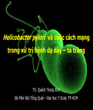
A REVIEW ON CURRENT TRENDS IN Helicobacter pylori
MANAGEMENT WITH MEDICINAL PLANTS AND ITS CONSTITUENTS
Huynh Anh Duy1*, Huynh Ngoc Thuy2, Tran Hung2
1Can Tho University
2University of Medicine and Pharmacy at Ho Chi Minh city
*Corresponding author: haduy@ctu.edu.vn
Received:08/04/2024
Reviewed:11/05/2024
Accepted:18/05/2024
ABSTRACT
Helicobacter pylori is a bacterium associated with gastric diseases and disorders of the
upper gastrointestinal tract. The gram-negative bacterium Helicobacter pylori is known as a
persistent colonizer of the human stomach, and this bacteria is also involved in extra-intestinal
diseases. In 1994, the International Agency for Research on Cancer, World Health Organization
classified H. pylori as a class 1 carcinogen, the only bacterium given this classification. Besides,
the emergence of H. pylori resistance to antibiotics has been a major clinical challenge in the field
of gastroenterology, and this concern has been shown an increasing tendency in many regions of
the world. To overcome the current circulating difficulties, new potential therapeutic targets were
uncovered to find active substances for the treatment of H. pylori infection. Several medicinal plants
and their isolated compounds have been reported for their antimicrobial activity against H. pylori.
It is demonstrated that they are efficacious against H. pylori strains that are resistant to drugs. The
mechanism of action of many of these plant extracts and plant-derived compounds is different from
that of conventional antibiotics. Therefore, natural compounds are emerging as a potential source
of raw materials with diverse mechanisms of action. Some commonly known mechanisms can be
listed as anti-urease activity, anti-adhesive activity, anti-inflammatory and gastroprotective activity,
and effects on the oxidative stress process. Recently, new classes of drugs with reasonable
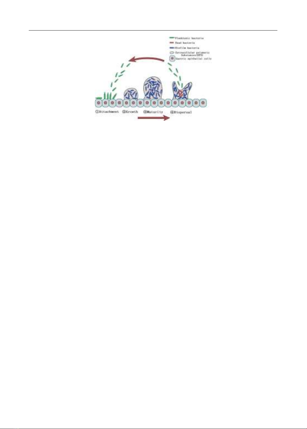
antibacterial mechanisms against H. pylori have also been mentioned, including (1) anti-biofilm
agents, (2) anti-virulence molecules (anti-VacA, anti-CagA agents, toxin BabA and LPS inhibitors,
anti-motility agents, Helicobacter pylori quorum sensing inhibitors), (3) mucolytic agents, and (4)
compounds that impact on essential proteins in the physiology of H. pylori such as inosin-5‘-
monophosphate dehydrogenase and HsrA inhibitors. This review article aims to summarize current
Can Tho Journal of Medicine and Pharmacy 10(7) (2024)
181
prospects, identify possible novel targets, and be considered as a complementary therapy in the
eradication treatment against Helicobacter pylori.
Keywords: anti-biofilm, anti-mucolytic, anti-virulence, Helicobacter pylori, HsrA inhibitors,
Inosin-5
-monophosphate dehydrogenase inhibitors.
I. INTRODUCTION
Helicobacter pylori (H. pylori) is a gram-negative, microaerophilic, and spiral
bacterium that was first cultured and identified by Marshall and Warren. This organism
mainly colonizes on the surface of the gastric mucosa and is associated with chronic
gastritis, gastric ulcers, gastric mucosa-associated tissue (MALT) lymphoma, gastric
cancer, and other disorders of the upper gastrointestinal tract [1]. In addition, resistance of
H. pylori to antibiotics has reached alarming levels worldwide, and the efficacy of the H.
pylori eradication treatment has decreased dramatically because of antibiotic resistance.
Historically, conventional antibiotics and proton pump inhibitors (PPIs) have been used in
triple therapy to treat H. pylori. However, due to widespread metronidazole and
clarithromycin resistance, H. pylori eradication rates have dropped to unacceptable levels.
Certain studies claim that typical triple therapy did not even come close to eliminating H.
pylori [2]. Researchers have explored a variety of strategies to mitigate the impacts of
antibiotic resistance, including changing medication regimens, increasing drug dosages, or
prolonging treatment periods [3]. According to the study of Li et al (2023), there is currently
no Helicobacter pylori vaccine for humans on the market [4].
Therefore, finding more new drugs derived from medicinal plants and their
compositions with unique mechanisms is always necessary in this day and age. With their
many modes of action in both in vitro, in vivo assay, and in small-scale clinical trials, natural
compounds are becoming more and more of a viable source of raw materials in Helicobacter
pylori treatment. Several widely recognized processes include anti-urease, anti-adhesive,
anti-inflammatory, and gastroprotective properties, in addition to their impact on the process
of oxidative stress in bacterial cells [5, 6]. To improve the rate of successful eradication of
H. pylori, various complementary therapies with novel targets have been uncovered such as
(1) anti-biofilm agents, (2) anti-virulence molecules (anti-VacA, anti-CagA agents, toxin
BabA and LPS inhibitors, anti-motility agents, H. pylori quorum sensing inhibitors), (3)
mucolytic agents, and (4) inosin-5’-monophosphate dehydrogenase and HsrA inhibitors.
Therefore, this review article aims to analyze current trends regarding these promising
mechanisms in Helicobacter pylori management.
II. CONTENTS
2.1. Natural anti-biofilm agents targeting H. pylori infection
2.1.1. Biofilm of Helicobacter pylori
One of the factors that affect the effectiveness of conventional eradication therapies
is the ability of H. pylori to form biofilms. Previously, most studies focused only on the
planktonic form (free-living) of H. pylori. However, several recent studies indicate that H.
pylori can develop biofilm structures when observed in both in vitro and in vivo models.
The presence of biofilm has been observed in the gastric mucosa, glands, and
gastrointestinal tract of mice or patients infected with H. pylori. The first evidence of the
presence of H. pylori biofilm during infection of the human gastric mucosa was reported in
articles by Carron and Coticchia in 2006. The studies using biopsy samples and SEM
Can Tho Journal of Medicine and Pharmacy 10(7) (2024)
182
analysis have demonstrated the dense presence of bacterial biofilm on the gastric mucosal
surface of patients positive for H. pylori with a rate of more than 97.3% [7], [8].
After penetrating the gastric mucosa, H. pylori releases proteins, polysaccharides,
extracellular DNA (eDNA), and other molecules to create extracellular polymeric substances
(EPS). These substances surround and adhere to each other by bacteria to form a biofilm.
Unlike the planktonic form, the bacteria make biofilm structures that are resistant to harsh
external environments, including exposure to antibiotics. Therefore, the formation of H. pylori
biofilms may be a major cause of persistent infections, antibiotic resistance, and treatment
failure [9, 10]. Notably, a biofilm of H. pylori contains virulence proteins such as AlpB, which
plays an important role in supporting bacterial adhesion to gastric mucosal cells and in
attaching bacterial cells together, the main steps in the biofilm formation process. H. pylori
strains lacking AlpB will face difficulties in developing biofilms [10], [11].
2.1.2. Stages of biofilm formation
Biofilm formation of H. pylori is divided into four stages (Figure 1), including (1)
attachment, (2) growth, (3) maturity, and (4) dispersal. At first, H. pylori adheres to the
gastric mucosa. This process is driven by cilia, pili structures, and lipopolysaccharides
(LPS), which are involved in the initial step of H. pylori infection. Next, the bacteria
multiply and produce extracellular polymeric substances (EPS), promoting the association
of bacteria with each other, and biofilm structure begins to form. The biofilm matures over
2-4 days and remains for a period. Then, when nutrients are depleted, and waste products
accumulate to a certain threshold, the biofilm will disintegrate. After that, there may be a
period of expansion of infection and biofilm formation at new locations [12].
Therefore, if the concentration or dosage of antibacterial agents is insufficient or if
only agents that destroy biofilm are used, it will not only be ineffective in eradicating all
bacteria but also break the biofilm. Hence, the dispersed bacterial flora can adhere to the
gastric epithelium, further expanding the biofilm size and increasing the scope of infection.
This may be the main reason for the phenomenon of biofilm size increasing instead of
decreasing after using subinhibitory doses of antibiotics. Therefore, agents should not be
used alone to only inhibit biofilm formation without antibacterial activity, antibiotics should
be combined to improve treatment effectiveness [9].
2.1.3. The role of H. pylori biofilm in antibiotic resistance
Biofilm formation reduces the susceptibility of bacteria to antibiotics, making it
difficult to eradicate H. pylori. Biofilm formation is considered a mechanism that helps H.
pylori survive and infect the gastric mucosa for a long time. Studies have also found a
correlation between biofilm formation and express increasing of many important virulence
genes. Because the bacteria in biofilm are exposed to a number of harsh conditions, such as
lack of nutrients, there can be an increase in mutation frequency and the emergence of
mutant strains causing antibiotic resistance [11], [12]. According to research by Fauzia et
al. (2020) in Indonesia, a higher antibiotic resistance rate on H. pylori strains can create
strong biofilm in all tested antibiotics, and the difference was the most significant for
clarithromycin (p = 0.002) [13].
Can Tho Journal of Medicine and Pharmacy 10(7) (2024)
183
Figure 1. Steps of H. pylori biofilm formation
(Source: Hou, C., et al., 2022)
2.1.4. Anti-biofilm agents
Bacteria carrying biofilm structures can cause 10-1000 times more antibiotic
resistance than bacteria in planktonic form. Therefore, developing agents aimed at
impacting biofilm to potentially remove or disperse biofilm may be an effective strategy to
reduce antibiotic resistance in Helicobacter pylori. Anti-biofilm agents can be considered
complementary therapy or alternative therapy in H. pylori eradication regimens [14]. Pure
compounds, extracts of natural origin, or some types of probiotics (Lactobacillus sp) are
demonstrated to have anti-biofilm effects and can inhibit bacteria on H. pylori strains from
being resistant to one antibiotic or multi-resistant, and data are presented in table 1. In the
in silico and in vitro screening tests by Spiegel et al. (2021), the dioscin compound (a
steroidal saponin) presents the most promising antibacterial, anti-biofilm formation
activities and shows the synergistic activity when combined with three conventional
antibiotics (clarithromycin, metronidazole, and levofloxacin) [15]. According to Xiao et al.
(2022), allicin is highly sensitive to AlpB and is believed to have anti-biofilm effects by
attaching to this protein [16].
2.2. Anti-virulence activity
Another approach to eradicating H. pylori is targeting the bacteria's virulence
factors. Inhibiting these factors can reduce the possibility of infection and increase the
effectiveness of antibiotic therapies. Furthermore, the structure of many bacterial
virulence factors has been clarified, so this treatment strategy has a number of
advantages over conventional antibiotic therapies that can be mentioned as follows: (i)
because it is not antibiotics so it does not affect the human gut microbiota; (ii) most
virulence factors are extracellular molecules so inhibitors can be easily exposed to them
[17].
The mechanism of action of anti-virulence agents can be divided into two categories:
(1) inhibition of a single virulence factor or (2) inhibition of processes involved in the
regulation of H. pylori virulence such as impacting on the quorum sensing process (signal
exchange pathway between bacterial cells), two-component regulatory system, the
flagellum system, and the bacterial virulence secretion systems or the process of post-
translational protein modifications because this process is related to protein maturation and
affects the biological function of bacterial cells. Among these, the inhibition of quorum-
sensing activity of H. pylori is most interesting [18],[19].
Can Tho Journal of Medicine and Pharmacy 10(7) (2024)
184
Table 1. Natural anti-biofilm agents targeting H. pylori infection
Anti-biofilm agents
Models
Tested strain of H. pylori
Chelidonium majus
and Corydalis cheilanthifolia
extracts
In vitro
HP 8064
Atractylodes lancea volatile oils
In vitro
NCTC 11637
Pistacia vera L. oleoresin
In vitro and
in vivo
30 clinical strains,
including multi-resistant strains
Hibiscus rosa sinensis Flower
In vitro
ATCC 43504, ATCC 51932
and 05 clinical strains
(OX.22, OX.63, OX.64,
OX.67, and OX.83)
Acorus calamus extract
In vitro
40 clinical strains
Vitex trifolia extract
In vitro
40 clinical strains
Casearia sylvestris
leaf derivatives
In vitro and
in vivo
ATCC 43504
Dihydrotanshinon I
(diterpenoid from
Salvia miltiorrhiza)
In vitro and
in vivo
ATCC 43504, HP G27, HP 26695,
HP NSH57 and BHKS159
Armeniaspirol A
(from Streptomyces armeniacus)
In vitro and
in vivo
HP G27, HP159, BHKS159
and 13 clinical strains
Curcumin
In vitro
ATCC43504, ATCC43526,
ATCC51932
Resveratrol (stilbenoids)
In vitro
ATCC 43629 and clinical strains
Myricetin (flavonoids)
In vitro
ATCC 700824, ATCC 51932
Nimbolid (Limonoid from
Azadirachta indica)
In vitro
ATCC 700684, HP G27,
HP 26695, HPAG1, HP SS1,
HP J99, HP 7.13 and USU101
Carvacrol and Thymol
(monoterpenoid)
In vitro
ATCC 43504
Dioscin (steroidal saponin)
In vitro
HP J99
Lactobacillus plantarum LN66
In vitro
ATCC 43504
Lactobacillus salivarius LN12
In vitro
ATCC43504, HP SS1
and clinical strains
2.2.1. Anti-VacA and anti-CagA agents
The key toxins of H. pylori are CagA and VacA, which play an important role in the
infection process. The toxin CagA is encoded by the cag pathogenicity island (cagPAI) and
translocated directly to gastric epithelial cells via the type IV secretion system (T4SS). Upon
entering target cells, CagA activates NF-kB, a main system in immune regulation and
inflammation, thereby increasing the secretion of proinflammatory cytokines (IL-8, TNF-α,
and IL-1β). Meanwhile, VacA is secreted from H. pylori, which is a vacuolating cytotoxin
causing apoptosis increase in host gastric mucosal cells. This toxin also increases the
production of the inflammatory cytokine IL-8 by activating the MAPK p38 molecule
through releasing intracellular Ca2+ ions [11].
Phenolic compounds, especially flavonoids, are important representatives of the