HUE JOURNAL OF MEDICINE AND PHARMACY ISSN 3030-4318; eISSN: 3030-4326
134
Hue Journal of Medicine and Pharmacy, Volume 14, No.6/2024
Isolation and structural determination of pentacyclic triterpenoids
from the leaves of Gymnosporia chevalieri tard
Doan Thi Ai Nghia1,4, Hoang Thi Nhu Hanh2, Le Tuan Anh3, Le Thi Hong Van4,
Vo Quoc Hung1, Nguyen Thi Hoai1, Ho Viet Duc1*
(1) Faculty of Pharmacy, University of Medicine and Pharmacy, Hue University
(2) Faculty of Engineering & Food Technology, University of Agriculture and Forestry, Hue University
(3) Mien trung Institute for Scientifc Research, Vietnam National Museum of Nature, VAST
(4) Faculty of Pharmacy, University of Medicine and Pharmacy at Ho Chi Minh city
Abstract
Background: The genus Gymnosporia, belonging to the Celastraceae family, which comprises
approximately 116 species globally, with 8 species identified in Vietnam. This work initially describes the
extraction, isolation, and structural identification of six triterpenoids from G. chevalieri collected in Vietnam.
Materials and methods: The leaves of G. chevalieri were subjected to extraction through immersion, followed
by a liquid-liquid partition process using organic solvents. Compounds were isolated using a combination of
thin-layer chromatography and column chromatography. Their structures were determined based on 1D-,
2D-NMR as well as by comparison with the reported spectroscopic data. Results & Conclusion: The chemical
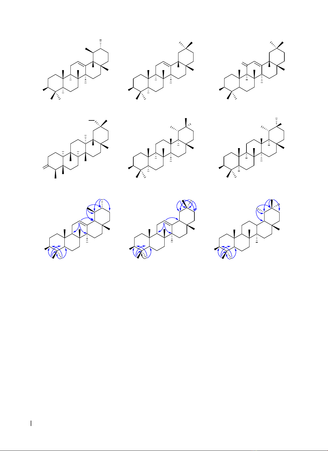
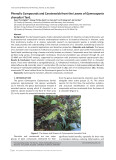
constituents of G. chevalieri was reported for the first time. Six pentacyclic triterpenoids have been isolated
and determined including mixture of α-amyrin (1a) and β-amyrin (1b), β-amyrenonol (2), 3-oxofriedelan-29-
ol (3), taraxastane-3β,20R-diol (4), and taraxastane-3β,20S-diol (5).
Keywords: Gymnosporia chevalieri, α-amyrin, β-amyrin, β -amyrenonol, 3-oxofriedelan-29-ol, taraxastane-
3,20-diol.
Corresponding Author: Ho Viet Duc. Email: hvietduc@hueuni.edu.vn
Received: 25/9/2024; Accepted: 14/11/2024; Published: 25/12/2024
DOI: 10.34071/jmp.2024.6.19
1. INTRODUCTION
The genus Gymnosporia (Celastraceae family)
comprises approximately 116 species worldwide [1].
Among these, eight species have been identified in
Vietnam, including G. diversifolia, G. stylosa, G. bonii,
G. chevalieri, G. gracilis, G. marcanii, G. mekongensis,
and G. tonkinensis [2]. Although phytochemical
studies on the genus Gymnosporia globally began
in the 1970s and have yielded impressive results,
domestic scientific interest in this resource has only
emerged in recent years, with a few publications
currently available on G. stylosa (commonly known
as “Dây lóp bóp”) [3], [4], [5].
Gymnosporia chevalieri (“Lõa châu” Chevalier)
is an endemic species in Vietnam. The chemical
constituents and biological activities of this species
remains relatively novel to scientific communities.
This article presents, for the first time, the extraction,
isolation, and structural determination of six
pentacyclic triterpenoids from the n-hexane extract
of the leaves of Gymnosporia chevalieri.
2. MATERIALS AND METHODS
2.1. Materials
The Gymnosporia chevalieri species was collected
in Đakrong District, Quảng Trị Province, in October
2023. The scientific name was identified by Dr. Anh
Tuan Le (Mien Trung Institute for Scientific Research,
Vietnam National Museum of Nature, VAST,
Vietnam). A specimen voucher (GC-01) has been
deposited at the Faculty of Pharmacy, University of
Medicine and Pharmacy, Hue University, Vietnam.
2.2. Methods
The powdered material was extracted with
methanol (MeOH) using maceration at room
temperature. The obtained crude extracts were
fractionated using liquid-liquid partitioning with
n-hexane, ethyl acetate (EtOAc). Pure compounds
were isolated by thin-layer chromatography
(TLC) and column chromatography (CC). TLC was
performed on pre-coated DC-Alufolien 60 F254 and
RP18 F254 plates (Merck, Germany). Compounds were
detected under UV light at wavelengths of 254 and
365 nm or by spraying the plates with 10% H2SO4
reagent followed by heating until color development.
Column chromatography was carried out using
various stationary phases, including normal silica gel
(40–63 µm, Merck, Germany), reverse-phase RP-18
(30–50 µm, Fuji Silysia Chemical, Japan), sephadex
LH-20 and MCI gel (Sigma-Aldrich, USA).
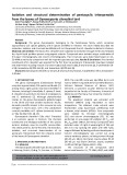
The structures of the compounds were
determined based on ¹H-, ¹³C-NMR, HSQC, and HMBC
HUE JOURNAL OF MEDICINE AND PHARMACY ISSN 3030-4318; eISSN: 3030-4326 135
Hue Journal of Medicine and Pharmacy, Volume 14, No.6/2024
spectra, combined with comparison to reference
spectral data. NMR spectra were recorded using
a Bruker Avance Neo 600 Spectrometer (Bruker,
Massachusetts, USA) at the Institute of Chemistry,
Vietnam Academy of Science and Technology. CDCl₃
was used as the solvent, and tetramethylsilane
(TMS) served as the internal standard.
3. RESULTS AND DISCUSSION
3.1. Extraction and Isolation
The leaves of Gymnosporia chevalieri were
cleaned, air-dried, and ground into powder (4 kg).
The powdered material was extracted by maceration
with MeOH (10 L x 3 times), and the solvent was
removed under reduced pressure to yield a crude
extract (GCM, 930 g). This extract was partitioned
into water (2 L) and successively extracted with
n-hexane and EtOAc (each, 4 L x 3 times). The
solvents were evaporated under reduced pressure
to obtain the corresponding extracts: n-hexane
(GCH; 201.8 g), EtOAc (GCE; 97.9 g), and aqueous
extract (GCW; 525 g).
The GCH extract was subjected to silica gel
column using a gradient elution of n-hexane/
acetone (100:0, 80:1, 40:1, 20:1, 10:1, 5:1, 2:1, 1:1,
and 0:100, v/v) to afford six fractions, GCH1-GCH6.
Fraction GCH3 (109.1 g) was repeatedly washed with
acetone to yield compound 1 (97.0 g). Fraction GCH5
(30.3 g) was separated by silica gel column, eluting
with a gradient of n-hexane/acetone (10:1, 5:1, 2:1,
1:1, v/v), yielding four fractions: GCH5.1-GCH5.4.
Fraction GCH5.1 (3.6 g) was subjected to silica gel
column with a gradient of n-hexane/EtOAc (5:1,
3:1, v/v), giving eight fractions: GCH5.1.1-GCH5.1.8.
Fraction GCH5.1.5 (31.3 mg) was purified by RP-18
column using acetone/water (20:1, v/v) to yield 3
(20 mg).
Fraction GCH5.1.4 (301.4 mg) was sequentially
separated by sephadex column using CH₂Cl₂/MeOH
(1:1, v/v), followed by RP-18 column using acetone/
water (10:1, v/v), yielding five fractions: GCH5.1.4.1-
GCH5.1.4.5. Fraction GCH5.1.4.5 (54.1 mg) was
purified by sephadex column using CH₂Cl₂/MeOH
(1:1, v/v), yielding compound 2 (6.7 mg).
Fraction GCH5.1.6 (650 mg) was separated by
MCI gel, eluting with a gradient of MeOH/water
(100:0, 8:2, and 6:4, v/v), yielding four fractions:
GCH5.1.6.1-GCH5.1.6.4. Fraction GCH5.1.6.1 (300
mg) was further separated by sephadex column using
CH₂Cl₂/MeOH (1:1, v/v), yielding three fractions:
GCH5.1.6.1.1-GCH5.1.6.1.3. Fraction GCH5.1.6.1.2
(209 mg) was further separated by silica gel column
using CH₂Cl₂/acetone (40:1, v/v), yielding five
fractions: GCH5.1.6.1.2.1-GCH5.1.6.1.2.5. Fraction
GCH5.1.6.1.2.3 (45.5 mg) was purified by RP-18
column using acetone/water (10:1, v/v), yielding
compound 4 (6.7 mg). Fraction GCH5.1.6.1.2.5 (19
mg) was purified by RP-18 column using acetone/
water (5:1, v/v), yielding compound 5 (3.5 mg).
3.2. Structural determination of isolates
Compound 1 was isolated as a white powder. The
¹H-NMR spectrum indicated characteristic signals of
two olefinic protons at δH 5.18/5.13 (t, J = 3.6 Hz);
two oxymethine groups at δH 3.23/3.22 (dd, J = 10.8,
4.8 Hz); and 16 methyl groups at δH 1.14, 1.07, 1.01,
1.00 (6H), 0.97, 0.95, 0.94, 0.92, 0.87 (6H), 0.83, 0.80,
and 0.79 (9H). These data suggest that compound 1
is a mixture of two triterpenoids (1a:1b) (Figure 1).
The ¹³C-NMR and HSQC spectra revealed the
presence of two oxymethine groups (δC 79.1/79.0)
and two trisubstituted double bonds (δC 139.6
(C)/124.5 (CH) and 145.2 (C)/121.8 (CH)), which were
attributed to 1a and 1b, respectively. The assignment
of NMR data for components 1a and 1b was based
on detailed analysis of the HMBC spectrum of
compound 1 (Table 1). Particularly, the position of the
OH group at C-3 of each compound was confirmed
by the correlations of H3-23 (δH 1.00)/H3-24 (δH 0.79)
with C-3 (δC 79.1/79.0), C-4 (δC 38.8/39.7), and C-5
(δC 55.2). The position of the double bond at Δ¹² was
established by HMBC correlations between H-12 (δH
5.13) and C-9 (δC 47.8)/C-14 (δC 41.6)/C-18 (δC 59.1)
for 1a, and between H-12 (δH 5.18) and C-9 (δC 47.7)/
C-14 (δC 42.1)/C-18 (δC 47.3) for 1b.
The correlation of two methyl doublets [δH
1.01 (H3-29), 0.92 (H3-30)] with C-19 (δC 39.6)/C-
20 (δC 38.8) confirmed the ursane skeleton for 1a.
Meanwhile, the correlation of two methyl singlets
[δH 0.87 (6H, H3-29 & H3-30)] with C-19 (δC 46.9)/C-
20 (δC 31.1)/C-21 (δC 34.8) suggested an oleanane
skeleton for 1b (Figure 2). Based on the above data,
compound 1 was identified as a mixture of α-amyrin
(1a) and β-amyrin (1b) [6]. The ratio of 1a:1b,
approximately 1.25:1, was determined from the
integration values of the characteristic H-12 signals
at δH 5.18 (for 1a) and 5.13 (for 1b).
HUE JOURNAL OF MEDICINE AND PHARMACY ISSN 3030-4318; eISSN: 3030-4326
136
Hue Journal of Medicine and Pharmacy, Volume 14, No.6/2024
35
1
7
914
12 18
HO
24 23
27
H
25
20
28
29
30
H
26
1a
35
1
7
914
12 18
HO
24 23
27
H
25
20
28
29 30
H
26
1b
3
5
1
7
914
12 18
HO
24 23
27
H
25
20
28
29 30
H
26
2
O
35
1
7
914
12 18
O
23
27
25
20
28
29 30
H
H
26
3
24
H
H H
HO
35
1
7
914
12 18
HO
24 23
27
H
25
20
28
29
30
H
26
4
H
35
1
7
914
12 18
HO
24 23
27
H
25
20
28
29
30
H
26
5
H
H
H
OH OH
Figure 1. Structures of pentacyclic triterpenoids isolated from Gymnosporia chevalieri
35
1
7
9
14
12 18
HO
23 24
27
20
28
29
30
1a
35
1
7
9
14
12 18
HO
23 24
27
20
28
29
30
1b
35
1
7
9
14
12 18
HO
23 24
27
20
28
29
30
4
OH
Figure 2. Selected HMBC correlations of 1a, 1b and 4
Compound 2 was isolated as a white powder,
readily soluble in CH₂Cl₂. The ¹H-NMR spectrum
displayed a characteristic olefinic proton signal at δH
5.58 (s) and a carbinol group signal at δH 3.22 (dd,
J = 11.4, 6.0 Hz). In addition, the high-field region
revealed eight singlet methyl group signals at δH
1.36, 1.14, 1.13, 1.00, 0.90, 0.89, 0.86, and 0.81.
The ¹³C-NMR and HSQC spectra of the compound
showed 30 carbon signals, including 8 CH3, 9 CH2, 5
CH, and 8 quaternary carbons (Table 2).
Notably, 1D-NMR data confirmed the presence
of several key functional groups, including a carbonyl
group (δC 200.3), a trisubstituted double bond (δC/
δH 170.6, 128.1/5.58), and a carbinol group (δC/δH
78.8/3.22). The strong downfield shift of the sp²
carbon (δC 170.6) and the singlet proton signal of
the olefin (δH 5.58) suggested the conjugation of
the double bond with a ketone group, forming an
enone structure. These spectral data indicated that
compound 2 is a pentacyclic triterpenoid with an
oleanane skeleton similar to β-amyrin (1b). Further
comparison with reference spectra [7] allowed the
identification of compound 2 as 3β-hydroxy-11-
oxo-olean-12-ene, also known as β-amyrenonol or
11-oxo-β-amyrin.
The ¹H-NMR signals of compound 3 mainly
appeared in the high-field region (δH 0.7-3.5), typical
for saturated terpenoids. Notable resonance signals
included an oxymethylene group [δH 3.26 (2H, q, J
= 10.2 Hz)], six methyl singlets [δH 1.22, 1.05, 1.04,
1.03, 0.87, and 0.73], and one methyl doublet [δH
0.88 (d, J = 6.6 Hz)].
The ¹³C-NMR spectrum revealed 30 carbon
signals, with notable features including a carbonyl
group (δC 200.3) and an oxymethylene group (δC
74.8). HSQC data allowed classification of the 30
HUE JOURNAL OF MEDICINE AND PHARMACY ISSN 3030-4318; eISSN: 3030-4326 137
Hue Journal of Medicine and Pharmacy, Volume 14, No.6/2024
Table 1. NMR data of mixture 1a:1b and reference compounds
C1a 1b
δC
#, a δC
a, b δH
a, c
(J, Hz) δC
*, a δC
a, b δH
a, c
(J, Hz)
138.8 38.8 t 1.63 m/0.98 m 38.6 38.6 t 1.63 m/0.98 m
227.3 27.3 t 1.60 m/1.57 m 27.2 27.3 t 1.60 m/1.57 m
379.1 79.1 d 3.23 dd (10.8; 4.8) 79.0 79.0 d 3.22 dd (10.8; 4.8)
4 38.8 38.8 s - 39.8 39.7 s -
5 55.2 55.2 d 0.74 m 55.2 55.2 d 0.74 m
618.4 18.4 t 1.55 m/1.40 m 18.4 18.4 t 1.55 m/1.40 m
732.9 33.0 t 1.54 m/1.35 m 32.5 32.5 t 1.54 m/1.35 m
840.0 40.0 s - 41.7 41.8 s -
947.7 47.8 d 1.52 m 47.6 47.7 d 1.55 m
10 36.9 36.9 s - 37.0 37.0 s -
11 23.3 23.3 t 1.63 m/0.98 m 23.7 23.7 t 1.88 m/1.85 m
12 124.4 124.5 d 5.13 t (3.6) 121.7 121.8 d 5.18 t (3.6)
13 139.6 139.6 s - 145.2 145.2 s -
14 41.5 41.6 s - 42.8 42.1 s -
15 28.1 28.1 t 2.00 m/0.86 m 26.9 27.0 t 2.00 m/0.80 m
16 26.6 26.6 t 1.83 m/0.96 m 26.2 26.2 t 1.77 td (13.2; 4.2)/0.96 m
17 33.8 33.8 s - 32.7 32.7 s -
18 59.1 59.1 d 1.32 m 47.2 47.3 d 1.95 m
19 39.7 39.6 d 1.32 m 46.8 46.9 t 1.67 m/1.00 m
20 39.6 38.8 d 0.87 m 31.1 31.1 s -
21 31.3 31.3 t 1.38 m/1.26 m 34.7 34.8 t 1.33 m/1.10 m
22 40.0 39.8 t 1.42 m/1.27 m 37.1 37.2 t 1.43 m/1.22 tt (10.2; 3.0)
23 28.1 28.1 q 1.00 s 28.1 28.1 q 1.00 s
24 15.7 15.7 q 0.79 s 15.6 15.6 q 0.79 s
25 15.6 15.6 q 0.95 s 15.5 15.5 q 0.94 s
26 17.4 17.5 q 0.79 s 16.8 16.8 q 0.97 s
27 23.4 23.4 q 1.07 s 26.0 26.0 q 1.14 s
28 28.8 28.8 q 0.80 s 28.4 28.4 q 0.83 s
29 16.9 16.9 q 1.01 d (6.0) 33.3 33.3 q 0.87
30 21.4 21.4 q 0.92 d (6.0) 23.5 23.6 q 0.87 s
#, *δC values of α-, β-amyrin [6], ameasured in CDCl3, b150 MHz, c600 MHz
carbons into 7 CH3 (δC 32.1, 25.8, 20.8, 18.5, 17.9,
14.7, 6.8), 12 CH2 (δC 74.8, 41.5, 41.3, 39.5, 35.9,
35.6, 32.7, 30.6, 30.5, 27.8, 22.3, 18.2), 4 CH (δC
59.5, 58.2, 53.4, 41.9), and 7 C (δC 213.2, 42.2, 40.0,
38.2, 37.4, 33.1, 29.8). The methyl doublet signal at
δC 6.8, which is diagnostic of triterpenoids with a
friedelane skeleton, further supports this structural
assignment. After comparison with reference
spectral data [8], compound 3 was identified as
3-oxofriedelan-29-ol.
HUE JOURNAL OF MEDICINE AND PHARMACY ISSN 3030-4318; eISSN: 3030-4326
138
Hue Journal of Medicine and Pharmacy, Volume 14, No.6/2024
Table 2. NMR data of 2, 3 and reference compounds
C2 3
δC
#, a δC
a, b δH
a, c
(J, Hz) δC
*, a δC
a, b δH
a, c
(J, Hz)
138.7 39.2 t 2.78 dt (13.2; 3.6)/0.98 m 22.3 22.3 t 1.95 m/1.67 m
223.4 27.3 t 1.66 m/1.62 m 41.6 41.5 t 2.38 m/2.29 m
380.5 78.8 d 3.22 dd (11.4; 6.0) 212.2 213.2 s -
4 38.0 39.2 s -58.3 58.2 d 2.24 ddd (7.2; 5.4; 1.8)
5 54.9 55.0 d 0.69 dd (12.0; 1.2) 42.2 42.2 s -
618.4 17.5 t 1.59 m/1.45 m 41.4 41.3 t 1.75 m/1.28 m
732.6 32.8 t 1.65 m/1.42 m 18.3 18.2 t 1.49 m/1.40 m
843.3 43.4 s -53.5 53.4 d 1.40 m
961.5 61.8 d 2.34 s 37.5 37.4 s -
10 36.9 37.1 s -59.6 59.5 d 1.52 m
11 201.4 200.3 s -35.7 35.6 t 1.45 m/1.26 m
12 127.9 128.1 d 5.58 s 29.8 30.5 t 1.35 m
13 170.2 170.6 s -40.0 40.0 s -
14 45.3 45.4 s -38.3 38.2 s -
15 26.4 26.5 t 2.06 td (13.2; 4.2)
1.82 td (13.2; 4.8)
32.8 32.7 t 1.55 m/1.32 m
16 26.4 26.4 t 1.17 m/0.96 m 36.0 35.9 t 1.59 m/1.33 m
17 32.3 32.4 s -29.8 29.8 s -
18 47.5 47.6 d 2.13 dd (13.8; 3.6) 42.0 41.9 d 1.61 m
19 45.0 45.2 t 1.67 m/1.06 m 30.6 30.6 t 1.35 m
20 31.0 31.1 s -33.2 33.1 s -
21 34.4 34.5 t 1.35 m/1.17 m 27.9 27.8 t 1.37 m
22 36.4 36.5 t 1.47 m/1.29 m 39.6 39.5 t 1.41 m/1.36 m
23 28.0 28.1 q 1.00 s 6.8 6.8 q 0.88 d (6.6)
24 16.6 16.4 q 1.14 s 14.7 14.7 q 0.73 s
25 15.7 15.6 q 0.81 s 17.9 17.9 q 0.87 s
26 17.3 18.7 q 1.13 s 18.4 18.5 q 1.05 s
27 23.5 23.5 q 0.89 s 20.8 20.8 q 1.04 s
28 28.7 28.8 q 0.86 s 32.1 32.1 q 1.22 s
29 33.0 33.1 q 0.90 s 74.8 74.8 t 3.26 q (10.2)
30 23.5 23.5 q 1.36 s 25.9 25.8 q 1.03 s
#δC values of β-amyrenyl acetate [7], *δC values of 3-oxofriedelan-29-ol [8], ameasured in CDCl3, b150 MHz,
c600 MHz