
e384 AACE CLINICAL CASE REPORTS Vol 5 No. 6 November/December 2019
Copyright © 2019 AACE
Case Report
PARATHYROMATOSIS: A RARE CASE OF
RECURRENT HYPERPARATHYROIDISM LOCALIZED BY
FOUR-DIMENSIONAL COMPUTED TOMOGRAPHY
Abraham E. Wei, DO1; Matthew R. Garrett, MD2; Ankur Gupta, MD, FACE1
Submitted for publication May 14, 2019
Accepted for publication August 8, 2019
From 1Wright State University Boonshoft School of Medicine, Dayton, Ohio,
and 2Southwest Ohio Ear, Nose, and Throat Specialists, Dayton, Ohio.
Address correspondence to Dr. Abraham Wei, Wright State University
Internal Medicine Residency, 1 Wyoming Street, Dayton, OH 45419.
E-mail: Abraham.wei@wright.edu.
DOI: 10.4158/ACCR-2019-0225
To purchase reprints of this article, please visit: www.aace.com/reprints.
Copyright © 2019 AACE.
ABSTRACT
Objective: To present a rare case of parathyromatosis.
Methods: We present the clinical, laboratory, and
imaging findings, along with a review of the literature.
Results: A 33-year-old man with a history of right
upper parathyroid adenoma removal 5 years prior due to
hyperparathyroidism was admitted for severe hypercalce-
mia (15.6 mg/dL; normal, 8.5 to 10.5 mg/dL) with elevated
plasma parathyroid hormone (PTH) (882 pg/mL; normal,
15 to 65 pg/mL). Ultrasound, computed tomography (CT),
sestamibi, and positron emission tomography scans were
unremarkable; however, a four-dimensional CT (4DCT)
of the neck showed an area of increased signal enhance-
ment and hypervascularity without discrete nodule in the
posterior right thyroid region. The patient underwent para-
thyroid surgical exploration with right hemithyroidectomy
and compartment neck dissection to remove the affected
tissue. PTH levels dropped to 208 pg/mL postoperatively;
calcium decreased but remained elevated at 12.7 mg/dL.
Pathology revealed the presence of several small nodular
foci of atypical hyperplastic parathyroid tissue in the right
thyroid and soft tissue in the left central neck compartment
consistent with parathyromatosis.
Conclusion: This case report represents the first-time
use of 4DCT to localize parathyromatosis. Parathyromatosis
is a rare but problematic cause of recurrent hyperparathy-
roidism. Ultrasound and 4DCT may represent the best
imaging modalities for identification and perioperative
management to remove all affected tissue without reseed-
ing. (AACE Clinical Case Rep. 2019;5:e384-e387)
Abbreviations:
CT = computed tomography; 4DCT = four-dimension-
al computed tomography; PTH = parathyroid hormone
INTRODUCTION
Parathyromatosis is a rare condition of multiple nests
of hyperfunctioning parathyroid tissue. It is likely either
the result of spillage and seeding of parathyroid tissue
around an operative area during parathyroid surgery or
the change in embryologic foci of parathyroid tissue
that become hyperplastic with physiologic stimuli (1).
Preoperative diagnosis and subsequent treatment can be
difficult. Imaging is important for identification and peri-
operative management to remove all affected tissue with-
out reseeding. This case report describes the diagnostic
challenges for this condition as well as the first-time use
of four-dimensional computed tomography (4DCT) in the
diagnosis of parathyromatosis.
CASE REPORT
A 33-year-old man first presented at 28 years of age
with epigastric pain and was found to have serum calcium
of 18.1 mg/dL (normal, 8.5 to 10.5 mg/dL) and an elevated
plasma parathyroid hormone (PTH) level of 1,199 pg/mL
(normal, 14 to 72 pg/mL). Computed tomography (CT) of
the neck revealed a 2 cm nodule posterior to the superior

Parathyromatosis and Imaging, AACE Clinical Case Rep. 2019;5(No. 6) e385 Copyright © 2019 AACE
right lobe of the thyroid gland and the patient underwent a
right upper parathyroidectomy. Repeat labs 2 weeks later
showed normalization of calcium at 9.2 mg/dL and of PTH
at 64 pg/mL (Table 1). Pathology was consistent with an
atypical parathyroid adenoma described as neoplastic para-
thyroid cells in trabecular arrangement with interspersing
fibrovascular bands without mitosis nor evidence of vascu-
lar or perineural invasion. Five years later, the patient
developed abdominal pain, nausea, and vomiting for 9 days
and was admitted for an elevated serum calcium of 15.6
mg/dL (normal, 8.5 to 10.5 mg/dL) with an elevated PTH
level of 882 pg/mL (normal, 15 to 65 pg/mL). Physical
exam was only significant for mild epigastric tenderness
on deep palpation. Serum creatinine was also noted to be
elevated at 1.6 mg/dL (normal, 0.5 to 1.4 mg/dL) attributed
to poor fluid intake by the patient. Urinary calcium was
noted to be high at 530 mg/24 hours (normal, 50 to 150
mg/24 hours) and serum 25-hydroxyvitamin D was low at
21 ng/mL (normal, >30 ng/mL). Intravenous normal saline
given during the hospital stay improved serum creatinine
to baseline level of 1.0 mg/dL. Serum calcium continued to
remain high and greater than 14 mg/dL. Cinacalcet 30 mg
twice-a-day was then initiated.
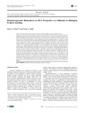
Ultrasound of the neck, technetium-sestamibi scan
with single photon emission CT, CT soft tissue neck with
contrast (Fig. 1 A) were all unremarkable. CT chest/abdo-
men/pelvis with contrast and positron emission tomog-
raphy (PET) scan did not show any evidence of ectopic
tumors or distant metastasis. A 4DCT of the neck was then
obtained that showed an area of increased signal enhance-
ment and hypervascularity without a discrete nodule in the
posterior right thyroid region near the site of prior parathy-
roid adenoma removal (Fig. 1 C). The patient then under-
went parathyroid surgical exploration with right hemithy-
roidectomy, and intraoperative frozen section analysis
showed the presence of abnormal parathyroid tissue inti-
mately involved with the right thyroid lobe. Intraoperative
biopsies of right inferior, left superior, and left inferior
parathyroid appeared normal and were not hyperplas-
tic. Central compartment neck dissection was completed
without additional abnormal parathyroid tissue identified.
Postoperatively, calcium level decreased but remained
elevated at 12.7 mg/dL. Cinacalcet, which had been held
before surgery, was restarted. Three days after surgery,
serum calcium improved and stabilized at 9.9 mg/dL and
PTH improved to 174 pg/mL. The patient was subsequent-
ly discharged. Pathology showed the presence of several
small nodular foci of atypical hyperplastic parathyroid
tissue without invasive growth in the right thyroid and soft
tissue in the left central neck compartment consistent with
parathyromatosis.
DISCUSSION
Parathyromatosis is a rare cause of recurrent hyper-
parathyroidism consisting of multiple nodules of benign
hyperplastic and hyperfunctioning parathyroid tissue after
spillage and seeding of parathyroid tissue during parathy-
roid surgery. Another cause of parathyromatosis describes
embryonic rests of parathyroid tissue that undergo hyper-
plasia under the influence of physiological stimuli such as
end-stage renal disease. Our patient’s past operative report
on his parathyroid adenoma removal makes no mention of
spillage of parathyroid tissue, but this can still be a possible
cause of his parathyromatosis. Otherwise, this may be some
form of disorder which allows local cells to differentiate
into parathyroid cells. Epidemiologic data reveal parathy-
romatosis affects people across a wide age range from 19
to 87 years old with the majority being female or having
end-stage renal disease, which makes our patient different
Table 1
Patient Laboratory Values
First presentation
(parathyroid adenoma with
parathyroidectomy)
Event
Calcium (mg/dL, normal
8.5-10.5 mg/dL)
PTH (pg/mL, normal
14-72 pg/mL)
On admission prior to surgery 18.1 1,199
1 day after surgery 9.0 NR
2 days after surgery (discharge day) 8.8 NR
2 weeks after surgery 9.2 64
Second presentation 5
years after first presentation
(parathyromatosis with
hemithyroidectomy)
Event
Calcium (mg/dL,
normal 8.5-10.5 mg/dL)
PTH (pg/mL,
normal 15-65 pg/mL)
On admission prior to surgery 15.6 882
1 day after surgerya12.7 NR
2 days after surgery 10.0 NR
3 days after surgery (discharge day) 9.9 174
Abbreviations: NR = not reported; PTH = parathyroid hormone.
Values in bold are out of normal range.
aStarted oral cinacalcet 30 mg twice a day.

e386 Parathyromatosis and Imaging, AACE Clinical Case Rep. 2019;5(No. 6) Copyright © 2019 AACE
from most cases (2,3,4). Parathyromatosis is often refrac-
tory to surgical intervention due to incomplete removal and
reseeding of hyperplastic parathyroid tissue (2). Mortality
from complications of parathyromatosis can be high with
one case series reporting a mortality rate of 40% (5).
Successful imaging of these small nodules of hyper-
plastic parathyroid tissue and obtaining a preoperative
diagnosis is difficult, but it enhances the chances of surgi-
cally removing all involved tissue. In a case series of 10
patients, only 40% of patients were successfully diagnosed
with parathyromatosis preoperatively (3). Ultrasound can
sometimes detect scattered hypoechoic, vascular small
nodules in thyroid tissue suggesting the diagnosis (6). CT,
sestamibi, and PET scans can reveal findings of parathyro-
matosis but are often negative (2). In our patient, all these
modalities failed to localize parathyromatosis.
We then obtained a 4DCT to localize the affected
tissue and optimize perioperative management and surgical
resection. 4DCT may be a popular imaging option to local-
ize and diagnose parathyromatosis in the future. 4DCT is
a relatively new imaging modality first introduced in 2006
that is utilized in the diagnosis and characterization of
suspected parathyroid adenoma as well as for brain aneu-
rysms, arterial-venous malformations, musculoskeletal
conditions such as joint instability, and internal impinge-
ment (7). Intravenous contrast is given and CT scans are
obtained at specific time-points corresponding to venous
and arterial circulations which enhances the quality of the
images. In a study of 45 patients who previously under-
went parathyroidectomy, sensitivity for localization of
hyperfunctioning parathyroid tissue for reoperation was
88% for 4DCT compared to 54% for sestamibi imaging
(8). The enhanced capability to image abnormal hypervas-
cular tissues with 4DCT makes it a good diagnostic test of
choice for localizing parathyromatosis.
Pathology of parathyromatosis often reveals multi-
ple nests of hyperplastic parathyroid cells with a lack of
real capsule of the parathyroid tissue and without lymph-
vascular invasion that is seen in an adenoma or cancer (9).
Definitive treatment is surgical removal of all offending
tissue; however, it is difficult to identify and remove all
the disseminated tiny nodules of hyperplastic parathy-
roid tissue that often adhere closely to scar tissue thereby
hindering excision. Even the more aggressive measure of
total thyroidectomy does not ensure cure as parathyromato-
sis tissue can remain in surrounding muscular or fat tissue
(3). Many cases are often refractory to or incompletely
cured with surgery, such as in our case where postoperative
PTH and serum calcium levels remained elevated, indicat-
ing incomplete removal of all affected parathyromatosis
tissue. Without localization via 4DCT, less parathyroma-
tosis tissue would have been removed or more tissue seed-
ing might have occurred if blind surgical manipulation was
done, likely leading to worse outcomes.
After failed surgical intervention, suppression of PTH
production with the calcium mimetic cinacalcet is the phar-
macologic treatment of choice. Bisphosphonates may also
be added to both decrease calcium levels and to minimize
the deleterious effect of PTH on the bone, especially if
there is concern for concomitant osteopenia or osteoporosis
(2). Several cases also successfully used vitamin D analogs
(e.g., paricalcitrol, doxercalciferol) to further inhibit PTH
synthesis and parathyromatosis activity, although at a cost
of increasing serum calcium level (6,10). Even with phar-
Fig. 1. A, Regular CT with contrast. B, 4DCT without contrast. C, 4DCT arterial phase with contrast enhance-
ment in right posterior thyroid (red arrow) where previous parathyroid adenoma was located and resected. D,
4DCT 90-second delay (venous) phase. CT = computed tomography; 4DCT = four-dimensional computed
tomography.
A B
C D

Parathyromatosis and Imaging, AACE Clinical Case Rep. 2019;5(No. 6) e387 Copyright © 2019 AACE
macologic interventions, parathyromatosis activity may
not be controlled. Ideally, this condition should be prevent-
ed in the first place by avoiding rupture of the parathyroid
capsule during parathyroidectomy.
CONCLUSION
This case represents the first reported use of 4DCT to
localize parathyromatosis tissue. 4DCT is very sensitive in
detecting hypervascular hyperparathyroid tissue. This may
become a popular imaging modality to localize and diag-
nose parathyromatosis. However, parathyromatosis may be
refractory to surgical and pharmacologic interventions in
several cases.
DISCLOSURE
The authors have no multiplicity of interest to disclose.
REFERENCES
1. Reddick RL, Costa JC, Marx SJ. Parathyroid hyperplasia and
parathyromatosis. Lancet. 1977;1:549.
2. Hage MP, Salti I, El-Hajj Fuleihan G. Parathyromatosis: a rare
yet problematic etiology of recurrent and persistent hyperparathy-
roidism. Metabolism. 2012; 61;762-775.
3. Matsuoka S, Tominaga Y, Sato T, et al. Recurrent renal hyper-
parathyroidism caused by parathyromatosis. World J Surg.
2007;31:299-305.
4. Fernandez-Ranvier GG, Khanafshar E, Jensen K, et al.
Parathyroid carcinoma, atypical parathyroid adenoma, or parathy-
romatosis? Cancer. 2007;110:255-264.
5. Stehman-Breen C, Muirhead N, Thorning D, Sherrard D.
Secondary hyperparathyroidism complicated by parathyromatosis.
Am J Kidney Dis. 1996;28:502-507.
6. Tublin ME, Yim JH, Carty SE. Recurrent hyperparathyroid-
ism secondary to parathyromatosis: clinical and imaging findings.
J Ultrasound Med. 2007;26:847-851.
7. Kwong Y, Mel AO, Wheeler G, Troupis JM. Four-dimensional
computed tomography (4DCT): a review of the current status and
applications. J Med Imaging Radiat Oncol. 2015;59:545-554.
8. Mortenson MM, Evans DB, Lee JE, et al. Parathyroid explo-
ration in the reoperative neck: improved preoperative localiza-
tion with 4D-computed tomography. J Am Coll Surg. 2008;206:
880-895.
9. Aksoy-Altinboga A, Akder Sari A, Rezanko T, Haciyanli M,
Orgen Calli A. Parathyromatosis: critical diagnosis regarding
surgery and pathologic evaluation. Korean J Pathol. 2012;46:
197-200.
10. Daphnis E, Stylianou K, Katsipi I, et al. Parathyromatosis and
the challenge of treatment. Am J Kidney Dis. 2006;48:502-505.

![Phác đồ sử dụng MMF trong dị ghép tế bào gốc [chuẩn nhất]](https://cdn.tailieu.vn/images/document/thumbnail/2025/20250508/antrongkim0609/135x160/7241746691763.jpg)
![Hướng dẫn chẩn đoán và điều trị ung thư tế bào gan [chuẩn nhất]](https://cdn.tailieu.vn/images/document/thumbnail/2025/20250508/antrongkim0609/135x160/4691746691992.jpg)


%20--%3e%3cdefs%3e%3cstyle%3e%20.st0%20{%20fill:%20%23fff;%20}%20.st1%20{%20fill:%20%237800fa;%20}%20%3c/style%3e%3c/defs%3e%3cpath%20class='st1'%20d='M117.78,12.18H43.11c2.9,3.47,4.65,7.94,4.65,12.82,0,5.6-2.3,10.66-6.01,14.29h76.02l7.22-13.56-7.22-13.56Z'/%3e%3cg%3e%3cpath%20class='st0'%20d='M53.58,26.17h-.59v-1.46h.59v-4.96h2.83c1.78,0,2.67.94,2.67,2.82v5.76c0,1.87-.89,2.81-2.67,2.81h-2.83v-4.96ZM55.36,21.37v3.34h1.1v1.46h-1.1v3.34h1.01c.61,0,.91-.37.91-1.1v-5.93c0-.74-.3-1.1-.91-1.1h-1.01Z'/%3e%3cpath%20class='st0'%20d='M65.99,31.14h-1.8l-.31-2.07h-2.19l-.31,2.07h-1.64l1.82-11.39h2.62l1.82,11.39ZM65.28,18.04c-.25.46-.51.77-.75.94-.21.15-.47.22-.79.22-.26,0-.57-.07-.92-.22l-.38-.15c-.14-.05-.26-.07-.37-.07-.3,0-.53.18-.71.54l-.91-.68c.25-.46.51-.77.75-.94.21-.14.48-.21.79-.21.26,0,.57.07.92.21l.38.15c.14.05.26.07.37.07.3,0,.53-.18.71-.54l.91.68ZM61.91,27.52h1.73l-.87-5.76-.87,5.76Z'/%3e%3cpath%20class='st0'%20d='M74.53,26.89v1.52c0,1.91-.89,2.86-2.67,2.86s-2.67-.95-2.67-2.86v-5.93c0-1.91.89-2.86,2.67-2.86s2.67.95,2.67,2.86v1.11h-1.69v-1.22c0-.75-.31-1.12-.93-1.12s-.93.37-.93,1.12v6.15c0,.74.31,1.11.93,1.11s.93-.37.93-1.11v-1.63h1.69Z'/%3e%3cpath%20class='st0'%20d='M81.4,31.14h-1.8l-.31-2.07h-2.19l-.31,2.07h-1.64l1.82-11.39h2.62l1.82,11.39ZM75.9,19.2l1.52-1.91h1.71l1.51,1.91h-1.61l-.76-.95-.75.95h-1.61ZM77.32,27.52h1.73l-.87-5.76-.87,5.76ZM83.1,15.99l-1.76,1.91h-1.26l1.17-1.91h1.86Z'/%3e%3cpath%20class='st0'%20d='M84.86,19.75c1.78,0,2.67.94,2.67,2.82v1.48c0,1.87-.89,2.81-2.67,2.81h-.85v4.28h-1.79v-11.39h2.64ZM84.01,21.37v3.86h.85c.58,0,.87-.36.87-1.08v-1.71c0-.71-.29-1.07-.87-1.07h-.85Z'/%3e%3cpath%20class='st0'%20d='M93.51,19.75c1.78,0,2.67.94,2.67,2.82v1.48c0,1.87-.89,2.81-2.67,2.81h-.85v4.28h-1.79v-11.39h2.64ZM92.66,21.37v3.86h.85c.58,0,.87-.36.87-1.08v-1.71c0-.71-.29-1.07-.87-1.07h-.85Z'/%3e%3cpath%20class='st0'%20d='M98.8,31.14h-1.79v-11.39h1.79v4.88h2.03v-4.88h1.83v11.39h-1.83v-4.88h-2.03v4.88Z'/%3e%3cpath%20class='st0'%20d='M105.36,24.55h2.46v1.62h-2.46v3.34h3.09v1.63h-4.88v-11.39h4.88v1.63h-3.09v3.18ZM108.17,17.29l-1.76,1.91h-1.26l1.17-1.91h1.86Z'/%3e%3cpath%20class='st0'%20d='M112.2,19.75c1.78,0,2.67.94,2.67,2.82v1.48c0,1.87-.89,2.81-2.67,2.81h-.85v4.28h-1.79v-11.39h2.64ZM111.35,21.37v3.86h.85c.58,0,.87-.36.87-1.08v-1.71c0-.71-.29-1.07-.87-1.07h-.85Z'/%3e%3c/g%3e%3ccircle%20class='st1'%20cx='25'%20cy='25'%20r='20'/%3e%3cpath%20class='st0'%20d='M32.78,19.27c2.92,0,4.43,2.55,5.28,5.33l.71,2.17c.14.38-.33.75-.71.75h-5.61c.19-.33.24-.71.09-1.08l-.75-2.45c-.43-1.32-.99-2.64-1.79-3.77.75-.57,1.65-.94,2.78-.94h0ZM25,18.38c3.25,0,4.9,2.78,5.89,5.89l.76,2.45c.14.42-.33.8-.8.8h-11.69c-.42,0-.94-.38-.8-.8l.75-2.45c.99-3.11,2.64-5.89,5.89-5.89h0ZM25,11.35c1.74,0,3.11,1.37,3.11,3.11s-1.37,3.11-3.11,3.11-3.11-1.41-3.11-3.11,1.41-3.11,3.11-3.11h0ZM17.27,19.27c1.08,0,1.98.38,2.73.94-.8,1.13-1.37,2.45-1.74,3.77l-.8,2.45c-.14.38-.05.75.09,1.08h-5.56c-.42,0-.9-.38-.75-.75l.71-2.17c.9-2.78,2.41-5.33,5.33-5.33h0ZM17.27,12.91c1.51,0,2.78,1.27,2.78,2.83s-1.27,2.83-2.78,2.83-2.83-1.27-2.83-2.83,1.27-2.83,2.83-2.83h0ZM32.78,12.91c1.56,0,2.78,1.27,2.78,2.83s-1.23,2.83-2.78,2.83-2.83-1.27-2.83-2.83,1.27-2.83,2.83-2.83h0ZM27.07,28.56v.09c0,.57-.24,1.08-.61,1.46h0v.05c-.38.33-.9.57-1.46.57s-1.08-.24-1.46-.61h0c-.38-.38-.61-.9-.61-1.46v-.09h1.41v.09c0,.19.05.38.19.47v.05c.09.09.28.19.47.19s.38-.09.47-.19v-.05c.14-.09.24-.28.24-.47t-.05-.09h1.41ZM30.99,28.56v.09c0,1.65-.66,3.16-1.74,4.24-1.08,1.08-2.59,1.79-4.24,1.79s-3.16-.71-4.24-1.79l-.05-.05c-1.04-1.08-1.7-2.55-1.7-4.2v-.09h1.41v.09c0,1.27.47,2.4,1.27,3.25h.05c.85.85,1.98,1.37,3.25,1.37s2.4-.52,3.25-1.37c.85-.8,1.37-1.98,1.37-3.25v-.09h1.37ZM34.99,28.56v.09c0,2.78-1.13,5.28-2.92,7.07-1.79,1.79-4.29,2.92-7.07,2.92s-5.23-1.13-7.07-2.92c-1.79-1.79-2.92-4.29-2.92-7.07v-.09h1.41v.09c0,2.4.94,4.53,2.5,6.08,1.56,1.56,3.72,2.5,6.08,2.5s4.52-.94,6.08-2.5c1.56-1.56,2.5-3.68,2.5-6.08v-.09h1.41Z'/%3e%3c/svg%3e)