HUE JOURNAL OF MEDICINE AND PHARMACY ISSN 3030-4318; eISSN: 3030-4326
178
Hue Journal of Medicine and Pharmacy, Volume 14, No.6/2024
Research of the normal reference values of right ventricular
longitudinal strain indices in healthy adults using 2D speckle-tracking
echocardiography
Tran Thanh Dat1, Hoang Anh Tien1, Nguyen Thi Thuy Hang1*
(1) University of Medicine and Pharmacy, Hue University
Abstract
Background: Although right ventricular strain indices obtained from speckle-tracking echocardiography
have prognostic value in cardiovascular diseases, normal reference values remain unclear and vary between
different manufacturers and software. In Vietnam, there have been no studies addressing this issue. This study
aims to determine the reference values of the two strain indices, RVFWLS and RVGLS, and to explore their
relationship with age and gender. Method: A total of 132 healthy adults (>18 age) with no medical history
of cardiovascular diseases and other internal diseases, who met all inclusion criteria were fully assessed,
including clinical assessment, laboratory measurements, and ECG examination. In addition, all participants in
our study underwent transthoracic echocardiographic examination, including both conventional parameters
and right ventricular strains. Results: The reference values for right ventricular free wall longitudinal strain
(RVFWLS) and right ventricular four-chamber longitudinal strain (RV4CLS) are -28.09 ± 3.47 and -24.90 ± 3.05,
respectively. The absolute values of RVFWLS and RV4CLS are higher in females than in males, with values of
-29.35 ± 3.37 and -26.10 ± 3.04 for females, compared to -26.58 ± 2.97 and -23.47 ± 2.39 for males, respectively,
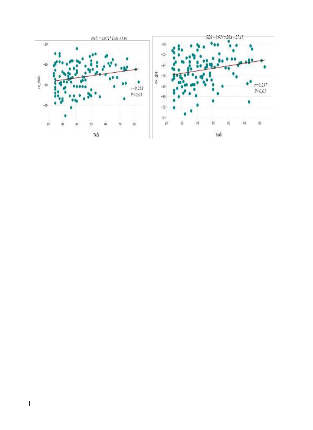
which are statistically significant (p<0.001). Age shows a positive correlation with both RVFWLS and RV4CLS,
with correlation coefficients of r = 0.218 and r=0.237, respectively, and statistical significance of p<0.05 and
p<0.01. Conclusion: The index RVFWLS and RV4CLS using speckle-tracking echocardiography are significantly
higher in females than in males (p<0.001). Therefore, caution should be exercised when using these values in
clinical practice.
Keywords: right ventricular strain, Reference values, Speckle tracking echocardiography.
Corresponding Author: Nguyen Thi Thuy Hang. Email: ntthang@huemed-univ.edu.vn
Received: 25/9/2024; Accepted: 24/11/2024; Published: 25/12/2024
DOI: 10.34071/jmp.2024.6.25
1. INTRODUCTION
Right ventricular function was previously
overlooked, and research on the right ventricle was
limited. However, in recent years, with advancements
in interventional cardiology and imaging diagnostics,
the structure and function of the right ventricle have
received increased attention [1-2].
The assessment of right ventricular systolic
function plays a crucial role in the diagnosis,
prognosis, and treatment of various cardiovascular
diseases. Common parameters used to evaluate
right ventricular function, such as right ventricular
strain (RVS), right ventricular fractional area change
(RV FAC), tricuspid annular plane systolic excursion
(TAPSE), and pulmonary artery systolic pressure
(PAPs), measured by 2D echocardiography and
Doppler tissue imaging, are influenced by right
ventricular overload pathologies and are dependent
on the imaging angle, which can lead to inaccuracies
[1-3]. Speckle-tracking echocardiography (STE) can
mitigate these limitations by being less affected
by imaging angles and right ventricular load
abnormalities. It also helps overcome the challenges
posed by the right ventricle’s complex structure and
passive movement.
Strain is a dimensionless parameter calculated
from the change in length between two points
before and after movement. With some technical
advancements, myocardial strain can be measured
using ultrasound imaging and has been integrated
into clinical practice to provide a non-invasive and
objective indicator of myocardial contractility.
Myocardial strain reflects both the regional and
global systolic function of the myocardium. Estimated
strain values from 2D echocardiography are strong
prognostic factors for various cardiovascular diseases.
They can detect subclinical myocardial changes at an
early stage and may serve as prognostic indicators for
multiple cardiovascular conditions [2].
Numerous studies have demonstrated that
longitudinal strain indices of the right ventricle
are crucial for diagnosing and prognosticating
cardiovascular conditions such as pulmonary
hypertension, arrhythmogenic right ventricular
HUE JOURNAL OF MEDICINE AND PHARMACY ISSN 3030-4318; eISSN: 3030-4326 179
Hue Journal of Medicine and Pharmacy, Volume 14, No.6/2024
cardiomyopathy, congenital heart disease, and heart
failure. Research has observed that in heart failure
patients, impaired right ventricular longitudinal
strain (RVLS) correlates with myocardial fibrosis
and decreased cardiac function [3-5]. The European
Association of Cardiovascular Imaging (EACVI) and the
American Society of Echocardiography (ASE) (2018)
recommend that the Right Ventricular Free Wall
Longitudinal Strain (RVFWLS) and Right Ventricular
Four-Chamber Longitudinal Strain (RV4CLS) are
essential indices for assessing right ventricular
function with high sensitivity and flexibility [5].
However, the normal values for right ventricular
longitudinal strain indices remain limited due to
the focus of studies on left ventricular longitudinal
strain indices and the lack of standardization across
different echocardiographic software platforms
[1-2], [5], This impacts the assessment of right
ventricular dysfunction. In Vietnam, there is a lack
of research on normal values for right ventricular
longitudinal strain. Therefore, we conducted a
study titled Research of the normal reference
values of right ventricular longitudinal strain
indices in healthy adults using 2D speckle-tracking
echocardiography to determine reference values
for the two indices, RVFWLS and RVGLS, and their
relationship with age and gender.
2. MATERIALS AND METHODS
2.1. Study population
- Inclusion criteria
All healthy subjects (healthcare staff and their
relatives) undergoing routine health check-ups
at Ba Ria Hospital from December 2023 to August
2024, with no history or symptoms of cardiovascular
or pulmonary disease, and with normal health
assessments and ECGs, will be included. Participants
must be aged 18 or older and consent to participate
in the study.
- Exclusion criteria
Smoking, hypertension, diagnosed diabetes or
fasting blood glucose levels >100 mg/dl, body mass
index >30 kg/m², creatinine >1.3 mg/dl, glomerular
filtration rate <60 ml/min/1.73 m², and a history of
dyslipidemia.
Abnormal findings on echocardiography (such
as abnormal myocardial motion, any degree of
valvular stenosis, or more than mild valvular
regurgitation, congenital heart disease, acquired
left and right heart disease); acute and chronic lung
disease, professional sports activity, pregnancy, and
arrhythmias.
Subjects with unclear echocardiographic images.
Individuals who decline to participate in the
study will be excluded.
2.2. Study method
Study Design: cross-sectional descriptive study.
Sample Size and Sampling Method: the sample
size is 132, selected using a convenience sampling
method from individuals undergoing routine health
check-ups at Ba Ria Hospital.
Specific Research Methods:
Conventional 2D echocardiography was
performed using a Philips Affinity 70G machine (USA)
by an experienced echocardiographer. All subjects
underwent echocardiographic assessment of left
and right ventricular morphology and function. This
process was used to exclude individuals who did not
meet the study criteria.
Subsequently, a 2D echocardiographic four-
chamber view focusing on the right ventricle was
performed and measured across three consecutive
cardiac cycles. The image depth and sector width
were adjusted to ensure resolution and 50-80 frames
per second (fps) [1-2], [5].
Right ventricular strain indices were assessed
using 2D myocardial strain echocardiography. The
four-chamber view focusing on the right ventricle
was used to measure longitudinal strain indices of
the right ventricle. These indices were calculated
semi-automatically using the Autostrain RV software
(version 15.0, Philips Healthcare, Andover, MA, USA)
(Figure 1) [5]. The right ventricular free wall and
the interventricular septum were divided into three
segments (basal, mid, and apical). RV4CLS represents
the average value obtained from all six segments,
while RVFWLS is the average value from the three
segments of the right ventricular free wall.
The automated analysis software divides the
right ventricle free wall and interventricular septum
into three regions (basal, mid, and apical) [2-5]. The
tracking quality is confirmed by the software and
visually verified using 2D images. Right ventricular
free wall strain and RV4CLS are measured fully
automatically. The software will process the image
and calculate, the recorded results will be displayed,
and can be adjusted.
Subjects with more than two segments showing
incomplete tracking, despite attempts to adjust the
position and width of the ROI, will be excluded from
the analysis.
The analysis results will record the longitudinal
strain of the entire right ventricle four-chamber view
(RV4CLS) and the longitudinal strain of the right
ventricular free wall (RVFWLS). These two indices
will be collected and used for the study data.
HUE JOURNAL OF MEDICINE AND PHARMACY ISSN 3030-4318; eISSN: 3030-4326
180
Hue Journal of Medicine and Pharmacy, Volume 14, No.6/2024
Figure 1. Right ventricular longitudinal strain indices using Autostrain RV software, QLAB version 15.0
(Philips Healthcare, Andover, MA, USA) [5]
Other variables, such as age and gender, were also recorded and analyzed for their relationship
and correlation with the right ventricular longitudinal strain indices measured by 2D speckle-tracking
echocardiography.
2.3. Statistical analysis
All collected data were processed using medical statistical algorithms on a computer with SPSS version
20.0 data analysis software.
3. RESULTS
3.1. Baseline characteristics of research participants
Table 1. Baseline characteristics
Characteristics n (%) Median (Quartiles)
Male 60 (45.5%)
Female 72 (54.5%)
Age (year) 43.29 ± 15.02 Minimum: 24; Maximum:83
Age ≥ 60 23 (17.4%)
Body Surface Area (BSA, m2)1.62 ± 0.19 1.59 (1.49-1.78)
The table shows that males constitute 45.5% and females 54.5%. The average age is 43.29 ± 15.02 years,
with the youngest being 24 years old and the oldest 83 years old, only 17.4% (23 subjects) were in the 60-85
age group. The average body surface area (BSA) is 1.62 m² ± 0.19.
3.2. Reference values for the two strain indices, RVFWLS and RVGLS
Table 2. Values of the right ventricular strain indices
Index Mean (%) Median (Quartiles)
Right Ventricular Free Wall Longitudinal
Strain (RV-FWLS) -28.09 ± 3.47 -27.65 (-30.45-25.30)
Right Ventricular Four-Chamber
Longitudinal Strain (RV-4CLS) -24.90 ± 3.05 -24.45 (-27.10-22.80)
The mean value of RVFWLS is -28.09% ± 3.47, and the mean value of RV4CLS is 24.90% ± 3.05.
HUE JOURNAL OF MEDICINE AND PHARMACY ISSN 3030-4318; eISSN: 3030-4326 181
Hue Journal of Medicine and Pharmacy, Volume 14, No.6/2024
Table 3. Values of the right ventricular strain indices when grouped by gender
Males (n = 60) Females (n = 72) p
Right Ventricular Free Wall Longitudinal Strain
(RVFWLS) (%) -26.58 ± 2.97 -29.35 ± 3.37 <0.001
Right Ventricular Free Wall Longitudinal Strain
(RVFWLS), (Median, Quartiles)
-26.20
(-28.60-24.45)
-29.30
(-32.05-27.05) <0.001
Right Ventricular Four-Chamber Longitudinal
Strain (RV4CLS) (%) -23.47 ± 2.39 -26.10 ± 3.04 <0.001
Right Ventricular Four-Chamber Longitudinal
Strain (RV4CLS), (Median, Quartiles)
-23.25
(-24.65-21.70)
-26.20
(-27.80-24.00) <0.001
Quartiles 25% and 75%.
There is a statistically significant difference between males and females in the free wall strain and overall
longitudinal strain indices of the right ventricle, with p<0.001.
3.3. Relationship between these indices
Figure 2. Correlation between the right ventricular free wall longitudinal strain (RV-FWLS) and the right
ventricle global longitudinal strain (RV-GLS)
There is a strong correlation between RV-FWLS and RV-GLS, with a correlation coefficient of r=0.898,
p<0.0001.
Figure 3. Agreement between RV-FWLS and RV-GLS
There is a high level of agreement between the two strain indices, with a mean difference of -3.19 and a
95% confidence interval of -3.46 to -2.92.
HUE JOURNAL OF MEDICINE AND PHARMACY ISSN 3030-4318; eISSN: 3030-4326
182
Hue Journal of Medicine and Pharmacy, Volume 14, No.6/2024
Figure 4. Linear regression equations between age and the two right ventricular strain indices
Age is positively correlated with RVFWLS and RV4CLS, with correlation coefficients of r = 0.218 (p = 0.0121)
and r = 0.237 (p = 0.0062), respectively (negative value decrease).
4. DISCUSSION
4.1. Baseline characteristics of research
participants
From November 2023 to August 2024, we
collected a total of 132 eligible subjects for inclusion
in the study, of which 54.5% were female and 45.5%
were male, with a mean age of 43.29 ± 15.02 years.
The NORMAL study (2018) reported a mean age
of 47 years in its study population, with females
accounting for 53% [6]; The study by Fine NM et
al. (2014) is a meta-analysis of 10 studies, involving
a total of 486 healthy subjects with a mean age
ranging from 43 to 57 years, of whom 59% were
female [7]; The WASE (2021) study by the World
Alliance of Societies of Echocardiography on right
ventricular function reported a mean age of 47
years, with females accounting for 49% of the study
population [8]. Although the proportion of females
in these studies is quite similar to our study, it is
evident that the mean age of our study population is
considerably lower. This can be explained by the fact
that individuals aged 60 and above often suffer from
cardiovascular conditions, leading to a significant
portion of this age group not meeting the selection
criteria. As a result, in our study, only 17.4% (23
subjects) were in the 60-85 age group. Similarly,
other authors have also noted that older age groups
are affected by comorbidities, and advanced age
may influence right ventricular strain values [5].
4.2. Reference values for the two strain indices,
RVFWLS and RVGLS
Our study reported mean values of right
ventricular free wall longitudinal strain (RVFWLS)
at -28.09 ± 3.47% and right ventricular global
longitudinal strain (RV4CLS) at -24.90 ± 3.05%. In
contrast, the NORMAL study (2018) found mean
RVFWLS values of -26.4% ± 4.2 and RV4CLS values
of -21.5% ± 3.2 [6]; The WASE study (2021) reported
RVFWLS and RV4CLS values of -28.3 ± 4.3 and
-25.4 ± 3.8, respectively [8]. It can be seen that the
results from our study are quite similar to those
of the WASE study (2021) and higher than those
reported in the NORMAL study (2018) [6-8]. This
can be explained by the fact that the studies used
ultrasound machines from different manufacturers
and corresponding processing software. Specifically,
our study and the WASE study (2021) used Philips
echocardiography machines with Auto Strain RV
QLAB 15.0 software, while the NORMAL study
(2018) used GE machines and EchoPAC software
[6]. Therefore, when referencing these parameters
in research, it is important to consider the software
used. Alternatively, a control group could be
employed in studies involving patients to obtain
more reliable results.
4.3. Relationship between these indices
Our study recorded gender and age differences in
right ventricular strain values. Specifically, the RV4CLS
in males was -23.47 ± 2.39, while in females it was
-26.10 ± 3.04. The RVFWLS in males was -26.58 ±
2.97, compared to -29.35 ± 3.37 in females. These
differences were statistically significant with p < 0.001.
In other words, the absolute values of right ventricular
strain indices (RVFWLS and RV4CLS) were higher in
females than in males. Similarly, several other studies
have also shown statistically significant gender