19
Journal of Medicine and Pharmacy, Volume 10, No.7/2020
Endocardial 2D speckle-tracking echocardiography in patients with
coronary artery disease
Dang Quoc Y2, Nguyen Gia Binh1, Nguyen Le Hoang Minh1, Nguyen Anh Vu1
(1) Hue University of Medicine and Pharmacy Hospital, Vietnam
(2) Minh Thien Hospital, Quang Nam Province
Abstract
Objectives: To study the left ventricular myocardial function with two-dimensional speckle tracking
echocardiography and the concordance of endocardial 2DSTE and coronary angiography on the localization
of coronary artery stenosis. Subjects and methods: A cross-sectional study was conducted in 60 patients with
coronary artery disease at Hue University of Medicine and Pharmacy Hospital. All of them were examinated
2DSTE (using endocardial layer strain analysis) and coronary angiography. Results: 60 patients (34 men, 26
women, 69.08 ± 12.44 yrs), statistically significant 2D-STE reduction of the deformation parameters: global
longitudinal strain (GLS) (−8.84% ± 4.74, p < 0.05); global circumferential strain (GCS) (−12.49% ± 6.02, p <
0.05). The agreement of the GLS segment and coronary artery stenosis by coronary angiography were k=0.34
(p < 0.05) at anterior wall, k = 0.53 (p < 0.05) at lateral wall, k = 0.24 (p < 0.05) at inferior wall. Conclusions:
The study using strain on 2DSTE shows the left ventricular systolic function reduced in patients with CAD.
There is a various agreement (not good) about the location of coronary lesions between 2D STE (endocardial
strain analysis) and coronary angiography.
Keywords: Ischemia heart disease, Digital Subtraction Angiography, two-dimensional speckle tracking
echocardiography
Corresponding author: Nguyen Anh Vu, email: navu@huemed-univ.edu.vn
Received: 12/7/2020; Accepted: 10/9/2020
1. OBJECTIVES
Coronary artery disease (CAD) is a common
disease in developed countries and tends to increase
rapidly in developing countries. Recently, myocardial
deformation parameters have been used as a tool
to assess early decline in cardiac function [10].
2DSTE helps to assess cardiac function in different
axis regardless of the angle and also to identify
the local motion abnormalities. Ischemic heart
disease causes regional myocardial disorders. The
endocardial layer often used to measure the strain
for the early discovery of reduced function of the
left ventricle but we don’t know it’s agreement with
angiography to indentify the lesions of myocardial
segments. Therefore, we conducted this study with
the aims:
1. To study the parameters of endocardial strain
on 2DSTE in patients with ischemic heart disease;
2. To find out the agreement between 2DSTE and
coronary angiography on the coronary lesions.
2. MATERIALS AND METHODS
The cross-sectional description study with
60 patients undergoing treatment at the Center
of Cardiology, Hue University of Medicine and
Pharmacy Hospital with ischemic heart disease.
Selection criteria: CAD patients confirmed by invasive
coronary angiography. Exclusion criteria: Patients
disagreeing to participate the study, patients with
severe heart failure, malignancy, blood disease,
renal failure with glomerular filtration rate < 60
ml/min/1.73m2, anemic patients, hyperthyroidism,
COPD, pregnant women.
Data were processed by the statistics softwares
SPSS 20.0. t test to compare 2 averages. The
correlation between two quantitative variables:
using Pearson correlation coefficient and linear
regression, p < 0.05 is considered statistically
significant. Cohen’s kappa statistic measures
agreement between 2DSTE and angiography on the
location of coronary stenosis.
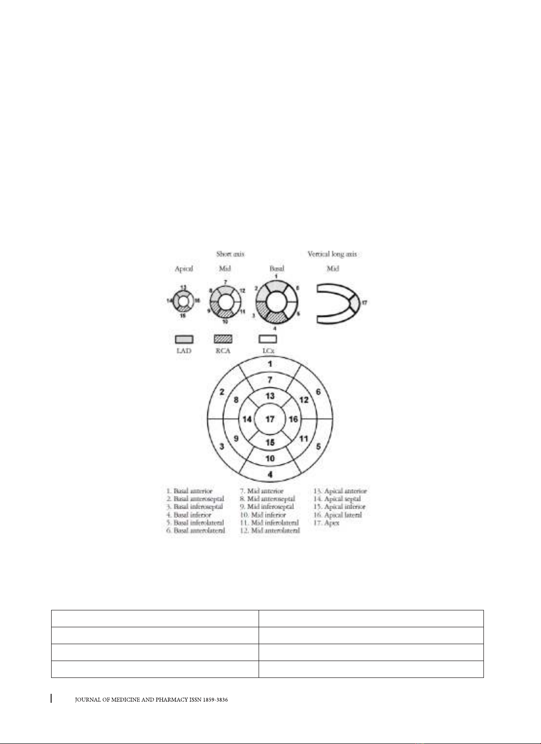
The study variables: risk factors, heart rate,
blood pressure, echocardiography parameters:
LVDs, LVDd, LVPWs, LVPWd, IVSd, IVSs GLS, GLSR,
GCS, GCSR GRS.
Echocardiography: The system Philips Afinity
70 with probe 1-5 MGh. From echocardiographic
grayscale images, offline analysis using two-
dimensional speckle tracking with commercially
available software (Qlab12) were performed
by a single investigator blinded to other clinical
information and imaging results of the patient.
DOI: 10.34071/jmp.2020.7.3