Binding areas of urokinase-type plasminogen activator– plasminogen activator inhibitor-1 complex for endocytosis receptors of the low-density lipoprotein receptor family, determined by site-directed mutagenesis Sune Skeldal1, Jakob V. Larsen1, Katrine E. Pedersen1, Helle H. Petersen1, Rikke Egelund1, Anni Christensen1, Jan K. Jensen1,2, Jørgen Gliemann3 and Peter A. Andreasen1,2
1 Department of Molecular Biology, University of Aarhus, Denmark 2 Interdisciplinary Nanoscience Center (iNANO), University of Aarhus, Denmark 3 Department of Medical Biochemistry, University of Aarhus, Denmark
Keywords low-density lipoprotein receptor-related protein; plasminogen activator inhibitor 1; sorting protein-related receptor; urokinase plasminogen activator; very-low-density lipoprotein receptor
Correspondence P. A. Andreasen, Department of Molecular Biology, University of Aarhus, Gustav Wied’s Vej 10C, 8000 Aarhus C, Denmark Fax: +45 86 12 31 78 Tel: +45 89 42 50 80 E-mail: pa@mb.au.dk
(Received 17 July 2006, revised 20 Septem- ber 2006, accepted 22 September 2006)
doi:10.1111/j.1742-4658.2006.05511.x
Some endocytosis receptors related to the low-density lipoprotein receptor, including low-density lipoprotein receptor-related protein-1A, very-low- density lipoprotein receptor, and sorting protein-related receptor, bind pro- tease-inhibitor complexes, including urokinase-type plasminogen activator (uPA), plasminogen activator inhibitor-1 (PAI-1), and the uPA–PAI-1 com- plex. The unique capacity of these receptors for high-affinity binding of many structurally unrelated ligands renders mapping of receptor-binding surfaces of serpin and serine protease ligands a special challenge. We have mapped the receptor-binding area of the uPA–PAI-1 complex by site-direc- ted mutagenesis. Substitution of a cluster of basic residues near the 37-loop and 60-loop of uPA reduced the receptor-binding affinity of the uPA–PAI-1 complex approximately twofold. Deletion of the N-terminal growth factor domain of uPA reduced the affinity 2–4-fold, depending on the receptor, and deletion of both the growth factor domain and the kringle reduced the affinity sevenfold. The binding affinity of the uPA–PAI-1 complex to the receptors was greatly reduced by substitution of basic and hydrophobic resi- dues in a-helix D and a-helix E of PAI-1. The localization of the implicated residues in the 3D structures of uPA and PAI-1 shows that they form a continuous receptor-binding area spanning the serpin as well as the A-chain and the serine protease domain of uPA. Our results suggest that the 10–100-fold higher affinity of the uPA–PAI-1 complex compared with the free components depends on the bonus effect of bringing the binding areas on uPA and PAI-1 together on the same binding entity.
very-low-density
lipoprotein
hormone carriers. In mammals, this receptor family includes LDLR itself, low-density lipoprotein receptor- protein-1A (LRP-1A), LRP-1B, megalin related receptor or LRP-2, (VLDLR), and apolipoprotein E receptor-2. These
The low-density lipoprotein receptor (LDLR) family of endocytosis receptors has been implicated in binding and endocytosis of a large number of structurally un- including apolipoproteins, protease– related proteins, inhibitor complexes, extracellular matrix proteins, and
Abbreviations a1-PI, a1-antiproteinase inhibitor; CTR, complement type repeat; HEK293T, human embryonic kidney cell line 293T; LDLR, low-density lipoprotein receptor; LRP, low-density lipoprotein receptor-related protein; PAI-1, plasminogen activator inhibitor 1; RAP, receptor-associated protein; RCL, reactive centre loop; sorLA, sorting protein-related receptor; SPD, serine protease domain; tPA, tissue-type plasminogen activator; uPA, urokinase-type plasminogen activator; uPAR, uPA receptor; VLDLR, very-low-density lipoprotein receptor.
FEBS Journal 273 (2006) 5143–5159 ª 2006 The Authors Journal compilation ª 2006 FEBS
5143
S. Skeldal et al.
uPA–PAI-1 complex receptor binding
receptors are constructed with the same types of domains, but with variable numbers of each type of domain. The domains include complement-type repeats (CTRs), YWTD-repeat-containing b-propellers, and epidermal growth factor precursor domains. The recep- tors also all have a transmembrane a-helix and a cyto- plasmic C-terminal domain mediating endocytosis via clathrin-coated pits. Generally, the CTRs are believed to mediate ligand binding. The related receptor sorting protein-related receptor (sorLA), which in addition to other types of domains also contains CTRs, have a lig- and repertoire overlapping that of the LDLR family. these receptors is the One ligand common to all 40-kDa receptor-associated protein (RAP) [1].
The crystal structures of the third domain of RAP in complex with a CTR pair from LDLR and of human rhinovirus serotype 2 in complex with CTRs from VLDLR have recently been solved [2,3]. Com- mon to all of these structures is the fact that binding to the CTRs is mediated through basic and hydropho- bic residues in the ligand. Also, some inferences can be made from an X-ray structure analysis of LDLR crys- tals obtained at pH 5, in which the b-propeller bends back and makes contact with the CTR cluster in a way believed to mimic ligand binding [4].
relatively unstable active
three b-sheets and nine a-helices [15] (Fig. 1). Three- dimensional structures of a covalently coupled a1-anti- proteinase inhibitor (a1-PI)–trypsin complex [16], a covalently coupled a1-PI–elastase complex [17], and several reversible complexes between serpins and pro- teases with the active-site Ser replaced by Ala [18–20] have been reported. The structures support biochemi- cal and biophysical evidence that complex formation is initiated by formation of a reversible docking complex, in which the P1–P1¢ bond in the surface-exposed react- ive centre loop (RCL) interacts with the active site of the protease. Next, the P1–P1¢ bond is cleaved, the P1 residue coupled to the active-site Ser of the protease by an ester bond, the N-terminal part of the RCL inserted as strand 4 in b-sheet A, and the protease translocated to the opposite pole of the serpin [15]. Serpins are thus attacked by the proteases as sub- strates, but the normal catalytic cycle stops at the acyl- enzyme intermediate stage. From the available 3D structures of stable protease–serpin complexes [16,17], it was inferred that the catalytic mechanism is halted because of distortion of the active site of the protease. The energy needed for the distortion stems from stabil- ization of the serpin in the ‘relaxed’ conformation by insertion of the RCL into b-sheet A, as opposed to the conformation. ‘stressed’, RCL insertion can also occur after abortive complex formation as the result of complete cleavage of the P1–P1¢ bond or by insertion of the uncleaved RCL in latent PAI-1 [15].
The fact that these receptors exhibit high-affinity binding of so many structurally unrelated ligands makes their ligand-binding potential a unique case of molecular recognition. Particularly interesting ligands are the serine protease urokinase-type plasminogen activator (uPA), its primary serpin inhibitor, plasmino- gen activator inhibitor-1 (PAI-1), and the correspond- ing protease-serpin complex, which have been shown to bind to LRP-1A [5], megalin [6,7], VLDLR [8,9], LRP-1B [10], and sorLA [11]. These receptors mediate endocytosis of uPA–PAI-1 complex accumulated on the cell surface by binding to the urokinase-type plasminogen activator receptor (uPAR), whereas endo- cytosis directly from the fluid phase is negligible [5]. Although both free uPA and free PAI-1 exhibit a dis- tinct affinity for these receptors, the uPA–PAI-1 com- plex binds to LRP-1A with an affinity much higher than that of the free components. Thus, the Kd value for binding of the uPA–PAI-1 complex to these recep- tors is reported to be (cid:2) 1 nm [6,8,11,12], whereas that for binding of free PAI-1 is reported to be (cid:2) 30 nm [11–13], and Kd values of 7–200 nm have been reported for binding of free u-PA [11,12,14].
inhibitor
the
of
The molecular recognition between the receptors and the uPA–PAI-1 complex should therefore be seen in relation to the mechanism of protease–serpin com- plex formation. X-ray crystal structure analyses have shown that serpins are globular proteins consisting of
Unfortunately, there is no X-ray crystal structure analysis of any receptor–protease–inhibitor complexes. However, on the basis of the established inhibitory mechanism for serpins, there are several possible expla- nations of the increased receptor-binding affinity of the uPA–PAI-1 complex compared with the affinity of the individual components, including increased affinity associated with a conformational change in PAI-1, increased affinity associated with a conformational change in uPA, and ⁄ or an avidity effect of two or more binding sites being brought together on the same ligand. We have now addressed this problem using introducing mutations into site-directed mutagenesis, both uPA and PAI-1, and studying binding to VLDLR, LRP-1A, and sorLA. We initially chose basic residues for mutation on the basis of evidence for receptor binding involving interactions between basic residues in the ligands and acidic residues in the recep- tor [21]. Binding of the uPA–PAI-1 complex to LRP- 1A and VLDLR was previously shown to require basic residues in a-helix D and a-helix E in the flexible joint-region [13,22,23]. We therefore mutated residues adjacent to this region in
FEBS Journal 273 (2006) 5143–5159 ª 2006 The Authors Journal compilation ª 2006 FEBS
5144
S. Skeldal et al.
uPA–PAI-1 complex receptor binding
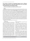
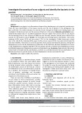
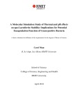
Fig. 1. The 3D structures of PAI-1 and uPA. The figure shows ribbon diagrams of the structure of the active conformation of PAI-1 [53] (pdb file 1B3K) and the structure of the SPD of uPA [27] (pdb file 1LMW). The localization of several a-helices and b-strands are indicated. The diagrams were constructed with the use of SWISSPDBVIEWER.
the uPA–PAI-1 complex, and we provide evidence that the complex has an extended receptor-binding area spanning uPA as well as PAI-1.
serum-free
Results
Screening of the effects of Ala substitutions in PAI-1 on uPA–PAI-1 complex–receptor binding in microtiter wells
bacterially expressed PAI-1. The source of PAI-1 for the preparation of the complexes was not purified PAI-1, but conditioned medium from human embryonic kidney cell line 293T (HEK293T) transfected with the corresponding cDNA. A well-established pro- cedure [24] was used to re-activate the latent PAI-1 in the medium by the use of denaturation with SDS and refolding by removing the SDS by addition of an excess of BSA. 125I-uPA–PAI-1 complexes were prepared by adding 125I-uPA to the medium and purifying the com- plexes by immuno-affinity chromatography [24].
As a control for the integrity of the 125I-uPA–PAI-1 complexes during the binding assay, we measured the binding of all Ala-substituted complexes to parallel wells coated with 20 lgÆmL)1 monoclonal antibody to PAI-1 (mAb2). This antibody coat bound 64 ± 10% of all complexes, and the binding of variant complexes always varied less than 20% from the binding of the wild-type complex.
For the initial screening, our strategy was to substitute basic and hydrophobic residues in PAI-1 with Ala, prepare complexes between the variants and 125I-uPA, measure the binding of 20 pm radioactive complexes to VLDLR, LRP-1A, or sorLA immobilized in microtiter wells, and express the binding of the Ala-substituted complexes relative to the binding of the wild-type com- plex. This relatively simple strategy was based on several facilitating arrangements.
Binding of the uPA–PAI-1 complex to the receptors
We used PAI-1 expressed in a human cell
was assumed to follow the equation:
line, because of solubility problems with nonglycosylated,
FEBS Journal 273 (2006) 5143–5159 ª 2006 The Authors Journal compilation ª 2006 FEBS
5145
S. Skeldal et al.
uPA–PAI-1 complex receptor binding
½RL(cid:3) ¼ ½R(cid:3)T½L(cid:3)=ðKd þ ½L(cid:3)Þ
each residue
separately. For
values (cid:2) 1 nm (see
and Kd
the
in which [RL] is receptor-bound ligand, [R]T is total receptor concentration, [L] is the concentration of free ligand, and Kd is the equilibrium dissociation constant. With the 20 pm concentrations of 125I-labeled ligands above), used here [L] << Kd, and the above equation is reduced to:
ð½RL(cid:3)=½L(cid:3)Þ ¼ ½R(cid:3)T=Kd
three residues to Ala. If a triple mutation resulted in more than a twofold reduction in binding of the uPA–PAI-1 complex to the receptor compared with the wild-type complex, we investigated the effect of single substituting mutants, a receptor-binding reduction of more than 1.5-fold was residues considered to define involved in receptor binding. Residues for Ala substi- tutions were selected on the basis of their proximity to the previously implicated residues in a-helix D and a-helix E, namely Lys71, Arg78, Lys82, Lys90, Arg120 and Lys124 [13,22,23].
(Fig. 2). However,
the
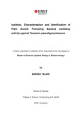
With this strategy, out of a total of 41 PAI-1 resi- dues tested, we identified 11 of importance for binding of the uPA–PAI-1 complex to one, two, or all three receptors involvement of Lys124 in receptor binding could not be confirmed.
Screening of effects of Ala substitutions in uPA on uPA–PAI-1 complex–receptor binding in microtiter wells
Hence, the fractional uPA–PAI-1 complex–receptor binding is expected to be inversely proportional to the Kd value. Control experiments (not shown) confirmed the linear relationship between [RL] and [L]. We there- fore used the amount of receptor-bound complex between 125I-labelled uPA and mutated PAI-1, as com- pared with the amount of receptor-bound complex between 125I-labeled uPA and wild-type PAI-1, as a measure of the effect of the substitution on the Kd value. With the accuracy and background binding of this assay, we expected that binding of up to 10-fold less than the control value would be different from background.
For analyses of the binding of complexes of uPA with PAI-1 mutants, we first mutated groups of
Besides the C-terminal, (cid:2) 30-kDa serine protease domain (SPD), uPA contains an N-terminal, (cid:2) 25- kDa A-chain, consisting of a growth factor domain
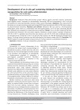
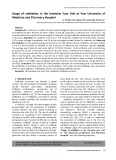
Fig. 2. Effect of Ala substitutions of PAI-1 residues on the binding of the uPA–PAI-1 complex to VLDLR, LRP-1A and sorLA in a solid-phase assay. The binding of the variant complexes was expressed relative to the binding of wild-type complex in the same experiment. Mean ± SD values are shown for at least three independent experiments. As compared with the binding of wild-type complexes, the binding to the receptors of the complexes between uPA and the following PAI-1 mutants were reduced less than twofold when tested as triple mutants or less than 1.5-fold when tested individually, and these mutated residues were therefore considered to be unimportant in the binding: H4A, H5A; P6A; P7A; Y9A; Q58A; K67A; D69A; D70A; P75A; L77A; M85A; P87A; W88A; E92A; T96A; R103A; D104A; K106; L107A; Q109A; P113A; H114; F119A; S121A; K124A; Q125A; W141A; H145A; K178A. (cid:2), Significantly different from binding observed with the correspond- ing wild-type (P < 0.01).
FEBS Journal 273 (2006) 5143–5159 ª 2006 The Authors Journal compilation ª 2006 FEBS
5146
S. Skeldal et al.
uPA–PAI-1 complex receptor binding
an
epitope
encompassing
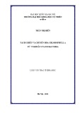
and a kringle domain [25]. In agreement with previous reports on LRP-1A [12] and sorLA [11], we now dem- onstrate that a truncated version of uPA without the A-chain (LMW-uPA) has a sevenfold reduced affinity for VLDLR (Fig. 3). Moreover, we demonstrated that a uPA variant with deletion of the growth factor domain (DGF-uPA) had 2–4-fold reduced binding to VLDLR, LRP-1A, and sorLA (see Fig. 3). Suspecting the involvement of basic residues in the contact with the receptors, we measured the effect on receptor bind- ing of substituting a cluster of three arginines (R108, R109, and R110) in the uPA kringle, but found no these mutations change in receptor binding after (Fig. 3). At the moment, possible endocytosis receptor- binding residues in the kringle is not known. We did not perform a mutational analysis of the growth factor domain, as this, under physiological conditions at the cell surface, is shielded from contact with the endo- cytosis receptors by binding to uPAR [5,12].
We previously reported that a number of mono- clonal antibodies against the SPD of uPA have epi- localized in the 37-loop and 60-loop [26] topes (Fig. 1). To preliminarily investigate whether a recep- in the SPD of uPA, we tor-binding site
exists
studied the effect on uPA–PAI-1 complex–receptor binding of one such monoclonal antibody, mAb3689, with Arg179(36), His180(37), and Arg181(37a) in the 37-loop of uPA (amino-acid residues in the SPD of uPA will be referred to by a double numbering system, based on numbering from the N-terminus of the native protein with the chymotrypsin template numbering system in parentheses; residues in the N-terminal A-chain of uPA will be referred to by numbering from the N-terminus of the native protein [27]). The presence of the antibody reduced the binding significantly (Fig. 3). We therefore studied the effect of Ala sub- stitution of clusters of residues in the 37-loop and 60-loop on receptor binding and demonstrated that both sets of mutations reduced receptor binding 2–4- fold (Fig. 3). As a control, we found that mAb3689 did not reduce the binding of the variant with the mutations in its epitope in the 37-loop (Fig. 3). Sub- stitution of a number of other residues was without effect on receptor binding (Fig. 3). We therefore con- cluded that two or more residues in the 37-loop and 60-loop of the SPD of uPA are part of the ligand– receptor interface.
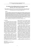
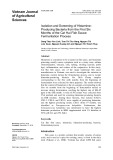
Fig. 3. Effect of Ala substitutions of uPA residues on the binding of the uPA–PAI-1 complex to VLDLR, LRP-1A, and sorLA in a solid-phase assay. The binding of the variant complexes was expressed relative to the binding of the wild-type complex in the same experiment. Mean ± SD values are shown for at least three independent experi- ments. Compared with binding of wild-type complexes, binding to the receptors of the complexes between PAI-1 and the following uPA mutants were reduced less than 1.5-fold, and the mutated residues were therefore considered unimportant for bind- ing: K212(62)A; E213(62a)A; D194(63)A; I216(65)A; Y218(67)A; N227(76)A; Q229(78)A; E235(84)A; K264(110a)A; E265(110b)A; R267(110d)A; H402(241)A; K404(243)A. (cid:2), Significantly different from binding observed with the corresponding wild-type complex (P < 0.01). ND, not deter- mined.
FEBS Journal 273 (2006) 5143–5159 ª 2006 The Authors Journal compilation ª 2006 FEBS
5147
S. Skeldal et al.
uPA–PAI-1 complex receptor binding
Screening of effects of Ala substitutions in both uPA and PAI-1 on uPA–PAI-1 complex–receptor binding in microtiter wells
59 ± 7% of
resulting
complex
To extend the results obtained by the microtiter well- binding assays by Biacore binding analysis and assays of receptor-mediated endocytosis, we used a complex between a PAI-1 variant with low receptor affinity and a uPA variant with low receptor affinity. We used a PAI-1 variant with a quadruple mutation in a-helix D, i.e. K71A-R78A-Y81A-K82A. This variant did not dif- fer significantly from wild-type PAI-1 with respect to specific inhibitory activity (wild-type 69 ± 4% and theoretical maximum), mutant the second-order the reaction with for rate constant uPA (wild-type 3.3 ± 0.7 · 106 m)1Æs)1 and mutant 3.7 ± 0.5 · 106 m)1Æs)1), and relative vitronectin bind- ing (mutant 1.03 ± 0.11 times that of wild-type). We used the uPA variant with the triple mutation in the 37-loop, i.e. R178(35)A-R179(36)A-R181(37a)A. This variant did not differ significantly from wild-type uPA with respect to Km for hydrolysis of S-2444 (pyro-Glu- Gly-Arg-p-nitroanilide), the Km values for wild-type and variant being 87 ± 11 lm and 85 ± 0.01 lm, respectively. The showed the expected more than 10-fold reduction in affinity for VLDLR and LRP-1A in microtiter well-binding assays (Fig. 4).
Surface plasmon resonance analysis of receptor binding of uPA–PAI-1 complexes
the
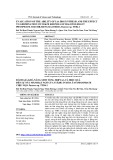
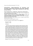
Fig. 4. Effect of Ala substitutions of both uPA and PAI-1 residues on binding of the uPA–PAI-1 complex to VLDLR, LRP-1A, and sor- LA in a solid-phase assay. Binding of wild-type and variant com- plexes to the receptors was estimated. Binding of the variant complexes is expressed relative to that of the wild-type complex in the same experiment. Mean ± SD values are shown for at least three independent experiments. (cid:2), Significantly different from bind- ing observed with the corresponding wild-type (P < 0.01). ND, not determined.
uPA–mutant PAI-1 complex. Fitting the binding data to a Langmuir 1 : 1 binding model by the use of the biaevaluation 3.0 software (global fitting) resulted in a Kd value for binding of the wild-type uPA–wild-type PAI-1 complex to either of the three receptors of (cid:2) 1 nm, while Kd for the mutant uPA–mutant PAI-1 complex was increased more than 10-fold. However, the fit to the simple 1 : 1 binding model was poor. As appears from Fig. 5, the rapid first phase of the associ- ation was followed by a second, slower phase, and a corresponding rapid dissociation phase, amounting to 20% of total binding, particularly with relatively high ligand concentrations. We therefore carried out a more
We analysed receptor binding of complexes between the quadruple a-helix D PAI-1 mutant (K71A-R78A-Y81A-K82A PAI-1) and the 37-loop uPA mutant [R178(35)A-R179(36)A-R181(37a)A uPA] by surface plasmon resonance. In this case, nonradio- active wild-type and mutant uPA–PAI-1 complexes were prepared from PAI-1 purified by immuno-affinity chromatography and re-activated by denaturation with guanidinium chloride and refolding by dialysis, and uPA purified by immuno-affinity chromatography. The complexes were purified from unreacted uPA and PAI-1 by immuno-affinity chromatography. VLDLR, LRP-1A, or sorLA was immobilized on Biacore chips, and wild-type and mutant uPA–PAI-1 complexes injec- ted on to the chips at concentrations of 1.5–100 nm. The time course of the binding obtained with VLDLR is shown in Fig. 5. Similar results were found with the two other receptors. It is evident from the figure that the binding was reduced with the mutant uPA–wild- type PAI-1 complex, the wild-type uPA–mutant PAI-1 the mutant complex,
particular, with
and,
in
FEBS Journal 273 (2006) 5143–5159 ª 2006 The Authors Journal compilation ª 2006 FEBS
5148
S. Skeldal et al.
uPA–PAI-1 complex receptor binding
experiments
reliable estimation of the Kd values by determining the association rate constants from the initial rate of association and the dissociation rate constant from the slow phase of dissociation (see Experimental proce- dures) and calculated the Kd values as the ratio between the association and dissociation rate constants (Table 1). The Kd values obtained for the wild-type uPA–wild-type PAI-1 complex, (cid:2) 1 nm, were in good agreement with those reported previously [6,8,11,12]. The Kd values of the complexes between mutant uPA and wild-type PAI-1 and wild-type uPA and mutant PAI-1 were increased 1.5–5-fold, mostly as the result of increased dissociation rate constants. A particularly large change, corresponding to 10–30-fold increased Kd values, was observed for the complex with mutations in both uPA and PAI-1. This increase was the result of a decreased association rate constant and an increased dissociation rate constant. The fold reduction in the Kd values determined by surface plasmon reson- ance did not differ significantly from those expected from the microtiter well-binding assays, although the average fold reductions of the Kd value were smaller in the Biacore than expected from the microtiter well-binding assays.
Effects of Ala substitutions of uPA and PAI-1 on receptor-mediated endocytosis of the uPA–PAI-1 complex
uPA
mutant
We measured the receptor-dependent degradation of the complexes between the quadruple a-helix D PAI-1 (K71A-R78A-Y81A-K82A PAI-1) and the mutant 37-loop [R178(35)A-R179(36)A- R181(37a)A uPA] in cell lines expressing VLDLR and LRP-1A. For VLDLR-mediated endocytosis, we used U937 cells. These cells were previously shown to con- tain VLDLR-II mRNA, i.e. a VLDLR variant without exon 16 encoding the O-linked sugar domain [28]. Lig- and blot analysis of membrane fragments from U937 cells revealed a RAP-binding membrane protein co-migrating with VLDLR-II (Fig. 6). For LRP-1A- mediated endocytosis, we used COS-1 cells, in which the only RAP-binding receptor detectable by RAP lig- and blotting analysis is LRP-1A [29]. In both cell lines,
Fig. 5. Surface plasmon resonance analysis of binding of wild-type and variant uPA–PAI-1 complexes to VLDLR. Binding was meas- ured using chips with (cid:2) 50 fmol ⁄ mm2 immobilized VLDLR. The chips were superfused with the indicated complexes at concentra- tions of 12, 6, 3 and 1.5 nM, followed by buffer alone at 480 s. Mutant uPA ¼ R178(35)A-R179(36)A-R181(37a)A uPA. Mutant PAI-1 ¼ K71A-R78A-Y81A-K82A PAI-1.
FEBS Journal 273 (2006) 5143–5159 ª 2006 The Authors Journal compilation ª 2006 FEBS
5149
S. Skeldal et al.
uPA–PAI-1 complex receptor binding
d K
M n
e h T
i
- n e r a p
e h t
±
±
±
±
0 0 1
- i c o s s d
t n e r e f f i d
n
i
)
f o
d n a
3 5 1 . 0 1 4 2 . 0 9 3 . 0 2 7 . 3
e c n e h
) 0 1 (
M n (
a ) 9 (
b ) 9 (
c ) 0 1 (
d K
r e t t a
l
e t a r
. e c n a n o s e r
i
5 . 1
d n a
i
e h t
e h t
y l t n a c fi n g s
2 0 . 1 6 6 . 5 0 8 4 . 0 4 0 7 . 0
d n a
l
l
n o m s a p
n e e w t e b
i
i
Table 2. Effect of substitutions of PAI-1 residues on the accumula- tion of uPA–PAI-1 on U937 cell surface. U937 cells were allowed to bind the respective uPA–PAI-1 mutant complexes for 1 h on ice and subsequently washed before incubation for 8 min at either 0 or 37 (cid:2)C. Cell surface-associated complex was then released with a low-pH buffer. Cell surface-associated and internalized complexes are expressed as percentage of total amount of cell-associated complex. Mean ± SD values are given for experiments performed in triplicate.
, d e s y a n a
4 0 1
, d e t a c d n
) 1 )
i
i
) 0 1 (
s (
1 ) k
) 0 1 (
a ) 9 (
b ) 9 (
e b
0 (cid:2)C, 8 min
37 (cid:2)C, 8 min
e r a
e c a f r u s
, 1 - I
t o n
n o i t a c o s s a
A P
i
y b
l
f o
i
e r a 7 3 . 1 ± 3 5 . 0 ± 0 0 . 1 ± 9 9 . 0 ± h c h w 1 0 . 3 · 6 9 . 5 3 4 . 8 0 0 . 4
d u o c
Inside
Cell surface associated Inside
Cell surface associated
PAI-1 variant
) 1 )
s n o i t a r t n e c n o c
i
±
±
±
±
5 )
e t a r
d e t a m
l
1 - I
s Æ 1 )
A P u
0 2 . 4 1 3 . 3 4 2 . 3 4 1 . 3 d n a
. ) 5 2 0 . 0
M
) 0 1 (
(
1 k
) 0 1 (
) 0 1 (
) 9 (
i t s e
<
a i t i n
s n o i t a n m r e t e d
i
P
s e s r u o c
(
s a
f o
i
,
5.7 ± 0.1 94.3 ± 0.1 39.2 ± 1.1 60.8 ± 1.1 Wild-type H5A-P6A-Q109A 5.0 ± 0.2 95.0 ± 0.2 35.8 ± 1.8 64.2 ± 1.8 H79A-F116A-R117A 4.5 ± 0.2a 95.5 ± 0.2a 20.6 ± 0.2a 79.4 ± 0.2a
A P – A P u
e h t
l
e m
l
i t
0 1 g n d n b A L r o S 5 0 . 7 · 3 7 . 6 2 1 . 3 3 5 . 6 h t o b n
A L r o s
m o r f
x e p m o c
±
±
±
±
a r e v e s
i
s r e b m u n
a Significantly different from the corresponding number for wild- type PAI-1 (P < 0.01).
)
d n a
t a
,
A P
e v r u c
s n o i t a t u m h t i
d n a
) 4 (
c ) 3 (
M n (
d K
a ) 4 (
b ) 4 (
A
i
n o i t a c o s s d
i
r o
l
4 5 0 . 0 1 5 0 . 0 6 2 . 0 9 6 . 2 1 - I 2 0 . 1 9 7 . 8 6 2 2 . 0 7 6 3 . 0
i
l i
1 - P R L
g n d n b
i
,
d e m r o f r e p
w x e p m o c
i
, s n o i t a v e d
±
±
±
±
e p y t - d 5 1 . 1 0 3 . 2 4 7 . 0 4 . 5
the receptor-mediated degradation of wild-type and variant complexes was reduced largely in parallel with receptor binding (Fig. 6).
w – A P u
4 0 1
) 1 )
R L D L V
n o i t a c o s s a
) 4 (
) 3 (
a ) 4 (
s (
1 ) k
b ) 4 (
e t a r a p e s
r o f
o t
e h t
i
l i
d r a d n a t s
l
l
l
h c a e
i
s e u a V
e h t 9 5 . 4 3 . 0 1 7 2 . 5 · 3 . 7 2 e p y t - d
e r e w s e s y a n a
r o f
b
w e h t
A
) 1 )
x e p m o c
±
±
±
±
, s t n e m
5 )
i
, s n a e M
s Æ 1 )
g n d n b 0 . 6 4 5 . 2 3 4 . 1 6 8 . 3
m o r f
1 - I
i
. ) 1 0 . 0
M
) 0 1 (
(
1 k
) 0 1 (
) 0 1 (
b ) 9 (
d e t a m
g n d n B
. c t e
<
i r e p x e
l
P
(
i t s e
l
A P – A P u
i
t n e r e f f i d
l
i
i
0 1 5 . 1 1 1 - P R L · 1 5 . 7 0 8 . 1 0 3 . 9
Using another PAI-1 mutant, i.e. a triple mutant con- taining three different substitutions each resulting in reduced receptor binding, we also showed that reduced complex–receptor binding was associated with increased accumulation of the complexes on the cell surface, compared with the wild-type complex. Another triple mutant without reduced receptor binding did not result in accumulation on the cell surface (Table 2).
. e r o c a B
f o
, s e b b u b
a u d v d n
i
x e p m o c
y b
r i a
)
i
i
l
y b
) 8 (
b ) 6 (
M n (
a ) 9 (
a ) 8 (
d K
i
e r e w s e u a v
i
g n d n b
e m o s
A P
y l t n a c fi n g S
d e t a m
d K
c
n I
e h t
7 4 . 0 ± 9 . 8 1 ± 2 1 . 1 ± 0 6 . 0 ± 1 - I 0 6 . 1 2 . 7 2 7 0 . 4 5 3 . 2
It is not possible to study the endocytosis of variant complexes with truncations of the A-chain of uPA. These variants do not bind to uPAR and their endo- cytosis is therefore negligible.
i t s e
d e s u a c
d n a
n o
l i
. ) 1 0 . 0
±
±
±
±
<
, 1 ) k
P
(
4 0 1
) 1 )
i
s e p a h s
e p y t - d 7 1 . 1 6 9 . 1 2 2 . 1 3 6 . 3
Discussion
w – A P u
, 1 k
a ) 9 (
) 9 (
a ) 9 (
s (
1 ) k
) 6 (
. ) s e r u d e c o r p
l
s e u d s e r
l
e h T
1 - I
e v r u c
s a w s r o t p e c e r
0 9 . 5 7 9 . 7 5 3 . 4 6 7 . 9 ·
x e p m o c
l i
i
A P
a t n e m
l
e h t
i
. s y a d
w h t i
A P
d n a
) 1 )
o t
r a u g e r r i
i r e p x E
5 )
s Æ 1 )
f o
A P u
M
-
) 0 1 (
(
R L D L V
1 k
) 0 1 (
) 9 (
b ) 9 (
d n a
l
t n e r e f f i d
f o
w d e v r e s b o
o w
-
t
s e x e p m o c
-
-
e s u a c e b
i
s t l u s e R
t a
n
i
i
-
t n a i r a v
i
-
-
-
-
l i
l
t n a i r a v
s p h c
e p y t - d 1 - I g n d n b 2 2 . 1 ± 3 8 . 0 ± 6 5 . 0 ± 8 6 3 . 0 ± 0 1 3 7 . 2 2 6 . 2 3 0 . 2 · 4 9 4 . 0
l i
l i
s n o i t u t i t s b u s
l
a t e d
A P
A 8 7 R A 1 7 K
A 2 8 K A 1 8 Y
A 8 7 R A 1 7 K
A 2 8 K A 1 8 Y
l
d n a
, d e t a u c a c
n
i
a A
A 2 8 K A 1 8 Y A 8 7 R A 1 7 K – A P u
e b
f o
r o t p e c e r
e p y t - d
t n e r e f f i d
t o n
l i
1 - I g n d n b m o r f e p y t - d W e p y t - d W
w
l
l i
-
-
t c e f f E
d e b i r c s e d
e m a s
f o
d u o c
. 1
i
s a (
e h t
w e h t
-
-
i
i
l
l i
l i
h t i
y l t n a c fi n g S
i
n o i t a
e p y t - d
A ) a 7 3 ( 1 8 1 R A ) 6 3 (
a
g n d n b
e l b a T
w
s e u a v
t n a i r a v A P u
A ) a 7 3 ( 1 8 1 R A ) 6 3 (
. s e s e h t
m o r f
9 7 1 R A ) 5 3 ( 8 7 1 R e p y t - d W e p y t - d W 9 7 1 R A ) 5 3 ( 8 7 1 R
In this work, we used site-directed mutagenesis to map the VLDLR, LRP-1A, and sorLA binding surfaces of the uPA–PAI-1 complex. The mapped interaction sur- face spans both PAI-1 and uPA. In PAI-1, residues His79, Tyr81, Met112, Phe116, Arg117, and Arg270 are implicated in the interaction surface. Also, the pre- viously reported reduced binding of the double mutants K82A-R120A and R78A-K124A [22] could be tracked back to reveal the importance of Arg120, as well as Lys82 and Arg78, in receptor binding, and con- firm the previous reports of the importance of Lys71, Lys82, and Lys90 [13] and Arg78 [23] in a-helix D. Of the 41 PAI-1 residues tested because of their proximity to a-helix D, 12 were Arg or Lys. Of the 11 residues implicated in binding, seven were Arg or Lys. Thus, basic residues constituted a much higher percentage of
FEBS Journal 273 (2006) 5143–5159 ª 2006 The Authors Journal compilation ª 2006 FEBS
5150
S. Skeldal et al.
uPA–PAI-1 complex receptor binding
Fig. 6. Receptor-mediated degradation of uPA–PAI-1 wild-type and variant complexes in U937 and COS-1 cells. U937 cells were incubated 125I-uPA–PAI-1 complex for 45 min at 37 (cid:2)C, by which time the amount of degraded complex was determined as the fraction with 10 pM of complex soluble in 7% trichloroacetic acid. The degradation of wild-type 125I-uPA–PAI-1 complex was set equal to 1, and the degradation of mutant complexes expressed relative to that. The figure shows mean ± SD for triple determinations in a typical experiment out of a total of three with U937 cells and two with COS-1 cells. Insert, 125I-RAP ligand blotting analysis of a membrane preparation from U937. Lane 1, purified VLDLR type II [44]; lane 2, membranes from U937 cells. The migration of molecular mass markers is indicated on the right.
site-directed mutagenesis of CTRs
[30]
the PAI-1 residues implicated in binding than of all the residues tested. This finding agrees well with the ligand recognition by this receptor hypothesis that class relies on electrostatic interactions between basic residues in the ligands and acidic residues in the recep- tors [21]. However, X-ray crystal structure analysis of LDLR at pH 5 [4] and receptor–ligand complexes [2,3], (see below) and our results presented here (Fig. 2) suggest that hydrophobic interactions may also be important. Almost all the residues implicated in binding were localized in and around a-helix D and a-helix E, but, interestingly, localized in substitution of Arg270, b-strand 2C, (cid:2) 2.7 nm from a-helix D, also affected binding, suggesting an even more extended binding surface (Fig. 7).
binding. In addition, we could confirm the previously implicated importance of the A-chain of uPA in the binding of the uPA–PAI-1 complex to the receptors. Moreover, we demonstrated that deletion of the uPA growth factor domain resulted in reduced binding. As the reduction was smaller than that caused by deletion it seems likely that both the of the entire A-chain, growth factor domain and the kringle contribute to the binding, but putative endocytosis receptor-binding resi- dues in the kringle remain unknown. The involvement of the growth factor domain readily explains the previously reported reduction of the affinity of the uPA–serpin complexes for LRP-1A and SorLA in the presence of the cellular receptor for uPA, uPAR [11,12,14], as uPAR binds to the growth factor domain of uPA [31]. Binding to uPAR would thus shield endo- cytosis receptor-binding residues.
In the model of the complex between PAI-1 and the uPA SPD, constructed from the 3D structure of the a1-PI–trypsin complex, as determined by X-ray crystal structure analysis [16], the residues of the 37-loop and 60-loop of uPA studied here are relatively close to
In uPA, we have shown that substitutions in and near the 37-loop and 60-loop caused a substantial reduction in binding. Thus, our findings for the first time implicate the SPD of uPA in the binding of the complex to the receptors. Obviously, we cannot exclude the possibility that additional residues in the SPD contribute to the
FEBS Journal 273 (2006) 5143–5159 ª 2006 The Authors Journal compilation ª 2006 FEBS
5151
S. Skeldal et al.
uPA–PAI-1 complex receptor binding
Fig. 7. The receptor binding surface in the uPA–PAI-1 complex. The uPA–PAI-1 complex shown is a SWISSPDBVIEWER surface display of a model constructed from the structure of the a1-PI–trypsin complex [16] (pdb file 1EZX), by overlayering the a1-PI part of that structure with the structure of cleaved PAI-1 [54] (pdb file 9PAI) and the trypsin part of that structure with the structure of the SPD of uPA [27] (pdb file is realistic, it does not allow any predictions of exact distances 1LMW). Although the relative orientation of uPA and PAI-1 in the model 125I-uPA–PAI-1 complex to VLDLR, LRP-1A, and sor- between amino-acid residues. The effects of Ala substitutions on the binding of 20 pM LA individually are depicted in the model. Ala substitution of residues colored red resulted in a more than fivefold reduction in binding. Ala substitution of residues colored pink resulted in a 1.5–5-fold reduction in binding. PAI-1 residue Tyr81 and uPA residue Arg178(35), which would have been colored pink, are not visible with the orientation of the structure used. Ala substitution of residues colored blue resulted in a less than 1.5-fold reduction in binding when tested individually or less than twofold reduction when tested as part of triple mutants. One structure is shown for binding to each receptor. In addition, the complex model is also shown as a ribbon diagram in which b-strand 2C of PAI-1 is colored yellow and a-helix D and E of PAI-1 are colored green. The 37-loop and 60-loop of uPA are shown in red and pink, respect- ively. Also indicated is a ribbon diagram of the pH 5 structure of the CTRs from LDLR [4] (pdb file 1 N7D).
FEBS Journal 273 (2006) 5143–5159 ª 2006 The Authors Journal compilation ª 2006 FEBS
5152
S. Skeldal et al.
uPA–PAI-1 complex receptor binding
lead to the conclusion that conformational changes in PAI-1 are unlikely to be the only cause of the increased endocytosis receptor affinity upon complex formation.
PAI-1, and it may be argued that the changed endocy- tosis receptor binding following substitution of these residues is due to a change in the relative orientations of uPA and PAI-1. However, in the two available crys- tal structure complexes [16,17], the corresponding resi- dues the remain fully surface-exposed. Secondly, substitution of these residues did not lead to changes in the stability of the complex, as evaluated by anti- body-binding assays performed in parallel with micro- titer well receptor-binding assays (data not shown). Thirdly, monoclonal antibodies with epitopes in the 37-loop and 60-loop remain accessible in the complex [26]. Fourthly, in contrast with monoclonal antibodies binding near a-helix F of PAI-1, a monoclonal anti- body to PAI-1 with an epitope near the expected inter- action phase with uPA did not induce lability of the complex [32]. Fifthly, the absence of any large struc- tural effects of the triple 37-loop uPA mutation was established by demonstrating unchanged enzyme activ- ity. The most obvious interpretation of the observed effects of the substitutions of the residues in the 37 and 60 loops is that these residues are directly situated at the surface of the interaction between the uPA– PAI-1 complex and the endocytosis receptors.
In the model of the complex between PAI-1 and the uPA SPD, constructed from the 3D structure of the a1-PI–trypsin complex, as determined by X-ray crystal structure analysis [16], the residues involved in binding form a continuous, extended interaction surface span- ning both PAI-1 and uPA (Fig. 7). Together with the involvement of the uPA A-chain, these findings argue for the possibility that the high affinity of the complex, compared with that of the individual components, is due to an extended interaction surface assembled by complex formation. Our results do not exclude the possibility that conformational changes or exposure of ‘cryptic’ sites on complex formation [13,23] contribute readily increased affinity, but are more to the explained by an avidity effect caused by two or more binding sites being brought together on the same lig- and. Thus, it should be noted that the 37-loop of uPA does not seem to be in the part of uPA that changes conformation on complex formation [33]. Another argument against the ‘cryptic’ site theory is that the 3D structures of the regions of cleaved a1-PI and tryp- sin-complexed a1-PI corresponding to the regions of PAI-1 implicated in endocytosis receptor binding are superimposable [16,34]. Assuming that cleaved and complexed PAI-1 are as similar as cleaved and com- plexed a1-PI, the observation of a Kd for uPA–PAI-1 complex–LRP-1A binding of 0.4 nm, a Kd for active PAI-1–LRP-1A binding of 55 nm, and a Kd for cleaved PAI-1–LRP-1A binding of 41 nm [12] must
Ligand binding by receptors of the LDLR family is critically dependent on the CTR clusters. VLDLR and sorLA have only one such cluster, with eight or 11 CTRs, respectively. LRP-1A has four clusters, of two, eight, 10, and 11 CTRs. In general, high affinity and high specificity in ligand recognition by receptors of the LDLR receptor family is believed to rely on several indi- vidual CTRs making contact with several receptor recognition patches on the same ligand. Each of the 217 known human CTRs comprises (cid:2) 40 amino acids. A conserved scaffold motif of only 12 amino acids is required for this common structure, with 28 noncon- served positions. Receptors from the LDLR family alone accounts for 132 different CTRs. Crystallographic and NMR studies of individual repeats have revealed that the sequence variability in short loop regions of each repeat results in a unique contour surface and charge density for each repeat [35]. Several observations suggest that the binding sites for plasminogen activator– PAI-1 complexes are also assembled from combinations of CTRs. Thus, although the interpretation of our pre- sent results with LRP-1A is hampered by the fact that both its CTR cluster II and IV bind to the uPA–PAI-1 complex [36–39], the results do demonstrate that the uPA–PAI-1 complex has slightly different interaction surfaces with each of the three receptors tested (Fig. 7). This observation is in agreement with conclusions from previous studies that VLDLR and LRP-1A have over- lapping but not identical binding specificities in a series of serine protease–serpin complexes [40]. By expression of VLDLR variants lacking specific complement-type repeats, it was found that the second CTR in the cluster of eight in this receptor was required for maximal affin- ity for the uPA–PAI-1 complex [41]. The tPA–PAI-1 complex and PAI-1 were reported to bind to a fragment encompassing CTR3-CTR4-CTR5-CTR6-CTR7 of CTR cluster II of LRP-1A (the CTRs of the most N-ter- minal CTR cluster being numbered 1 and 2) [38,42,43]. The minimal functional unit capable of binding the uPA–PAI-1 complex to LRP-1A CTR cluster II was reported to be a two-CTR fragment, and the CTR5– CTR6 pair to be the pair mainly responsible for binding the uPA–PAI-1 complex [30]. From the dimensions of the CTRs emerging from the X-ray crystal structure analysis of LDLR [4], the uPA SPD and the PAI-1 region around a-helix D and a-helix E would each make contact with a CTR. To also accommodate the A-chain of uPA and the more distantly localized R270 of PAI-1, two additional CTRs would be needed. Thus, whereas
FEBS Journal 273 (2006) 5143–5159 ª 2006 The Authors Journal compilation ª 2006 FEBS
5153
S. Skeldal et al.
uPA–PAI-1 complex receptor binding
the individual components uPA and PAI-1 would each only be able to make contact with one or two CTRs, we propose that the high affinity of the complex is related to contacts with three or four CTRs.
medium containing soluble VLDLR was poured on to an RAP-coupled Sepharose column, recirculated for 24 h, and washed. The equilibration and washing buffer was Hepes- buffered saline (10 mm Hepes, pH 7.4, 0.14 m NaCl) with 2 mm CaCl2 and 1 mm MgCl2. Elution was carried out with 0.1 m acetic acid (pH 3.5) ⁄ 0.5 m NaCl ⁄ 10 mm EDTA. Fractions were neutralized immediately with 0.1 vol. 1 m Tris (pH 9) ⁄ 25 mm CaCl2. Relevant fractions were selected on the basis of 125I-RAP-blots, and pooled. The receptor preparations were concentrated with a Centriplus YM-10 spin column, dialysed into NaCl ⁄ Pi (10 mm sodium phos- phate, pH 7.4, 0.14 m NaCl), and stored at )80 (cid:2)C.
In conclusion, we here add several new details to the map of the endocytosis receptor-binding surface of ser- ine protease–serpin complexes and for the first time implicate the SPD of uPA in the binding. The residues that mediate the binding affinity of the A-chain of uPA remain to be established. In the long term, exact mapping of the interaction surface will require X-ray structure analysis of cocrystals between the uPA–PAI-1 complex and one of the receptors.
Experimental procedures
Full-length LRP-1A was purified from human placentas as described [45]. Mature, truncated soluble human sorLA (residues 54–2107), lacking the N-terminal propeptide and the C-terminal cytoplasmic and transmembrane part, was produced in CHO cells and purified as described [46].
Cell lines
Proteases, inhibitors, and their complexes
U937 cells were cultured in RPMI 1640 medium. COS-1 and HEK293T cells were cultured in Dulbecco modified Eagle’s medium. Both media were supplemented with 10% fetal bovine serum, 100 UÆmL)1 penicillin and 100 lgÆmL)1 streptomycin. The medium for HEK293T cells also con- tained 1% nonessential amino acids. Cells were maintained in a humidified atmosphere with 5% CO2 at 37 (cid:2)C. CHO cells were cultured as described [44].
Human wild-type uPA and uPA mutants were expressed recombinantly in and purified from conditioned media of HEK293T cells, as previously described [26]. Recombinant uPA N-terminally truncated at Lys136 (LMW-uPA) was a gift from W Gu¨ nzler, Gru¨ nenthal Company, Aachen, Ger- many. uPA variants were characterized with respect to Km for hydrolysis of S-2444 (pyro-Glu-Gly-Arg-p-nitroanilide) as previously described [47].
Receptors
Human wild-type PAI-1 and PAI-1 mutants were expressed recombinantly in and purified from conditioned media of HEK293T cells, as previously described [24]. Some PAI-1 variants were characterized with respect to spe- cific inhibitory activity, second-order rate constant for the reaction with uPA, and relative vitronectin affinity, as des- cribed previously [26,48]. The numbering of PAI-1 residues was as described previously [49].
A stop codon succeeding the codon encoding amino acid 772 was introduced into human VLDLR type-I cDNA, i.e. cDNA containing all exons of the human VLDLR gene (kindly provided by A. Soutar, University College London, UK) [44]. The resulting cDNA, encoding soluble VLDLR-I without the cytoplasmic domain and transmembrane region (residues 1–772, sVLDLR), was cloned into the plasmid pcDNA3.1- and controlled by DNA sequencing. Transient transfection by the calcium phosphate precipitation method and expression of soluble VLDLR were carried out by standard methods [26]. Briefly, HEK293T cells were grown to full confluence and split 1 : 2 and allowed to grow for an additional 24 h. Cells were then split 1 : 3 and seeded on gelatine-coated dishes. After 24 h, chloroquine was added directly to the cultures to a final concentration of 25 lm. The cultures were then incubated for 1 h. Then 30 lg DNA and water were mixed to a final volume of 1752 lL, and 248 lL 2 m CaCl2 was added. This mixture was added dropwise to 2 mL 42 mm Hepes, pH 7.05, con- taining 274 mm NaCl, 10 mm KCl, 1.5 mm Na2HPO4, and 11 mm d-(+)-glucose. This solution was then added drop- wise to the conditioned medium of one 15-cm culture dish with HEK293T cells. After 7–11 h of incubation, the med- ium was replaced with serum-free medium, which was har- vested 48 h later. This was repeated twice. The conditioned
125I-labeling of uPA, LMW-uPA, and RAP was per- formed as described [12]. Complexes between 125I-uPA and purified PAI-1 were prepared as described [40]. In some cases, complexes between wild-type 125I-uPA and PAI-1 variants were prepared by the use of conditioned media from cells transfected with the corresponding PAI-1 cDNA. In these cases, the conditioned media were incubated for 1 h at room temperature with 0.1% SDS, followed by an eightfold dilution with ice-cold 0.114 m Tris ⁄ HCl, pH 7.4, containing 1.14% BSA, in order to re-activate the latent PAI-1. 125I-uPA was then added in an amount correspond- ing to a 15-fold molar excess of PAI-1 over 125I-uPA. In experiments performed to compare different PAI-1 variants, aliquots of one and the same preparation of 125I-uPA were added to separate portions of conditioned medium with the different PAI-1 variants. The 125I-uPA–PAI-1 complexes incubation at were allowed to form during 30 min of 37 (cid:2)C. The 125I-uPA–PAI-1 complexes were purified by pouring the uPA–PAI-1 mixture on to an anti-uPA
FEBS Journal 273 (2006) 5143–5159 ª 2006 The Authors Journal compilation ª 2006 FEBS
5154
S. Skeldal et al.
uPA–PAI-1 complex receptor binding
by the factor by which antibody binding of each variant complex differed from that of the wild-type complex.
Ligand degradation and internalization experiments
mAb6-coupled Sepharose column followed by an anti-PAI- 1 mAb2-coupled Sepharose column. Both columns were equilibrated and washed in washing buffer (0.1 m Tris, 1 m NaCl, 1% BSA) and eluted with 0.1 m acetic acid (pH 2.9) ⁄ 1 m NaCl, and fractions immediately neutralized with 0.1 vol. 1 m Tris (pH 9) ⁄ 1 m NaCl. 125I-LMW-uPA– PAI-1 complex was produced and purified in the same way as uPA–PAI-1 except that an anti-uPA mAb2-coupled Sepharose column was used instead of an anti-uPA mAb6- coupled Sepharose column.
To prepare nonradioactive wild-type and mutant uPA– PAI-1 complex, recombinant PAI-1 expressed in and puri- fied from HEK293T cells in the latent form (see above) were denatured and refolded by the use of guanidinium chloride [24]. The re-activated PAI-1 was mixed with puri- fied uPA also expressed in and purified from HEK293T cells (see above). The complexes formed were purified by immuno-affinity chromatography on two successive col- umns coupled with monoclonal antibodies against PAI-1 and uPA, respectively [12].
Antibodies and other proteins
acid-precipitable
versus
total
Anti-PAI-1 mAb2, anti-uPA mAb2, anti-uPA mAb6, and anti-uPA mAb3689 have previously been described [26,50]. Recombinant RAP was a gift from M Etzerodt, Depart- ment of Molecular Biology, University of Aarhus [5].
Solid-phase binding assays
For ligand degradation experiments with U937 cells, the cells were washed three times with serum-free medium sup- plemented with 0.5% BSA and incubated for 45 min at 37 (cid:2)C in 0.5 mL of the same buffer with 10 pm 125I-labeled ligand and a cell density of 10 · 106 cellsÆmL)1. The cultures were then made 7% with respect to trichloroacetic acid and centrifuged. Supernatants and pellets were c-coun- ted separately. For experiments with COS-1 cells, the cells were cultured in 24-well cell culture dishes to a confluence (cid:2) 80%, washed three times with serum-free medium supple- mented with 0.5% BSA, and incubated for 4 h at 37 (cid:2)C with 0.3 mL of the same buffer with 10 pm 125I-labeled lig- and. The medium was then removed, and the cells solubi- lized with 1% SDS. BSA was added to the cell extracts to a final concentration of 1%. Cell medium and cell extracts were then made 7% with respect to trichloroacetic acid and centrifuged. Supernatant and pellets were c-counted sepa- rately. Ligand degradation was defined as nontrichloroace- tic added radioactivity radioactivity. The degradation in incubations with 100 nm RAP was scored as nonspecific degradation, not mediated by RAP-binding receptors, and subtracted from all other data [51]. Separate control experiments showed that there was linearity between time and amount of ligand degraded with the incubation times used here (not shown).
To evaluate the distribution of 125I-uPA–PAI-1 com- plexes between the cell surface and intracellular vesicles, we washed U937 cells three times with serum-free medium con- taining 0.5% BSA and cell density adjusted to 10 · 106 cell- sÆmL)1 in the same buffer. Then 0.5 mL cell suspension was incubated with 10 pm 125I-uPA–PAI-1 for 1 h on ice with gentle rocking and then washed three times in ice-cold washing buffer. Cells where then incubated at 37 (cid:2)C or kept on ice for 8 min and subsequently quickly placed on ice. Cell surface-associated ligand was released by washing cells three times with an ice-cold buffer comprising 0.2 m acetic acid, pH 2.6, and 0.1 m NaCl. The remaining radioactivity was defined as internalized ligand. Internalized and cell sur- face-associated ligand were expressed as a percentage of the total radioactivity.
125I-RAP blotting analysis of membrane fragments from U937 cells
Membrane fragments from U937 cells were purified as described previously [44]. Ligand blotting analysis with 125I-RAP was carried out as previously described [52], using VLDLR type-II as a molecular mass marker [44].
Receptors were coated on to microtiter wells overnight in 50 mm NaHCO3, pH 9.6, with a receptor density adjusted to obtain 5–10% binding of wild-type 125I-uPA–PAI-1 com- plex, corresponding to receptor concentrations in the coating buffer of (cid:2) 1 lgÆmL)1. The wells were blocked with 10% BSA in binding buffer (10 mm Hepes, pH 7.8, 140 mm NaCl, 2 mm CaCl2, 1 mm MgCl2) for 2 h, washed, and incubated overnight at 4 (cid:2)C with 20 pm 125I-ligand. Washing and incu- bation with ligands were performed in binding buffer with 1% BSA. When the effect of monoclonal antibodies on binding was investigated, the antibodies were added at a con- centration of 20 lgÆmL)1. After the incubation, the super- nantants were removed for c-counting and the wells washed. Bound ligand was released from the wells with 10% SDS and taken for c-counting. Ligand binding was expressed as bound divided by free ligand and corrected for nonspecific binding, i.e. the radioactivity recovered from wells without receptor. As a control for the integrity of the complexes, the binding of all Ala-substituted complexes to parallel wells coated with 20 lgÆmL)1 anti-PAI-1 mAb2 was shown not to differ more than 20% from that of the wild-type. The bind- ing of variant uPA–PAI-1 complexes to the receptors was corrected by the use of the small variations in antibody bind- ing, assumed to be caused by a dissociation of small fractions of the complexes, by dividing the actually observed binding
FEBS Journal 273 (2006) 5143–5159 ª 2006 The Authors Journal compilation ª 2006 FEBS
5155
S. Skeldal et al.
uPA–PAI-1 complex receptor binding
Surface plasmon resonance analysis
References
1 Bu G & Schwartz AL (1998) RAP, a novel type of ER
chaperone. Trends Cell Biol 8, 272–276.
2 Fisher C, Beglova N & Blacklow SC (2006) Structure of an LDLR-RAP complex reveals a general mode for ligand recognition by lipoprotein receptors. Mol Cell 22, 277–283.
3 Verdaguer N, Fita I, Reithmayer M, Moser R & Blaas D (2004) X-ray structure of a minor group human rhi- novirus bound to a fragment of its cellular receptor pro- tein. Nat Struct Mol Biol 11, 429–434.
4 Rudenko G, Henry L, Henderson K, Ichtchenko K,
containing immobilized receptor minus
Surface plasmon resonance measurements were performed on a Biacore 3000 instrument (Biacore, Uppsala, Sweden) equipped with CM5 sensor chips. The receptors were immobilized to densities of (cid:2) 50 fmolÆmm)2, and samples for binding (40 lL) were injected at 5 lLÆmin)1 at 25 (cid:2)C in 10 mm Hepes, pH 7.4, containing 150 mm NaCl, 1.5 mm CaCl2, 1 mm EGTA, and 0.01% P20. Various concentra- tions of uPA–PAI-1 complex were injected for 8 min. After discontinuation of the injection, dissociation of bound lig- and was followed for another 8 min. Binding was expressed in relative response units as the response obtained from the the flow cell response obtained when using an activated but uncoupled chip.
Brown MS, Goldstein JL & Deisenhofer J (2002) Struc- ture of the LDL receptor extracellular domain at endo- somal pH. Science 298, 2353–2358.
5 Nykjaer A, Petersen CM, Moller B, Jensen PH,
Moestrup SK, Holtet TL, Etzerodt M, Thogersen HC, Munch M, Andreasen PA, et al. (1992) Purified alpha 2-macroglobulin receptor ⁄ LDL receptor-related protein binds urokinase.plasminogen activator inhib- itor type-1 complex. Evidence that the alpha 2-macro- globulin receptor mediates cellular degradation of urokinase receptor-bound complexes. J Biol Chem 267, 14543–14546.
The Kd values were calculated as the ratio between the dissocation rate constants (k)1 values) and the association rate constants (k1 values). The dissocation rate constants were estimated from semilogarithmic plots of the relative amount of uPA–PAI-1 complex bound to the chip versus time during the dissociation phase. The data were fitted to biphasic dissociation kinetics, to distinguish between slowly dissociating, high-affinity binding (rate constant k)1), and low affinity, rapidly dissociating binding (rate constant k)2):
ð1Þ
Relative binding ¼ A expð(cid:4)k(cid:4)1tÞ þ B expð(cid:4)k(cid:4)2tÞ
6 Moestrup SK, Nielsen S, Andreasen P, Jorgensen KE, Nykjaer A, Roigaard H, Gliemann J & Christensen EI (1993) Epithelial glycoprotein-330 mediates endocytosis of plasminogen activator-plasminogen activator inhibi- tor type-1 complexes. J Biol Chem 268, 16564–16570.
7 Willnow TE, Goldstein JL, Orth K, Brown MS &
To estimate the association rate constants (k1 values), we estimated the initial rates of association by linear regression analysis. The initial rates were converted into k1 values by the following equation:
ð2Þ
Initial rate ¼ k1½RT(cid:3)½uPA (cid:4) PAI-1 complex(cid:3)
Herz J (1992) Low density lipoprotein receptor-related protein and gp330 bind similar ligands, including plas- minogen activator-inhibitor complexes and lactoferrin, an inhibitor of chylomicron remnant clearance. J Biol Chem 267, 26172–26180.
Here, the [RT] values were calculated from the response units bound to the chips when exposed to 100 nm wild-type uPA– wild-type PAI-1 complex, after correction for the rapidly dis- sociating fraction of (cid:2) 20%. In all cases, the initial associ- ation rates showed the expected relationship with the uPA– PAI-1 complex concentration. The Kd values were then cal- culated as k)1 ⁄ k1.
8 Heegaard CW, Simonsen AC, Oka K, Kjoller L, Chris- tensen A, Madsen B, Ellgaard L, Chan L & Andreasen PA (1995) Very low density lipoprotein receptor binds and mediates endocytosis of urokinase-type plasminogen activator-type-1 plasminogen activator inhibitor com- plex. J Biol Chem 270, 20855–20861.
Statistical analysis
Results were analysed by Student’s t-test.
9 Argraves KM, Battey FD, MacCalman CD, McCrae KR, Gafvels M, Kozarsky KF, Chappell DA, Strauss JF, 3rd & Strickland DK (1995) The very low density lipoprotein receptor mediates the cellular catabolism of lipoprotein lipase and urokinase-plasminogen activator inhibitor type I complexes. J Biol Chem 270, 26550–26557.
Acknowledgements
We acknowledge the Danish Cancer Research Founda- tion, The Interdisciplinary Nanoscience Centre of Aar- hus University (iNANO), the Danish Cancer Society, the Danish National Research Foundation, and Novo Nordisk Foundation.
10 Li Y, Knisely JM, Lu W, McCormick LM, Wang J, Henkin J, Schwartz AL & Bu G (2002) Low density lipoprotein (LDL) receptor-related protein 1B impairs urokinase receptor regeneration on the cell surface and inhibits cell migration. J Biol Chem 277, 42366– 42371.
FEBS Journal 273 (2006) 5143–5159 ª 2006 The Authors Journal compilation ª 2006 FEBS
5156
S. Skeldal et al.
uPA–PAI-1 complex receptor binding
11 Gliemann J, Hermey G, Nykjaer A, Petersen CM,
Jacobsen C & Andreasen PA (2004) The mosaic recep- tor sorLA ⁄ LR11 binds components of the plasminogen- activating system and platelet-derived growth factor-BB similarly to LRP1 (low-density lipoprotein receptor-rela- ted protein), but mediates slow internalization of bound ligand. Biochem J 381, 203–212.
22 Rodenburg KW, Kjoller L, Petersen HH & Andreasen PA (1998) Binding of urokinase-type plasminogen acti- vator-plasminogen activator inhibitor-1 complex to the endocytosis receptors alpha2-macroglobulin receptor ⁄ low-density lipoprotein receptor-related protein and very-low-density lipoprotein receptor involves basic resi- dues in the inhibitor. Biochem J 329, 55–63.
23 Stefansson S, Muhammad S, Cheng XF, Battey FD, Strickland DK & Lawrence DA (1998) Plasminogen activator inhibitor-1 contains a cryptic high affinity binding site for the low density lipoprotein receptor- related protein. J Biol Chem 273, 6358–6366. 24 Munch M, Heegaard CW & Andreasen PA (1993)
12 Nykjaer A, Kjoller L, Cohen RL, Lawrence DA, Garni- Wagner BA, Todd RF, 3rd van Zonneveld AJ, Gli- emann J & Andreasen PA (1994) Regions involved in binding of urokinase-type-1 inhibitor complex and pro- urokinase to the endocytic alpha 2-macroglobulin recep- tor ⁄ low density lipoprotein receptor-related protein. Evidence that the urokinase receptor protects pro-uro- kinase against binding to the endocytic receptor. J Biol Chem 269, 25668–25676.
Interconversions between active, inert and substrate forms of denatured ⁄ refolded type-1 plasminogen activa- tor inhibitor. Biochim Biophys Acta 1202, 29–37. 25 Andreasen PA, Kjoller L, Christensen L & Duffy MJ
(1997) The urokinase-type plasminogen activator system in cancer metastasis: a review. Int J Cancer 72, 1–22.
26 Petersen HH, Hansen M, Schousboe SL & Andreasen
13 Horn IR, van den Berg BM, Moestrup SK, Pannekoek H & van Zonneveld AJ (1998) Plasminogen activator inhibitor 1 contains a cryptic high affinity receptor bind- ing site that is exposed upon complex formation with tissue-type plasminogen activator. Thromb Haemost 80, 822–828.
14 Croucher D, Saunders DN & Ranson M (2006) The
PA (2001) Localization of epitopes for monoclonal anti- bodies to urokinase-type plasminogen activator: rela- tionship between epitope localization and effects of antibodies on molecular interactions of the enzyme. Eur J Biochem 268, 4430–4439.
27 Spraggon G, Phillips C, Nowak UK, Ponting CP, Saun- ders D, Dobson CM, Stuart DI & Jones EY (1995) The crystal structure of the catalytic domain of human uro- kinase-type plasminogen activator. Structure 3, 681–691.
urokinase ⁄ PAI-2 complex: a new high affinity ligand for the endocytosis receptor low density lipoprotein recep- tor-related protein. J Biol Chem 281, 10206–10213. 15 Ye, S & Goldsmith EJ (2001) Serpins and other covalent protease inhibitors. Curr Opin Struct Biol 11, 740–745. 16 Huntington JA, Read RJ & Carrell RW (2000) Struc- ture of a serpin-protease complex shows inhibition by deformation. Nature 407, 923–926.
17 Dementiev A, Dobo J & Gettins PG (2006) Active site distortion is sufficient for proteinase inhibition by ser- pins: structure of the covalent complex of alpha1-protei- nase inhibitor with porcine pancreatic elastase. J Biol Chem 281, 3452–3457.
28 Martensen PM, Oka K, Christensen L, Rettenberger PM, Petersen HH, Christensen A, Chan L, Heegaard CW & Andreasen PA (1997) Breast carcinoma epithelial cells express a very low-density lipoprotein receptor var- iant lacking the O-linked glycosylation domain encoded by exon 16, but with full binding activity for serine proteinase ⁄ serpin complexes and Mr-40,000 receptor- associated protein. Eur J Biochem 248, 583–591.
29 Kjoller L, Simonsen AC, Ellgaard L & Andreasen PA
18 YeS, Cech AL, Belmares R, Bergstrom RC, Tong Y, Corey DR, Kanost MR & Goldsmith EJ (2001) The structure of a Michaelis serpin-protease complex. Nat Struct Biol 8, 979–983.
19 Dementiev A, Simonovic M, Volz K & Gettins PG
(1995) Differential regulation of urokinase-type-1 inhibi- tor complex endocytosis by phorbol esters in different cell lines is associated with differential regulation of alpha 2-macroglobulin receptor and urokinase receptor expression. Mol Cell Endocrinol 109, 209–217.
(2003) Canonical inhibitor-like interactions explain reac- tivity of alpha1-proteinase inhibitor Pittsburgh and antithrombin with proteinases. J Biol Chem 278, 37881– 37887.
20 Dementiev A, Petitou M, Herbert JM & Gettins PG
30 Andersen OM, Petersen HH, Jacobsen C, Moestrup SK, Etzerodt M, Andreasen PA & Thogersen HC (2001) Analysis of a two-domain binding site for the urokinase-type plasminogen activator-plasminogen acti- vator inhibitor-1 complex in low-density-lipoprotein- receptor-related protein. Biochem J 357, 289–296.
(2004) The ternary complex of antithrombin-anhydro- thrombin-heparin reveals the basis of inhibitor specifi- city. Nat Struct Mol Biol 11, 863–867.
31 Ploug M, Gardsvoll H, Jorgensen TJ, Lonborg Hansen L & Dano K (2002) Structural analysis of the interac- tion between urokinase-type plasminogen activator and its receptor: a potential target for anti-invasive cancer therapy. Biochem Soc Trans 30, 177–183.
21 Andreasen PA, Sottrup-Jensen L, Kjoller L, Nykjaer A, Moestrup SK, Petersen CM & Gliemann J (1994) Receptor-mediated endocytosis of plasminogen activa- tors and activator ⁄ inhibitor complexes. FEBS Lett 338, 239–245.
FEBS Journal 273 (2006) 5143–5159 ª 2006 The Authors Journal compilation ª 2006 FEBS
5157
S. Skeldal et al.
uPA–PAI-1 complex receptor binding
42 Horn IR, van den Berg BM, van der Meijden PZ,
32 De Taeye B, Verbeke K, Compernolle G, Biesemans W, Gils A & Declerck PJ (2003) Structural determinants in the stability of the serpin ⁄ proteinase complex. Biochem Biophys Res Commun 307, 529–534.
Pannekoek H & van Zonneveld AJ (1997) Molecular analysis of ligand binding to the second cluster of complement-type repeats of the low density lipoprotein receptor-related protein. Evidence for an allosteric component in receptor-associated protein-mediated inhibition of ligand binding. J Biol Chem 272, 13608– 13613.
43 Vash B, Phung N, Zein S & DeCamp D (1998) Three
33 Egelund R, Petersen TE & Andreasen PA (2001) A ser- pin-induced extensive proteolytic susceptibility of uroki- nase-type plasminogen activator implicates distortion of the proteinase substrate-binding pocket and oxyanion hole in the serpin inhibitory mechanism. Eur J Biochem 268, 673–685.
complement-type repeats of the low-density lipoprotein receptor-related protein define a common binding site for RAP, PAI-1, and lactoferrin. Blood 92, 3277–3285.
44 Rettenberger PM, Oka K, Ellgaard L, Petersen HH,
34 Huntington JA, Pannu NS, Hazes B, Read RJ, Lomas DA & Carrell RW (1999) A 2.6 A˚ structure of a serpin polymer and implications for conformational disease. J Mol Biol 293, 449–455.
35 Silverman J, Liu Q, Bakker A, To W, Duguay A, Alba BM, Smith R, Rivas A, Li P, Le H, et al. (2005) Multi- valent avimer proteins evolved by exon shuffling of a family of human receptor domains. Nat Biotechnol 23, 1556–1561.
Christensen A, Martensen PM, Monard D, Etzerodt M, Chan L & Andreasen PA (1999) Ligand binding proper- ties of the very low density lipoprotein receptor. Absence of the third complement-type repeat encoded by exon 4 is associated with reduced binding of Mr 40,000 receptor-associated protein. J Biol Chem 274, 8973–8980.
45 Moestrup SK & Gliemann J (1991) Analysis of ligand
recognition by the purified alpha 2-macroglobulin recep- tor (low density lipoprotein receptor-related protein). Evidence that high affinity of alpha 2-macroglobulin- proteinase complex is achieved by binding to adjacent receptors. J Biol Chem 266, 14011–14017.
46 Jacobsen L, Madsen P, Jacobsen C, Nielsen MS,
36 Moestrup SK, Holtet TL, Etzerodt M, Thogersen HC, Nykjaer A, Andreasen PA, Rasmussen HH, Sottrup- Jensen L & Gliemann J (1993) Alpha 2-macroglobulin- proteinase complexes, plasminogen activator inhibitor type-1-plasminogen activator complexes, and receptor- associated protein bind to a region of the alpha 2- macroglobulin receptor containing a cluster of eight complement-type repeats. J Biol Chem 268, 13691– 13696.
Gliemann J & Petersen CM (2001) Activation and func- tional characterization of the mosaic receptor Sor- LA ⁄ LR11. J Biol Chem 276, 22788–22796.
37 Willnow TE, Orth K & Herz J (1994) Molecular dissec- tion of ligand binding sites on the low density lipopro- tein receptor-related protein. J Biol Chem 269, 15827– 15832.
47 Hansen M, Wind T, Blouse GE, Christensen A, Peter- sen HH, Kjelgaard S, Mathiasen L, Holtet TL & Andreasen PA (2005) A urokinase-type plasminogen activator-inhibiting cyclic peptide with an unusual P2 residue and an extended protease binding surface demonstrates new modalities for enzyme inhibition. J Biol Chem 280, 38424–38437.
48 Einholm AP, Pedersen KE, Wind T, Kulig P, Overg-
38 Neels JG, van Den Berg BM, Lookene A, Olivecrona G, Pannekoek H & van Zonneveld AJ (1999) The sec- ond and fourth cluster of class A cysteine-rich repeats of the low density lipoprotein receptor-related protein share ligand-binding properties. J Biol Chem 274, 31305–31311.
39 Obermoeller-McCormick LM, Li Y, Osaka H, Fitz-
Gerald DJ, Schwartz AL & Bu G (2001) Dissection of receptor folding and ligand-binding property with func- tional minireceptors of LDL receptor-related protein. J Cell Sci 114, 899–908.
aard MT, Jensen JK, Bodker JS, Christensen A, Charl- ton P & Andreasen PA (2003) Biochemical mechanism of action of a diketopiperazine inactivator of plasmino- gen activator inhibitor-1. Biochem J 373, 723–732. 49 Andreasen PA, Riccio A, Welinder KG, Douglas R, Sartorio R, Nielsen LS, Oppenheimer C, Blasi F & Dano K (1986) Plasminogen activator inhibitor type-1: reactive center and amino-terminal heterogeneity deter- mined by protein and cDNA sequencing. FEBS Lett 209, 213–218.
50 Wind T, Jensen MA & Andreasen PA (2001) Epitope
40 Kasza A, Petersen HH, Heegaard CW, Oka K, Chris- tensen A, Dubin A, Chan L & Andreasen PA (1997) Specificity of serine proteinase ⁄ serpin complex binding to very-low-density lipoprotein receptor and alpha2- macroglobulin receptor ⁄ low-density-lipoprotein-recep- tor-related protein. Eur J Biochem 248, 270–281.
mapping for four monoclonal antibodies against human plasminogen activator inhibitor type-1: implications for antibody-mediated PAI-1-neutralization and vitronectin- binding. Eur J Biochem 268, 1095–1106.
51 Jensen PH, Christensen EI, Ebbesen P, Gliemann J & Andreasen PA (1990) Lysosomal degradation of recep-
41 Mikhailenko I, Considine W, Argraves KM, Loukinov D, Hyman BT & Strickland DK (1999) Functional domains of the very low density lipoprotein receptor: molecular analysis of ligand binding and acid-dependent ligand dissociation mechanisms. J Cell Sci 112, 3269– 3281.
FEBS Journal 273 (2006) 5143–5159 ª 2006 The Authors Journal compilation ª 2006 FEBS
5158
S. Skeldal et al.
uPA–PAI-1 complex receptor binding
tor-bound urokinase-type plasminogen activator is enhanced by its inhibitors in human trophoblastic chor- iocarcinoma cells. Cell Regul 1, 1043–1056.
53 Sharp AM, Stein PE, Pannu NS, Carrell RW, Berken- pas MB, Ginsburg D, Lawrence DA & Read RJ (1999) The active conformation of plasminogen activator inhi- bitor 1, a target for drugs to control fibrinolysis and cell adhesion. Structure 7, 111–118.
52 Simonsen AC, Heegaard CW, Rasmussen LK, Ellgaard L, Kjoller L, Christensen A, Etzerodt M & Andreasen PA (1994) Very low density lipoprotein receptor from mammary gland and mammary epithelial cell lines binds and mediates endocytosis of M (r) 40,000 receptor asso- ciated protein. FEBS Lett 354, 279–283.
54 Aertgeerts K, De Bondt HL, De Ranter CJ & Declerck PJ (1995) Mechanisms contributing to the conforma- tional and functional flexibility of plasminogen activator inhibitor-1. Nat Struct Biol 2, 891–897.
FEBS Journal 273 (2006) 5143–5159 ª 2006 The Authors Journal compilation ª 2006 FEBS
5159