
BioMed Central
Page 1 of 9
(page number not for citation purposes)
Journal of Translational Medicine
Open Access
Research
Epigenetic control of the ubiquitin carboxyl terminal hydrolase 1 in
renal cell carcinoma
Barbara Seliger*1, Diana Handke1, Elisabeth Schabel1, Juergen Bukur1,
Rudolf Lichtenfels1 and Reinhard Dammann2
Address: 1Martin Luther University Halle-Wittenberg, Institute of Medical Immunology, Halle, Germany and 2Martin Luther University Halle-
Wittenberg, AWG Tumour Genetics of the Medical Faculty, Halle, Germany
Email: Barbara Seliger* - Barbara.Seliger@medizin.uni-halle.de; Diana Handke - dihandke@freenet.de;
Elisabeth Schabel - Elisabeth.Schabel@medizin.uni-halle.de; Juergen Bukur - juergen.bukur@medizin.uni-halle.de;
Rudolf Lichtenfels - rudolf.lichtenfels@medizin.uni-halle.de; Reinhard Dammann - reinhard.dammann@gen.bio.uni-giessen.de
* Corresponding author
Abstract
Background: The ubiquitin carboxyl-terminal hydrolase 1 (UCHL1) gene involved in the
regulation of cellular ubiquitin levels plays an important role in different cellular processes including
cell growth and differentiation. Aberrant expression of UCHL1 has been found in a number of
human solid tumors including renal cell carcinoma (RCC). In RCC, UCHL1 overexpression is
associated with tumor progression and an altered von Hippel Lindau gene expression.
Methods: To determine the underlying mechanisms for the heterogeneous UCHL1 expression
pattern in RCC the UCHL1 promoter DNA methylation status was determined in 17 RCC cell
lines as well as in 32 RCC lesions and corresponding tumor adjacent kidney epithelium using
combined bisulfite restriction analysis as well as bisulfite DNA sequencing.
Results: UCHL1 expression was found in all 32 tumor adjacent kidney epithelium samples.
However, the lack of or reduced UCHL1 mRNA and/or protein expression was detected in 13/32
RCC biopsies and 7/17 RCC cell lines and due to either a total or partial methylation of the UCHL1
promoter DNA. Upon 2'-deoxy-5-azacytidine treatment an induction of UCHL1 mRNA and
protein expression was found in 9/17 RCC cell lines, which was linked to the demethylation degree
of the UCHL1 promoter DNA.
Conclusion: Promoter hypermethylation represents a mechanism for the silencing of the UCHL1
gene expression in RCC and supports the concept of an epigenetic control for the expression of
UCHL1 during disease progression.
Background
The highly conserved ubiquitin-proteasome complex is in
addition to its general function in the protein turnover
process also associated with the regulation of cell growth,
differentiation, the modulation of membrane receptors
and cellular stress responses as well as the turnover of dif-
ferent cytoskeletal components. It is comprised of
enzymes involved in the protein ubiquitination/deubiq-
uitination as well as of the subunits of the 20S proteasome
that degrades ubiquitin-conjugated proteins [1,2]. Ubiq-
Published: 26 October 2009
Journal of Translational Medicine 2009, 7:90 doi:10.1186/1479-5876-7-90
Received: 31 July 2009
Accepted: 26 October 2009
This article is available from: http://www.translational-medicine.com/content/7/1/90
© 2009 Seliger et al; licensee BioMed Central Ltd.
This is an Open Access article distributed under the terms of the Creative Commons Attribution License (http://creativecommons.org/licenses/by/2.0),
which permits unrestricted use, distribution, and reproduction in any medium, provided the original work is properly cited.

Journal of Translational Medicine 2009, 7:90 http://www.translational-medicine.com/content/7/1/90
Page 2 of 9
(page number not for citation purposes)
uitination is a reversible biological process consisting of
enzymes, that attach single or multiple ubiquitin mole-
cules to protein substrates and deubiquinating enzymes
(DUB), e.g. ubiquitin carboxyl-terminal hydrolases
(UCH) and ubiquitin- specific proteases (USP) [3,4]. The
protein gene product 9.5 (PGP 9.5) also termed ubiquitin
carboxyl-terminal hydrolase-1 (UCHL1), a member of the
UCH protein family, represents a soluble 25 kD protein
with both ubiquitin hydrolase and dimerization-depend-
ent ubiquitin ligase activities [5,6]. As a member of the
ubiquitin-proteasome complex UCHL1 is involved in the
control of the intracellular proteolysis, protein turnover
and regulatory processes, which are important in main-
taining normal cellular homeostasis [7]. UCHL1 expres-
sion exhibits marked tissue specificity and is mainly
expressed in testis and neuronal tissues at various differ-
entiation stages [8,9]. In addition, UCHL1 expression was
detected during kidney development, in particular during
the differentiation of renal tubules representing the origin
of clear cell renal cell carcinoma (RCC) and in the regula-
tion of the cell cycle of parietal epithelial cells of the Bow-
man's capsule [10,11]. Since UCHL1 is expressed in
pathophysiological situations of the kidney such as acute
ischaemic renal failure, renal hypertrophy, von Hippel
Lindau (VHL) disease as well as neoplastic transformation
of renal cells it may play a fundamental role in the mech-
anisms controlling the protein turnover of the kidney.
There exists conflicting evidence concerning the role of
UCHL1 in tumorigenesis varying from anti-tumor to pro-
tumor properties depending on the tumor type analysed
[12-14]. Several studies demonstrated aberrant UCHL1
expression in acute lymphoblastic leukaemia, myeloma,
melanoma, neuroblastoma, pancreatic, esophageal, lung,
thyroid, colon and renal cell carcinoma (RCC). In certain
tumor types UCHL1 expression is even associated with
tumor progression and decreased survival rates of patients
[12,13,15-21]. However there is also evidence that
UCHL1 expression might be associated with suppression
of tumor growth in RCC [21]
DNA methylation at CpG dinucleotides within the pro-
moter region of genes is a common event in the pathogen-
esis of tumors including urological cancers and has been
explored as both mechanism and marker of tumor pro-
gression with potential application for diagnosis, classifi-
cation and prognosis of disease [22-29]. Using different
technologies UCHL1 has been identified as a frequently
silenced gene in a cancer-specific manner, in particular in
pancreatic, gastric, colon, ovarian, head neck squamous
cell and hepatocellular carcinoma [14,30-35]. Thus, in
order to understand the underlying molecular mechanism
of the aberrant UCHL1 expression in RCC lesions [21],
microarray analysis of the RCC cell line ACHN either left
untreated or treated with the demethylating agent 2'-
deoxy-5-azacytidine (DAC) was performed demonstrat-
ing an aberrant hypermethylation of the UCHL1 pro-
moter DNA and an association with UCHL1
downregulation in RCC lesions [36]. We here extended
these data and determined whether the promoter DNA
methylation also contributes to the lack of UCHL1 expres-
sion in 32 pairs of primary RCC lesions and correspond-
ing tumor adjacent kidney epithelium as well as 17 RCC
cell lines. The given methylation status of the UCHL1 pro-
moter DNA was further correlated with the UCHL1 mRNA
and protein expression levels in these samples. Moreover,
silenced UCHL1 expression could be restored in RCC cell
lines by treatment with the demethylating agent DAC.
Methods
Cell lines and tissue culture
The human RCC cell lines employed in this study were
established from patients with primary RCC of the clear
cell type [21,37,38]. All tumor cell lines were maintained
in high glucose Dulbecco's modified Eagles medium
(DMEM) supplemented with 10% fetal calf serum, 2 mM
glutamine, 100 U/ml penicillin/streptomycin, 1 mM non-
essential amino acids and 1 mM sodium pyruvate (Gibco/
BRL, Life Technologies, Karlsruhe, Germany).
Patients and tumor biopsies
This study used tumor specimens of RCC obtained from
patients undergoing nephrectomy at the Department of
Urology of the University Hospital in Mainz, Germany.
All cases had been reviewed by a pathologist according to
the WHO classification criteria. Clinicopathologic data
obtained from the patients included sex, age, TNM stage
and histological subtype. The study design was approved
by the Ethical committee of the Johannes Gutenberg Uni-
versity of Mainz and informed consent was obtained from
all RCC patients.
DAC treatment
To assess the ability of the DNA methyltransferase inhibi-
tor DAC to induce the expression of UCHL1, RCC cell
lines were treated for 5 days with 1, 5 and 10 μM DAC
(Sigma-Aldrich GmbH, Taufkirchen, Germany). Subse-
quently untreated and DAC treated cells were harvested,
lysed and total mRNA and/or total protein extracted. The
resulting samples were then subjected to qRT-PCR, West-
ern blot and methylation assays.
Semi-quantitative and real-time reverse transcription
polymerase chain reaction ((q)RT-PCR) analysis
Total RNA was extracted from the samples using the RNe-
asy Mini Kit (Qiagen, Hilden, Germany) according to the
manufacturer's instructions. cDNA was synthesized from
3 μg RNA treated with DNase I (Invitrogen GmbH, Karl-
sruhe, Germany) using oligo dT primers (Fermentas,
Mannheim, Germany) and Superscript II reverse tran-
scriptase (Invitrogen). Real time PCR was performed with

Journal of Translational Medicine 2009, 7:90 http://www.translational-medicine.com/content/7/1/90
Page 3 of 9
(page number not for citation purposes)
the UCHL1-specific primer set (sense: 5'-GCCAATGTCG-
GGTAGATG-3'; anti-sense: 5'-AGCGGACTTCTCCTTGTC-
3') using an annealing temperature of 62°C. β-actin
served as the reference gene (sense: 5'-GAAGCATTTGCG-
GTGGACGAT-3'; anti-sense: 5'-TCCTGTGGCATCCAC-
GAAACT-3'. All real time PCR analyses were performed in
a thermal cycler (Rotorgene, Corbett Life Science, Aus-
tralia) using the QuantiTect SYBR-Green PCR Kit (Qia-
gen). UCHL1 expression levels were normalized against β-
actin amplicons. The UCHL1 expression after 5-days DAC
treatment was calculated as x-fold expression of the
respective untreated sample, which was set to 1.
Western blot analysis
20 μg of total protein/lane from untreated or DAC-treated
RCC cell lines was subjected to Western blot analysis as
previously described [21]. The membranes were incu-
bated either with the anti-UCHL1-specific polyclonal rab-
bit antibody (PG 9500, BIOMOL, Hamburg, Germany) or
with the anti-β-actin-specific monoclonal antibody
(mAb) AC15 (ab6276, Abcam Ltd., Cambridge, UK) serv-
ing as a loading control. Horseradish peroxidase (HRP)-
conjugated swine anti-rabbit IgG (P0217, DAKO, Ham-
burg, Germany) or rabbit anti-mouse IgG (P0260, DAKO)
were used as secondary antibodies. The immunostaining
was visualized using a chemiluminescence detection kit
(LumiLight Western Blotting Substrate, ROCHE Diagnos-
tics GmbH, Mannheim, Germany) according to the man-
ufacturer's instructions.
DNA extraction and analysis of the methylation status of
the UCHL1 promoter
In order to investigate the methylation status of the
UCHL1 promoter DNA, a CpG islet within the UCHL1
promoter containing 22 CpG dinucleotides was mapped
using the CpGplot tool (EBI Tools, EMBOSS CpGPlot;
http://www.ebi.ac.uk/emboss/cpgplot). Subsequently,
bisulfite-specific primers flanking the transcription start
site of the CpG islet in the UCHL1 promoter were
designed with the Oligo 4.0 program relying on the refer-
ence sequence GI: 16949651 (National Bioscience, MN,
USA). Upon isolation of genomic DNA from established
RCC cell lines and/or biopsy specimens with the QIAamp
DNA Mini Kit (Qiagen), 1 μg of DNA sample was sub-
jected to bisulfite modification as previously described
[39]. The methylation status of the UCHL1 promoter was
determined using combined bisulfite restriction analysis
(COBRA) as well as sequencing [39]. Briefly, 100 ng
bisulfite treated DNA was amplified in 25 μl reaction
buffer containing 0.2 mM dNTP mix, 1.5 mM MgCl2, 2 U
Taq polymerase and 10 pmol of the primers 5'-GAG TTT
TAG AGT AAT TGG GAT GGT GAA-A-3' and 5'-CCA CTC
ACT TTA TTC AAC ATC TAA AAA ACA-3' using the follow-
ing conditions: denaturation at 95°C for 3 min and 20
sec, primer annealing at 56°C for 25 seconds (25×) and
primer extension at 72°C for 40 seconds and 5 min. The
resulting amplicon (536 bp) was subjected to a nested
PCR amplification with a set of internal primers (sense: 5'-
GGT TTT GTT TTT GTT TTT TTT GTA TAG GTT-3' and anti-
sense: 5'-AAA AAC AAA TAC AAA AAA AAA AAC AAA
ACC-3') using 1/5th of the first PCR product using the
same PCR conditions, but extended to 30 cycles. Subse-
quently, 20-50 ng of the resulting PCR products (265 bp)
were digested with 10 U BstU I and Taq I (New England
Biolabs, Beverly, MA, USA) prior to separation on 2% Tris-
acetate EDTA agarose gels.
For bisulfite genomic sequencing, the PCR products were
gel-purified employing the PCR Purification Kit (Qiagen)
according to the manufacturer's instructions and thereaf-
ter directly subjected to sequence analysis by a commer-
cially available service provider (MWG Biotech,
Martinsried, Germany). To analyse single sequences the
purified PCR products were cloned into the pCR II vector
using the TOPO TA Cloning Kit (Invitrogen) and subse-
quently the inserts of individual colonies subjected to
sequence analysis.
Results
Correlation of the UCHL1 expression level in RCC cell
lines of the clear cell type with the promoter DNA
methylation status
We have recently demonstrated a heterogeneous expres-
sion pattern of UCHL1 mRNA and/or protein in both
RCC cell lines and RCC lesions, which is associated with
the RCC subtype, VHL status and with tumor progression
[21]. In order to investigate the molecular mechanism(s)
involved in this heterogeneous expression pattern, the
DNA methylation status of the CpG islet in the UCHL1
promoter was determined in a series of 17 established pri-
mary RCC cell lines exhibiting heterogeneous UCHL1
expression levels. As determined by RT-PCR and Western
blot analysis, 3/17 RCC cell lines express neither UCHL1
mRNA nor protein, 4/17 RCC cell lines exhibit low
UCHL1 transcription, but no UCHL1 protein, whereas 9/
17 express high levels of UCHL1 mRNA and protein
(Table 1; [21]). Based on this screening we tested whether
the lack of UCHL1 expression in RCC cell lines could be
attributed to aberrant CpG islet methylation within its
promoter region, which represents a common mechanism
of gene silencing in various human cancers [31,34]. There-
fore, the DNA methylation status of a genomic 265 bp
DNA fragment containing 22 CpG dinucleotides next to
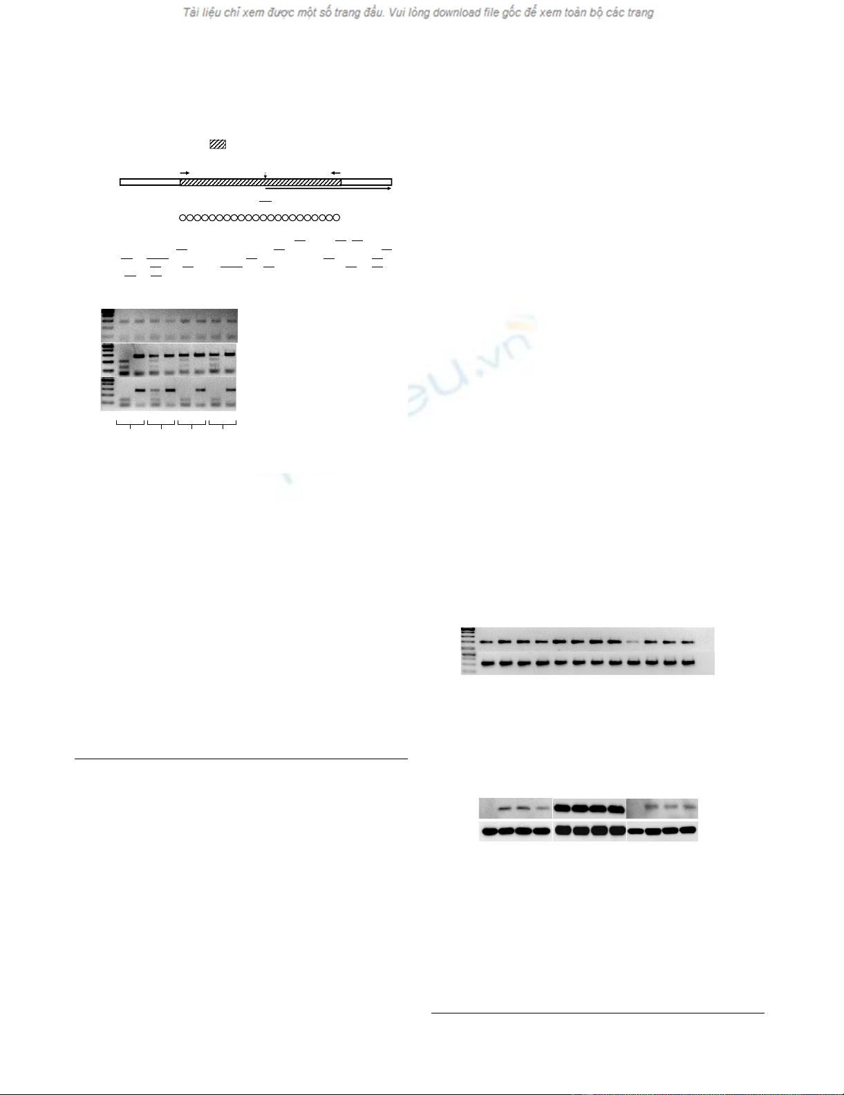
the transcriptional start site of the UCHL1 gene (Figure
1A) was investigated by both COBRA and direct bisulfite
sequencing. As representatively shown for 3 RCC cell lines
in Figure 1B, the methylation pattern of the UCHL1 pro-
moter DNA was highly heterogeneous varying from total
to partial to lack of methylation. In MZ1851RC cells for
example the UCHL1 promoter DNA was not methylated,
Journal of Translational Medicine 2009, 7:90 http://www.translational-medicine.com/content/7/1/90
Page 4 of 9
(page number not for citation purposes)
whereas the COBRA-based analysis indicated a partial
methylation of the UCHL1 promoter DNA in the RCC cell
line MZ2862RC, characterized by methylation of some of
the CpG dinucleotides within the core region of the
UCHL1 promoter while other CpG sites remain unmeth-
ylated. In addition strong methylation of the promoter
DNA core region, as defined by either methylation of all
CpG sites or only few unmethylated CpG sites within the
core region of the CpG islet, was found in the RCC cell
line MZ1851LN. The status of the methylation pattern
was directly associated with the response to DAC treat-
ment: RCC cell lines with a strongly methylated UCHL1
promoter DNA responded to low concentrations of DAC
(1 μM, MZ1851RC), whereas higher DAC doses were
required to efficiently demethylate partially methylated
promoters (10 μM, MZ2862RC). Based on the methyla-
tion status RCC cell lines could be classified into 3 differ-
ent subgroups. The first category consists of RCC cell lines
with a high to complete UCHL1 promoter DNA methyla-
tion predominantly lacking both UCHL1 mRNA and pro-
tein expression. The second exhibits a partially
methylated promoter, which corresponds to low to mod-
erate UCHL1 expression levels, whereas the third category
is represented by RCC cell lines with unmethylated pro-
moters expressing high levels of UCHL1 (Table 1). In
order to verify the COBRA results and to determine the
Table 1: Association of the UCHL1 mRNA and protein expression pattern with the methylation status
UCHL1 expression methylation pattern
RCC cell line mRNA protein BstU I Taq I sequencing
MZ1257RC + + U U U
MZ1774RC + + U U U
MZ1790RC (+) - M M P
MZ1851RC + + U U U
MZ1851LN* (+) - M M M
MZ1879RC - - M M M
MZ1940RC - - M M M
MZ1973RC + + U U U
MZ2175RC - - P P P
MZ2733RC + + U U U
MZ2789RC + - P P P
MZ2858RC + + U U U
MZ2861RC + + U U U
MZ2862RC (+) - P M P
MZ2885RC + n.d. U U U
MZ2904RC + + (pp) P P P
MZ2905RC + + U U U
*Cell line derived from a lymph node metastasis of a patient suffering from RCC. The methylation pattern of the UCHL1 promoter DNA was
determined by COBRA and/or sequencing.
(-): no expression detectable; ((+)) weak expression detectable; (+) expression detectable; (U) unmethylated UCHL1 promoter; (P): partially
methylated UCHL1 promoter (M): fully methylated UCHL1 promoter; (pp): expression verified by proteomic profiling of the corresponding RCC
lesion; n.d. not done
Journal of Translational Medicine 2009, 7:90 http://www.translational-medicine.com/content/7/1/90
Page 5 of 9
(page number not for citation purposes)
extent of methylation bisulfite DNA sequencing of the
respective UCHL1 promoter region was performed in rep-
resentative RCC cell lines [see Additional file 1]. As sum-
marized in Table 1, the bisulfite DNA sequencing data
confirmed the heterogeneous methylation pattern of the
UCHL1 promoter detected by COBRA in RCC cell lines,
but also stressed the point that there exists no strict homo-
geneity in regard to the methylation status of CpG oligo-
nucleotides. Even within a given cell line the efficacy of
the DAC treatment varied from the demethylation of 1 to
18 CpG dinucleotides within the UCHL1 promoter DNA
(data not shown). Nevertheless, the data suggest that
UCHL1 hypermethylation is tightly associated with the
transcriptional silencing of UCHL1 in RCC cell lines.
Restoration of UCHL1 expression in RCC by treatment
with DAC
To confirm that UCHL1 promoter DNA hypermethyla-
tion is responsible for the silencing of UCHL1, a selected
number of UCHL1- and UCHL1+ RCC cell lines were
treated with different concentrations of DAC (1, 5, 10 μM)
for 5 days. As shown in Figure 2, DAC treatment of RCC
cell lines displaying either partially (MZ2862RC) or fully
methylated (MZ1851LN) UCHL promoter DNA regions
led to the induction of UCHL1 mRNA (Figure 2A) restor-
ing protein expression (Figure 2B). However, as represent-
atively shown for MZ1851RC in RCC cell lines lacking
UCHL1 promoter DNA methylation DAC treatment did
neither alter the mRNA nor the protein expression levels
of UCHL1. In contrast, the restored UCHL1 expression
was associated with a partial or total demethylation of the
UCHL1 promoter DNA as determined by COBRA (Figures
2A and 2B). Based on qRT-PCR analyses the induction at
the mRNA level ranges from 1.1 - 1.4 fold in the RCC cell
line MZ1851RC (unmethylated UCHL1 promoter DNA)
to 11 - 13 fold in the RCC cell line MZ1851LN (strong
methylated UCHL1 promoter DNA) to 11 - 18 fold in the
RCC cell line MZ2862RC (partially methylated UCHL1
promoter DNA).
Methylation of UCHL1 in human primary RCC lesions, but
not of corresponding normal kidney epithelium
Since an impaired UCHL1 expression was not only found
in RCC cell lines, but also at a high frequency in primary
UCHL1 promoter in RCC cell linesFigure 1
UCHL1 promoter in RCC cell lines. A) Schematic dia-
gram of the UCHL1 core promoter DNA region with its
respective CpG islet. The sequence segment of interest
taken from the reference GI 16949651 as indicated is dis-
played below the scheme. The putative methylation sites
(CpG dinucleotides) are underlined in the sequence stretch.
(B) Representative COBRA pattern for RCC cell lines dis-
playing a distinct methylation status of the UCHL1 promoter
DNA (MZ1851RC: unmethylated; MZ1851LN: fully methyl-
ated; MZ2862RC: partially methylated) are shown. Genomic
DNA extracted from the given RCC cell lines upon treat-
ment with different DAC concentrations was treated with
bisulfite and amplified by nested PCR as described in Meth-
ods. The resulting 265 bp amplicons were either digested
with BstU I (+) or left untreated (-) and subsequently sepa-
rated in 2% agarose gels in TAE buffer. A 100 base pair DNA
ruler loaded in the first lane served as length standard.
A
MZ2862RC
MZ1851RC
+
|
+
|
+
|
+
|
MZ1851LN
100 bp ladder
BstUI
untreated
1 μM
5 μM
10 μM
DAC
B
22 CpG-dinucleotides
5‘- GTTTTGTTTTTGTTTTTTTTGTATAGGTTTTATAGTGCGTTTGGTCGGCGTTTTATA
GTTGTAGTTTGGGCGGTTTCGTTAGTTGTTTTTCGTTTTTTTTAGGTTATTTTTGTCG
GGCGTTTCGCGAAGATGTAGTTTAAGTCGATGGA GATTAATTTCGAGGTGAGCGTT
AGGTGTATCGTTATTCGGAGAGCGCGAGGTCGAGGGAGGGGGAGTCGAGTCGTT
GATCGGTTCGGTTTTGTTTTTTTTTTTGTATTTGTTTTT -3’
5'- -3'
CpG-Box (265 bp)
coding sequence
-130 +135
+1
ATG
Reference GI:16949651
Restoration of UCHL1 expression by DAC treatment in RCC cell linesFigure 2
Restoration of UCHL1 expression by DAC treat-
ment in RCC cell lines. The representative RCC cell lines
either left untreated or treated with 1, 5, 10 μM DAC for 5
days were subjected to UCHL1-specific semi-quantitative
RT-PCR (A) and Western blot analyses (B) as described in
the Methods section.
UCHL1
ß-actin
MZ2862RC MZ1851RC MZ1851LN
DAC
A
BMZ2862RC MZ1851RC MZ1851LN
UCH-L1
ß-actin
DAC
100 bp ladder
untreated
1μM
5μM
10μM
untreated
1μM
5μM
10μM
untreated
1μM
5μM
10μM
H
2
O control
untreated
1μM
5μM
10μM
untreated
1μM
5μM
10μM
untreated
1μM
5μM
10μM


![Báo cáo seminar chuyên ngành Công nghệ hóa học và thực phẩm [Mới nhất]](https://cdn.tailieu.vn/images/document/thumbnail/2025/20250711/hienkelvinzoi@gmail.com/135x160/47051752458701.jpg)


