
PROTOCOL Open Access
High activity of sequential low dose
chemo-modulating Temozolomide in
combination with Fotemustine in metastatic
melanoma. A feasibility study
Michele Guida
1*
, Antonio Cramarossa
2
, Ettore Fistola
1
, Mariangela Porcelli
1
, Giuseppe Giudice
3
, Katia Lubello
1
,
Giuseppe Colucci
1
Background
Metastatic melanoma (MM) is an incurable chemoresis-
tant cancer with poor prognosis. Until now, only few
drugs have shown some activity. So this tumor repre-
sents an opportunity to verify new and more effective
treatment strategies.
Presently, Dacarbazine (DTIC) remains the standard
chemotherapy for MM with an overall response rate of
approximately 10-15% with complete response in less
than 5% of patients and a survival about 8-10 months
[1,2]. No other agents have demonstrated better results
than DTIC in phase III studies also when utilized as poli-
chemotherapy or in association to immunotherapy [3-6].
Temozolomide (TMZ) has been recently utilized in
MM. It is a novel oral alkylating agent having a high
oral bioavailability and extensive tissue distribution,
including penetration through the blood-brain barrier.
Patients with MM achieved overall response rates of
nearly 20% with single-agent TMZ as similar as DTIC
[7-9]. Also nitrosureas are considered drugs of any activ-
ity in MM including patients with brain metastatic.
Among nitrosurea analogs, fotemustine (FM) has been
more extensively studied in MM, especially in Europe. It
is a third generation chloroethylnitrosourea that has
demonstrated significant antitumoral effects in MM
with a response rate averaging 20%. However, its use is
somewhat limited by its myelotoxic side effect, especially
when old schedules are utilized [10-12].
The activity of alkylating agents depends on their
capacity to form alkyl adducts with DNA, in some cases
causing cross-linking of DNA strands. However, the
antineoplastic activity of these agents is limited by cellu-
lar resistance principally induced by the DNA repair
enzyme O(6)-methylguanine DNA-methyltransferase
(MGMT), a DNA suicide enzyme which removes alkyl
groups from alkylated DNA strands [13-16]. In tumor
cell lines and xerografts an inverse correlation between
the level of this protein and the sensibility to the cyto-
toxic effects of nitrosureas including FM has been
demonstrated [17,18]. Moreover, studies evaluating the
tumor MGMT levels in patients with brain tumors
receiving nitrosureas reported a positive correlation
between low level content of MGMT and a better survi-
val [19,20].
Preclinical studies and recent clinical experiences
also support the concept that continuous exposure to
alkylating agent TMZ, streptozocin, procarbazine, and
DTIC, can effectively deplete cells of MGMT, which is
the primary mechanism of tumor resistance to nitro-
sureas, thus reversing the resistance to these che-
motherapeutic agents [21-23]. In particular, sequential
administration of TMZ and FM is able to induce
depletion of MGMT both in blood lymphocytes and in
tumoral tissue [24].
Recent clinical experiences have confirmed that con-
tinuous exposure to alkilating agent procarbazine in
association with FM is an active treatment in patients
with recurrent malignant gliomas [25]. At present, in
spite of numerous experimental experience, very few
data exist regarding the clinical use of TMZ as chemo-
modulating agent in MM patients. In particular, no
established doses, timing and schedules are known.
Thus, we planned this study in MM patients to verify
the hypothesis that depletion of MGMT induced by low
dose TMZ could render melanoma cells more susceptible
* Correspondence: micguida@libero.it
1
Department of Medical Oncology; National Institute of Cancer, Bari, Italy
Full list of author information is available at the end of the article
Guida et al.Journal of Translational Medicine 2010, 8:115
http://www.translational-medicine.com/content/8/1/115
© 2010 Guida et al; licensee BioMed Central Ltd. This is an Open Access article distributed under the terms of the Creative Commons
Attribution License (http://creativecommons.org/licenses/by/2.0), which permits unrestricted use, distribution, and reproduction in
any medium, provided the original work is properly cited.

to FM. We used two different schedules of sequential
combination of TMZ and FM in to assess their profile of
toxicity and efficacy.
Patients and methods
Patients
Fourteen patients with histologically confirmed stage IV
MM and chemotherapy-naïve were enrolled into two
consecutive cohorts of 7 pts each, treated with two dif-
ferent schedules.
The patients were required to have measurable lesions
(according to RECIST’s criteria), adequate renal, hepatic
and bone marrow functions, an adequate ECOG perfor-
mance status (0-2) and life expectancy of at least
12 weeks. Adjuvant immunotherapy, and previous radio-
therapy or locoregional treatments on non-target lesions
were permitted. Patients with asymptomatic brain
metastases were also enrolled if they had additional dis-
ease sites. Also patients with symptomatic brain metas-
tases were admitted on condition that they had
additional disease sites and brain disease stabilized by
previous locoregional treatments. Patients who had
received previous cytotoxic treatment for metastatic dis-
ease were not enrolled. The trial was approved by the
local ethical committee and written informed consent
was obtained from all patients before study entry.
The period of accrual was from April to December
2009. The main patient characteristic are listed in table
1. The median age was 64 years, range 38-76; ECOG PS
1, range 0-2. Disease sites included soft tissues/lymph
nodes 13, lung 7, liver 3, bone 3, brain 1, spleen 1, adre-
nal gland 1, endopelvic mass 1. Basal LDH was evalu-
ated in all patients (normal range 240-480 mg/dl). It
resulted elevated in 1 patient (about double of the up
limit of normal range) and near the upper normal limit
in 3 patients. According to AJCC melanoma staging [2],
2 patients had M1a staging, 4 patients had M1b staging,
and 8 patients had M1c staging. Two patients had only
1 metastatic site; 7 patients had 2 metastatic sites;
5 patients had 3 or more metastatic sites.
Treatment
Two different treatment schedules were used for the
two cohorts of patients. In the first cohort, TMZ was
administered orally at a single dose of 100 mg/m
2
on
days 1 and 2, 7 and 8; FM was given intravenously at a
dose of 100 mg/m
2
on days 2 and 8, 4 h after TMZ.
Treatment cycles were repeated every 4 weeks for 2
Table 1 Patient characteristics and clinical outcomes according to the two cohorts
N.
Pts
Age
(years)
Sex ECOG
PS
Primary
site
DFI
(months)
Basal
LDH
N. cycle of
chemotherapy
Disease sites Response
(duration)
Survival
(months)
Cohort A
1 69 F 1 Skin 4 360 7 Lung Soft tissue Lymph
nodes
PR (9 months) 19+
2 76 M 1 Skin 2 238 6 Soft tissue PR (7 months) 17+
3 49 M 1 Unknown _ 324 2 Lung Bowel PR (6 months) CR
(2 months)
14
4 73 M 2 Skin 96 379 1 Lung Brain SD (4 months) 4
5 62 F 1 Anal
mucosa
11 403 7 Endopelvic mass
Lymph nodes
PD 9
6 71 M 1 Skin 36 291 6 Soft tissue Lymph
nodes Bone
SD (5 months) 14
7 64 M 1 Scalp 7 251 2 Lung Bone PD 13+
Cohort B
1 64 M 1 Skin 10 474 3 Liver Lymph nodes
Spleen Lung
PD 5
2 38 F 2 Skin 11 309 7 Soft tissue Adrenal
gland Bone
PR (6 months) 10
3 76 F 1 Skin 25 289 7 Soft tissue Lymph
nodes
SD (5 months) 12
4 48 F 1 Skin 24 394 6 Lung SD (7 months) 14+
5 42 M 0 Skin 12 442 3 Liver Lung PD 13+
6 75 F 1 Skin 12 839 8 Soft tissue Lymph
nodes Liver
RP (11+ months) 13+
7 59 M 0 Skin 24 330 6 Lung Lymph nodes SD (4 months) 13+
Abbreviations: LDH: lactate dehydrogenase; CR: complete response; PR: partial response; SD: stable disease; PD: progressive disease.
Guida et al.Journal of Translational Medicine 2010, 8:115
http://www.translational-medicine.com/content/8/1/115
Page 2 of 8

consecutive cycles and then every 3 weeks for further
6 cycles. In the second cohort of patients, chemotherapy
was administered at the same dose but every 3 weeks
for a total of 9 cycles.
Toxicity was evaluated according to the NCI-Common
Toxicity Criteria grading system. Different grades of
toxicity and eventual reduction of dose were evaluated
before each cycle of therapy. Patients were assessable for
toxicity if they had received at least one cycle of treat-
ment. The FM dosage was reduced by nearly 25% of the
starting dose when the severe (grade 3 or 4) hematolo-
gic toxicity occurred. A 50% dose reduction was
required in case of severe (grade 3 or 4) new hematolo-
gic toxicity. Patients requiring more than two dose
reductions and for whom dosing was delayed for up to
3 weeks were removed from the study. Drug administra-
tion was postponed by 1 week if there was no full hema-
tologic recovery from the prior cycle of treatment.
Granulocyte Colony Stimulating Factors (G-CSFs) were
allowed after the patient experienced grade 3-4
neutropenia.
Patients with progressive disease (PD) at any time
were withdrawn from the study. Patients with stable dis-
ease (SD) or with partial response (PR) or complete
response (CR) continued the treatment according to the
protocol.
Evaluation
This study was designed to detect the toxicity and clinical
response of two different schedules of sequential TMZ
and FM association. The pre-study evaluation was com-
pleted within 2 weeks before receiving the study drugs.
On entry, all patients had a complete medical history and
physical examination. Complete blood cell count with
differential and platelet count, serum lactate dehydro-
genas and standard biochemical analysis were performed
before every treatment cycle. A complete blood cell
count was also performed every week to better studying
the myelotoxicity of the treatment that is known being its
principal dose-limiting toxicity. Before each cycle, com-
mon toxicity criteria, performance status and measure-
ment of clinically assessable disease were documented.
Patients were evaluated for response if they received one
or more cycles of treatment. Tumor response was evalu-
ated by physical examination, computed tomography
scan, or other tests according to the basal evaluation per-
formance or according to clinical requests.
Objective tumor response was evaluated according to
Response Evaluation Criteria In Solid Tumors (RECIST)
criteria. A complete response (CR) was defined as com-
plete disappearance of all lesions. A partial response
(PR) was defined as a ≥30% decrease in the sum of
longest diameter of all measured lesions. Stable disease
(SD) was defined as no significant change in measurable
and nonmeasurable disease. Progressive disease (PD)
was defined as a >20% increase in the product of the
two longest perpendicular diameters of any measurable
lesions or in the estimated size on nonmeasurable dis-
ease, the appearance of a new lesion, or the reappear-
ance of old lesions.
In cohort A, patients performed the first re-evaluation
after two cycles of therapy; then after every three cycles.
In cohort B, patients were evaluated every three cycles
of treatment.
Results
Safety and dose delivery
The toxicity profile was evaluated on 73 cycles of ther-
apy delivered, 31 cycles for Cohort A (schedule 1-28)
and 42 for Cohort B (schedule d1-21). The main side
effects are reported in table 2. The schedule d 1,8-28
was characterized by a heavier hematological toxicity
with respect to schedule 1-21, mainly in terms of
thrombocytopenia G3-4 (3 of 7 patients vs 1of7
patients). Nevertheless, platelet transfusions were not
necessary and no clinically significant bleeding compli-
cations occurred. G3-4 neutropenia occurred in 1
patient in cohort A and in none in cohort B. G1-2 ane-
mia frequently occurred in both cohorts of patients (in
4 and 5 patients respectively).
Other minor side effects included nausea-vomiting
involving about 50% of patient in both cohorts, transa-
minase increase in 1 patient in cohort A, and asthenia
in 1 patient in cohort B.
The median of delivered cycles was 5 (range 1-9).
Dose reduction was necessary in 4 patients in cohort A
and in 2 patients of cohort B due to severe thrombocy-
topenia. Chemotherapy was also delayed in 4 patients of
cohort A and in 2 patients of cohort B because of failure
of hematologic recovery prior next cycle of therapy.
Table 2 Main side effects in the two cohorts of patients
Cohort Toxicity G3-G4 Toxicity G1-G2
Neutropenia Thrombopenia Anemia Others Neutropenia Thrombopenia Anemia Others
Cohort A
Schedule 1,8,28
1/7 pts 3/7 pts 0/7 pts 0/7 4/7 4/7 4/7 4/7 (1 transaminase increasing; 3
nausea-vomiting)
Cohort B
Schedule 1, 21
0/7 pts 1/7 pts 0/7 pts 0/7 6/7 5/7 5/7 5/7 (1 asthenia; 4 nausea-
vomiting)
Guida et al.Journal of Translational Medicine 2010, 8:115
http://www.translational-medicine.com/content/8/1/115
Page 3 of 8

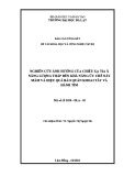
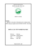
Response and survival
Globally, we obtained 1 complete response (CR) and 4
partial response (PR) with a global response rate of
35.7%. The response duration ranged from 6 to 11+
months (median 8 months). We also obtained stable dis-
ease (SD) in 5 patients (35.7%), 2 in cohort A and 3 in
cohort B. The unique CR lasting about 2 months
occurred in a Cohort A patient who had mediastinal
lymphopaty and bowel localizations (Figure 1). Than,
after 8 months from starting therapy, patient presented
an intestinal bleeding with a rapid anemization that
required a surgical resection of part of the small intes-
tine. The pathological analysis confirmed the diagnosis
of metastases from melanoma. Patient died about 6
months later because of a rapid disseminated brain and
meningeal spreading. The 2 PR occurring in Cohort A
regarded one patient with multiple and diffuse cuta-
neous and subcutaneous lesions, and another patient
with multiple disease sites including lung, lymph nodes
and soft tissue. Both are alive after 19 months and 17
months, respectively. We also reported 2 SD in this
group with a survival of 4 months in a patient with
brain metastases who died for a cerebral hemorrhagic
accident arising in the tumor metastasis. The other
patient is died after 14 months.
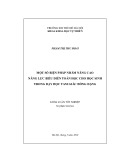
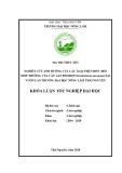
In the Cohort B we reported 2 PR and 3 SD. The PR
regarded one patient with subcutaneous, adrenal gland
and bone lesions. The duration of response was 6
months and the overall survival was 10 months. The
other PR occurred in a female with a disseminated dis-
ease including axillaries lymph nodes involvement, dif-
fuse subcutaneous localizations, multiple liver
metastases, and elevated LDH levels. After 2 cycles of
therapy patient showed a dramatic response in all meta-
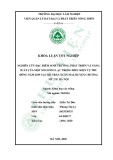
static sites (Figure 2) and a significant decrease of LDH.
The biopsy of a subcutaneous lesion performed after the
third cycle of therapy confirmed the diagnosis of meta-
static melanoma and revealed a diffuse regression of the
neoplastic cells with the presence of abundant melanoci-
tic pigment. Immunohistochemistry revealed an intense
staining of neoplastic component for S100 protein,
HMB 45 and MART 1. Moreover, an impressive lym-
phocytic (CD3+, CD4+, CD8+) and macrophage cells
(CD68+) infiltration was present (Figure 3, 4). At pre-
sent, after 13 months from starting therapy, this patient
is alive in PR. Regarding the 3 patients with SD, 1 died
after 10 months and the others are alive after 13 months
and 14 months. The median overall survival of the
entire group is more than 13 months. At a median fol-
low up of 13 months, 7 of 14 patients are alive.
Figure 1 Complete response in patient with mediastinal lymphopaty and bowel metastases treated in Cohort A (schedule d1,8-28).
Guida et al.Journal of Translational Medicine 2010, 8:115
http://www.translational-medicine.com/content/8/1/115
Page 4 of 8

Figure 2 Dramatic partial response in patient with liver, lymph nodes and subcutaneous metastases treated in Cohort B (schedule
d1-21).
Figure 3 Pathological features of a subcutaneous lesion biopsied after 3 cycles of therapy showing a diffuse regression of the
neoplastic cells with abundant melanocitic pigment.
Guida et al.Journal of Translational Medicine 2010, 8:115
http://www.translational-medicine.com/content/8/1/115
Page 5 of 8


%20--%3e%3cdefs%3e%3cstyle%3e%20.st0%20{%20fill:%20%23fff;%20}%20.st1%20{%20fill:%20%237800fa;%20}%20%3c/style%3e%3c/defs%3e%3cpath%20class='st1'%20d='M117.78,12.18H43.11c2.9,3.47,4.65,7.94,4.65,12.82,0,5.6-2.3,10.66-6.01,14.29h76.02l7.22-13.56-7.22-13.56Z'/%3e%3cg%3e%3cpath%20class='st0'%20d='M53.58,26.17h-.59v-1.46h.59v-4.96h2.83c1.78,0,2.67.94,2.67,2.82v5.76c0,1.87-.89,2.81-2.67,2.81h-2.83v-4.96ZM55.36,21.37v3.34h1.1v1.46h-1.1v3.34h1.01c.61,0,.91-.37.91-1.1v-5.93c0-.74-.3-1.1-.91-1.1h-1.01Z'/%3e%3cpath%20class='st0'%20d='M65.99,31.14h-1.8l-.31-2.07h-2.19l-.31,2.07h-1.64l1.82-11.39h2.62l1.82,11.39ZM65.28,18.04c-.25.46-.51.77-.75.94-.21.15-.47.22-.79.22-.26,0-.57-.07-.92-.22l-.38-.15c-.14-.05-.26-.07-.37-.07-.3,0-.53.18-.71.54l-.91-.68c.25-.46.51-.77.75-.94.21-.14.48-.21.79-.21.26,0,.57.07.92.21l.38.15c.14.05.26.07.37.07.3,0,.53-.18.71-.54l.91.68ZM61.91,27.52h1.73l-.87-5.76-.87,5.76Z'/%3e%3cpath%20class='st0'%20d='M74.53,26.89v1.52c0,1.91-.89,2.86-2.67,2.86s-2.67-.95-2.67-2.86v-5.93c0-1.91.89-2.86,2.67-2.86s2.67.95,2.67,2.86v1.11h-1.69v-1.22c0-.75-.31-1.12-.93-1.12s-.93.37-.93,1.12v6.15c0,.74.31,1.11.93,1.11s.93-.37.93-1.11v-1.63h1.69Z'/%3e%3cpath%20class='st0'%20d='M81.4,31.14h-1.8l-.31-2.07h-2.19l-.31,2.07h-1.64l1.82-11.39h2.62l1.82,11.39ZM75.9,19.2l1.52-1.91h1.71l1.51,1.91h-1.61l-.76-.95-.75.95h-1.61ZM77.32,27.52h1.73l-.87-5.76-.87,5.76ZM83.1,15.99l-1.76,1.91h-1.26l1.17-1.91h1.86Z'/%3e%3cpath%20class='st0'%20d='M84.86,19.75c1.78,0,2.67.94,2.67,2.82v1.48c0,1.87-.89,2.81-2.67,2.81h-.85v4.28h-1.79v-11.39h2.64ZM84.01,21.37v3.86h.85c.58,0,.87-.36.87-1.08v-1.71c0-.71-.29-1.07-.87-1.07h-.85Z'/%3e%3cpath%20class='st0'%20d='M93.51,19.75c1.78,0,2.67.94,2.67,2.82v1.48c0,1.87-.89,2.81-2.67,2.81h-.85v4.28h-1.79v-11.39h2.64ZM92.66,21.37v3.86h.85c.58,0,.87-.36.87-1.08v-1.71c0-.71-.29-1.07-.87-1.07h-.85Z'/%3e%3cpath%20class='st0'%20d='M98.8,31.14h-1.79v-11.39h1.79v4.88h2.03v-4.88h1.83v11.39h-1.83v-4.88h-2.03v4.88Z'/%3e%3cpath%20class='st0'%20d='M105.36,24.55h2.46v1.62h-2.46v3.34h3.09v1.63h-4.88v-11.39h4.88v1.63h-3.09v3.18ZM108.17,17.29l-1.76,1.91h-1.26l1.17-1.91h1.86Z'/%3e%3cpath%20class='st0'%20d='M112.2,19.75c1.78,0,2.67.94,2.67,2.82v1.48c0,1.87-.89,2.81-2.67,2.81h-.85v4.28h-1.79v-11.39h2.64ZM111.35,21.37v3.86h.85c.58,0,.87-.36.87-1.08v-1.71c0-.71-.29-1.07-.87-1.07h-.85Z'/%3e%3c/g%3e%3ccircle%20class='st1'%20cx='25'%20cy='25'%20r='20'/%3e%3cpath%20class='st0'%20d='M32.78,19.27c2.92,0,4.43,2.55,5.28,5.33l.71,2.17c.14.38-.33.75-.71.75h-5.61c.19-.33.24-.71.09-1.08l-.75-2.45c-.43-1.32-.99-2.64-1.79-3.77.75-.57,1.65-.94,2.78-.94h0ZM25,18.38c3.25,0,4.9,2.78,5.89,5.89l.76,2.45c.14.42-.33.8-.8.8h-11.69c-.42,0-.94-.38-.8-.8l.75-2.45c.99-3.11,2.64-5.89,5.89-5.89h0ZM25,11.35c1.74,0,3.11,1.37,3.11,3.11s-1.37,3.11-3.11,3.11-3.11-1.41-3.11-3.11,1.41-3.11,3.11-3.11h0ZM17.27,19.27c1.08,0,1.98.38,2.73.94-.8,1.13-1.37,2.45-1.74,3.77l-.8,2.45c-.14.38-.05.75.09,1.08h-5.56c-.42,0-.9-.38-.75-.75l.71-2.17c.9-2.78,2.41-5.33,5.33-5.33h0ZM17.27,12.91c1.51,0,2.78,1.27,2.78,2.83s-1.27,2.83-2.78,2.83-2.83-1.27-2.83-2.83,1.27-2.83,2.83-2.83h0ZM32.78,12.91c1.56,0,2.78,1.27,2.78,2.83s-1.23,2.83-2.78,2.83-2.83-1.27-2.83-2.83,1.27-2.83,2.83-2.83h0ZM27.07,28.56v.09c0,.57-.24,1.08-.61,1.46h0v.05c-.38.33-.9.57-1.46.57s-1.08-.24-1.46-.61h0c-.38-.38-.61-.9-.61-1.46v-.09h1.41v.09c0,.19.05.38.19.47v.05c.09.09.28.19.47.19s.38-.09.47-.19v-.05c.14-.09.24-.28.24-.47t-.05-.09h1.41ZM30.99,28.56v.09c0,1.65-.66,3.16-1.74,4.24-1.08,1.08-2.59,1.79-4.24,1.79s-3.16-.71-4.24-1.79l-.05-.05c-1.04-1.08-1.7-2.55-1.7-4.2v-.09h1.41v.09c0,1.27.47,2.4,1.27,3.25h.05c.85.85,1.98,1.37,3.25,1.37s2.4-.52,3.25-1.37c.85-.8,1.37-1.98,1.37-3.25v-.09h1.37ZM34.99,28.56v.09c0,2.78-1.13,5.28-2.92,7.07-1.79,1.79-4.29,2.92-7.07,2.92s-5.23-1.13-7.07-2.92c-1.79-1.79-2.92-4.29-2.92-7.07v-.09h1.41v.09c0,2.4.94,4.53,2.5,6.08,1.56,1.56,3.72,2.5,6.08,2.5s4.52-.94,6.08-2.5c1.56-1.56,2.5-3.68,2.5-6.08v-.09h1.41Z'/%3e%3c/svg%3e)