
BioMed Central
Page 1 of 8
(page number not for citation purposes)
World Journal of Surgical Oncology
Open Access
Research
Application of light microscopical and ultrastructural
immunohistochemistry in the study of goblet cell carcinoid in the
appendix
Maya V Gulubova*1, Yovcho Yovchev2, Tatyana Vlaykova3,
Philip Hadjipetkov2, Diana K Prangova1 and Angel Popharitov2
Address: 1Department of General and Clinical Pathology, Medical Faculty, Trakia University, Stara Zagora, 11 Armeiska Str., Stara Zagora, Bulgaria,
2Department of General Surgery, Medical Faculty, Trakia University, Stara Zagora, Bulgaria and 3Department of Chemistry and Biochemistry,
Medical Faculty, Trakia University, Stara Zagora, Bulgaria
Email: Maya V Gulubova* - mgulubova@hotmail.com; Yovcho Yovchev - yovtchev@abv.bg; Tatyana Vlaykova - tvlaykov@mf.uni-sz.bg;
Philip Hadjipetkov - philiphadjipetkov@abv.bg; Diana K Prangova - dianaprangova@hotmail.com; Angel Popharitov - popkharitov@abv.bg
* Corresponding author
Abstract
Background: Goblet cell carcinoids appear less frequently in the appendix than do other
carcinoids. In the presented work a case with a goblet cell carcinoid of the appendix is described.
Methods: Routine histological and histochemical methods were employed, with a combination of
histochemistry and immunohistochemistry on one section and light and electron microscopical
immunohistochemisty on paraffin-embedded material, were applied to identify the type of the
carcinoid and to reveal the fine structure of cell types in the tumour nests of the appendix.
Results: During the biopsy of a patient who had undergone appendectomy, an infiltration with
clusters of goblet cells in the submucosa of the appendix was found. After a second operation of
right-sided hemicolectomy, similar clusters of goblet cells were detected in the muscle layers of the
caecum. After 18 months the patient died from cirrhosis and had not developed metastases or any
recurrence. Immunohistochemically the serotonin-, somatostatin-, chromogranin A- and
synaptophysin-positive endocrine cells were basally attached to mucin-secreting cells. The
combined staining revealed simultaneously present endocrine cells (chromogranin-A-positive) and
mucin-secreting cells (PAS- or alcian blue-positive). The ultrastructural immunohistochemistry
showed that chromogranin A-positive cells had discoid and pleomorphic granules and were located
in tumour nests or as single cells in the appendiceal wall.
Conclusion: The combined histochemical and immunohistochemical procedure and the
ultrastructural immunohistochemistry on archival material could contribute in clarifying the
diagnosis of goblet cell carcinoid.
Background
In the last 30 years, histochemical, immunohistochemical
and electron microscopic techniques were applied in the
study of carcinoids of the appendix. With the aid of previ-
ously mentioned techniques an endocrine cell compo-
nent has been detected in these tumours. In clinical
Published: 6 February 2008
World Journal of Surgical Oncology 2008, 6:15 doi:10.1186/1477-7819-6-15
Received: 23 May 2007
Accepted: 6 February 2008
This article is available from: http://www.wjso.com/content/6/1/15
© 2008 Gulubova et al; licensee BioMed Central Ltd.
This is an Open Access article distributed under the terms of the Creative Commons Attribution License (http://creativecommons.org/licenses/by/2.0),
which permits unrestricted use, distribution, and reproduction in any medium, provided the original work is properly cited.

World Journal of Surgical Oncology 2008, 6:15 http://www.wjso.com/content/6/1/15
Page 2 of 8
(page number not for citation purposes)
aspect, published articles have predominantly addressed
the diagnostic procedures, progression of, and therapy for
the entity [1-4].
Goblet cell carcinoids appear in the appendix less fre-
quently than other carcinoids (and constitute approxi-
mately 5% of all appendicle primary tumours) [1,3,5,6].
The goblet cell carcinoid is characterized histologically by
goblet cells or signet ring-like cells arranged in clusters,
separated by smooth muscle or stroma [2,3]. The endo-
crine cells are arranged basally in tumour glands [5]. Gob-
let cell carcinoids were considered more aggressive than
classical carcinoids [2,3].
In order to determine the clinical behaviour for this
tumour there existed several criteria such as low grade of
differentiation, increased mitotic activity, invasion in the
caecum, lymph nodes metastases and tumour size larger
than 2 cm [7]. The right hemicolectomy was prevalent in
a number of patients with goblet cell carcinoid [7,8]. In
the last years an adjuvant chemotherapy was applied in
the treatment of this type of carcinoid [9].
Almost all of the studies concerning precise diagnosis of
goblet cell carcinoids, were histological and histochemical
[1,6], or immunohistochemical [2,3,10]. The endocrine
component of that carcinoid was shown to be positive for
chromogranin A, serotonin, glucagon and pancreatic
polypeptide [2,3,10]. The data about the ultrastructural
studies were scarce [11]. We did not find an ultrastructural
immunohistochemical study on this type of carcinoid
published in English. Our report describes a combined
histochemical and immunohistochemical technique and
simultaneously presents the mucinous and the endocrine
cell components of the goblet cell carcinoid on light
microscopical paraffin sections. Ultrastructural immuno-
histochemistry on a paraffin-embedded specimen from
goblet cell carcinoid was applied to reveal the fine struc-
ture of cell types in the tumour nests of the appendix.
Methods
Pathology
A 60 year old man diagnosed as having an acute perfora-
tive appendicitis and periappendicular abscesses, was
treated with surgery. The pathological diagnosis was a
goblet cell carcinoid of the appendix (WHO histological
classification 8243/3), infiltrating the mesoappendix. The
macroscopic finding consisted in a slightly tight, oval area in
the submucosa of the appendix, located near the caecum
and measuring about 0.3 cm in diameter. Concomitant
liver cirrhosis (proven hostologically) was observed. Light
microscopical finding was present in many groups of gob-
let cells, separated by fibrous stroma in the submucosa
and the muscle layer of the appendix. Small pools of
mucin were found between the cell nests. Some tumour
nests had central lumens, mimicking normal crypts.
After four months the patient was treated with a second
operation, right-sided hemicolectomy. The macroscopic
appearance of the colon was almost normal. Only slight
induration was observed in the submucosa of the caecum,
at the place of the previous appendiceal resection. Histo-
logically, the muscle layer and the submucosa of the cae-
cum were diffusely infiltrated by goblet cells arranged in
clusters and separated by fibrous stroma. In the wall of the
caecum single tumour cells and nests infiltrated the mye-
nteric plexus. Nuclear atypism and mitoses were visible.
After the right-sided hemicolectomy the patient was
treated with six courses of 5 fluorouracil and leucovorin.
In the 18 month period image analysis did not reveal
metastases or recurrence. The patient was admitted to the
hospital where he died from decompensated liver cirrho-
sis resulting in variceal oesophageal bleeding and with an
autopsy confirming no recurrence of tumour.
Methods
Routine histology
The sections were stained with hematoxyllin and eosin.
Histochemistry
Mucins in the lumen of tumour nests and in the goblet
cells stained positively with PAS reaction and alcian blue.
Light and electron microscopical immunohistochemistry
Earlier the floating section immunohistochemistry meth-
odology was described [12]. The two procedures were car-
ried out simultaneously and according to the method of
De Vos et al. [13] on samples embedded in paraffin. In
brief: paraffin sections 5 µm thick for light microscopical
immunohistochemistry mounted on slides and 40 – 60
µm thick for electron microscope immunohistochemistry
were prepared. They were dewaxed twice in xylene for 30
minutes at 56°C, followed by descending ethanol series.
The sections were then soaked overnight in 10% sucrose
solution at 4°C. The sections were also incubated in 1.2%
hydrogen peroxide in methanol for 30 min, and rinsed in
phosphate balanced solution (PBS), pH 7.4, for 15 min.
The sections were then blocked for 30 min with normal
mouse serum (DAKO). After incubating with the primary
mouse (rabbit) anti-human antibodies overnight, the cry-
ostat sections were washed in PBS and incubated with a
secondary anti-mouse (rabbit) biotinylated antibody
(DAKO) for 4 h, and subsequently with the streptavidin-
HRP complex (DAKO) for 4 h, rinsed in PBS, and then in
0.05 M Tris-HCl buffer, pH 7.5, for 10 min. The reaction
was made visible by using a mixture of 3 mg 3,3'-diami-
nobenzidine (DAB) (DAKO), in 15 ml 0.05 M Tris-HCl
World Journal of Surgical Oncology 2008, 6:15 http://www.wjso.com/content/6/1/15
Page 3 of 8
(page number not for citation purposes)
buffer, pH 7.5, and 36 µl 1% hydrogen peroxide for 10–
20 min, and rinsed in PBS.
After dehydration the paraffin sections were mounted
with entellan for light microscopy. For better visualization
of the DAB reaction product the sections were not coun-
terstained. Sections incubated with non-immune sera
instead of the primary antibodies were used as negative
controls.
The free floating sections (40–60 µm thick) were postfixed
in PBS containing 2% osmium tetroxide for 30 min at
2°C, followed by a rinse in PBS. Finally, sections were
dehydrated in graded concentrations of ethanol and pro-
pylene oxide, and flat-embedded with Durcupan,
between celophane sheets. Ultrathin sections were cut
from areas with immune reactive endocrine cells visible
on cellophane preparations. For better visualization of the
DAB reaction product they were not counterstained with
uranyl acetate. Ultrathin sections were examined and pho-
tographed with an OPTON EM 109 electron microscope
at 50 kV.
A combined histochemical and immunohistochemical
staining
After deparaffinization the 5 µm thick sections were
stained first with PAS-reaction or with toluidine blue.
Then, the preparations were not mounted with Kanada
balsam. They were hydrated in PBS, pH 7.4 for 10 min.
Endogenous peroxidase was quenched with 1.2% hydro-
gen peroxide in methanol for 30 min, and rinsed in PBS,
pH 7.4, for 15 min. Then, the sections were incubated
overnight with the rabbit anti-human chromogranin A, or
with the mouse anti-human serotonin. After washing
them in PBS, pH 7.4, incubation with a secondary anti-
rabbit (mouse) biotinylated antibody (DAKO) for 4 h was
done, and subsequently with the streptavidin-HRP com-
plex (DAKO) for 4 h. They were rinsed in PBS, pH 7.4, and
then in 0.05 M Tris-HCL buffer, pH 7.5, for 10 min.
Finally the reaction was developed with DAB solution as
was described above. The sections were mounted with
entellan. The pink or blue colour of mucins (PAS or
alciane blue) remained visible. Brown endocrine cells
could be observed at the basement membrane, beneath
goblet cells in the nests.
Immunochemicals
The antibodies used were: rabbit anti-human chrom-
ogranin A (N1535), rabbit anti-human synaptophysin
(N1566), mouse anti-human synaptophysin (U0037),
rabbit anti-human somatostatin (N1551), and mouse
anti-human serotonin (N1530), all obtained from DAKO
A/S Denmark. The rabbit anti-human gastrin (PA019-5P),
rabbit anti-human bombesin (PA062-5P), rabbit anti-
human secretin (PA067-5P) and rabbit anti-human β-
endorphin (PA063-5P) were obtained from BioGenex
Laboratories, San Ramon, CA, USA. The detection system
used was DAKO LSAB®2 System, HRP (K0675), and
DAKO®DAB Chromogen tablets (S3000) (DAKO A/S
Denmark).
Results
Histology
The submucosa and the muscle layer of the appendix were
diffusely infiltrated by goblet cells, arranged in clusters,
and separated by fibrous stroma (Figure 1). Small pools of
mucin were found between the cell nests. Tumour nests
had central lumens, mimicking normal crypts. In the wall
of the caecum single tumour cells and nests infiltrated the
myenteric plexus, the muscle layer, and its submucosa.
Nuclear atypism was visible.
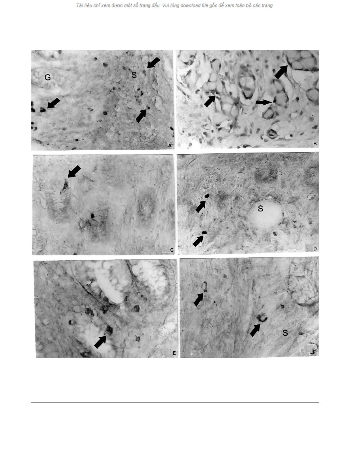
Light microscopic immunohistochemistry
Dispersed endocrine cells or endocrine cells in nests con-
taining 3–4 goblet cells were observed in the submucous
and muscle layer of the appendix. The endocrine cells in
appendiceal tumour nests were chromogranin A- (Figure
2a,b), somatostatin- (Figure 2c,d), synaptophysin- (Fig-
ure. 2e,f) and serotonin-positive. The endocrine cells,
invading the wall of the caecum were all chromogranin A-
, synaptophysin- and serotonin- (Figure 3) positive. The
endocrine cells in the appendix and caecum tumour sam-
ples were bombesin-, endorphin-, gastrin- and secretin-
negative.
A combined histochemical and immunohistochemical
staining
PAS-positive mucous cells were surrounded by brown
chromogranin A-positive endocrine cells (Figure 4a,b).
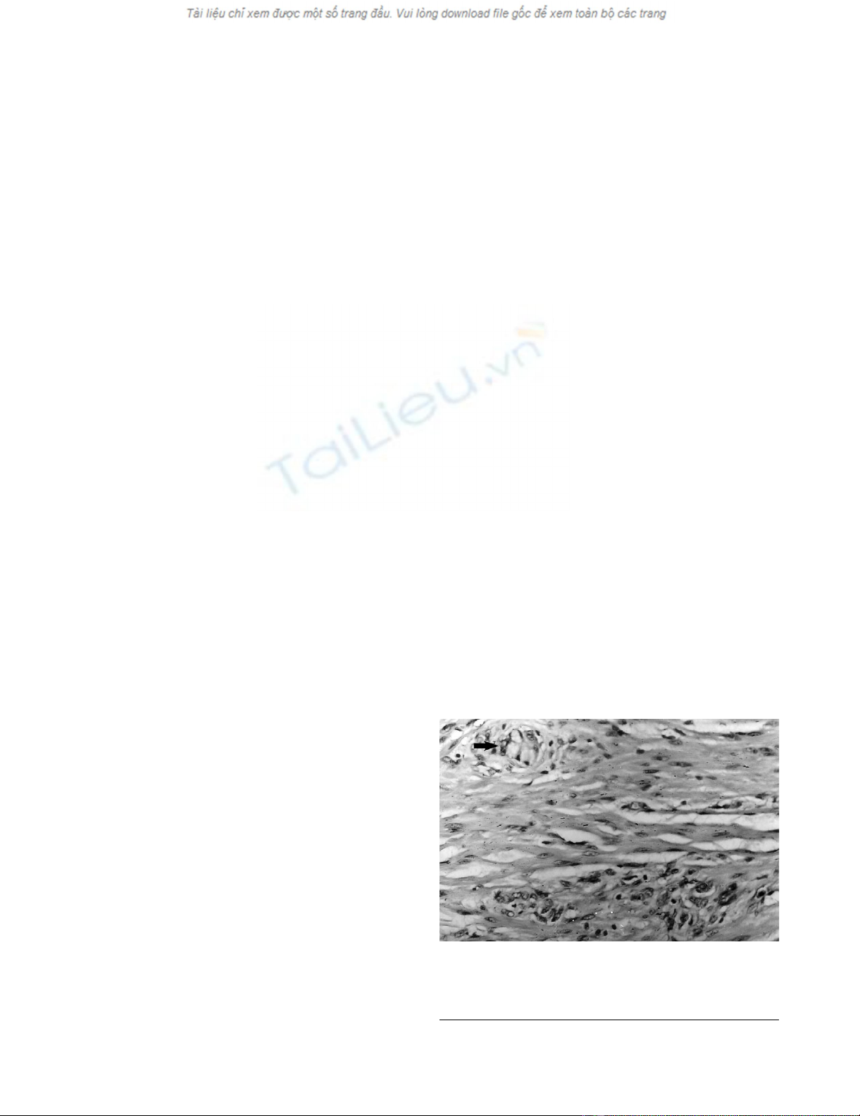
Clusters of goblet cells (arrow) infiltrated the muscle layer of the appendixFigure 1
Clusters of goblet cells (arrow) infiltrated the muscle layer
of the appendix. (Hematoxyllin and eosin). Magnification ×
300.
World Journal of Surgical Oncology 2008, 6:15 http://www.wjso.com/content/6/1/15
Page 4 of 8
(page number not for citation purposes)
a. Chromogranin A-positive cells (arrows) in the normal appendiceal glands (G) and in submucosa (S)Figure 2
a. Chromogranin A-positive cells (arrows) in the normal appendiceal glands (G) and in submucosa (S), b. Chromogranin A-
positive endocrine cells (arrows) delineate the tumour nests of goblet cells, c. Somatostatin-positive endocrine cells (arrows)
in the normal appendiceal mucosa, d. Somatostatin-positive cells (arrows) in the appendiceal submucosa (S), e. Synapto-
physin-positive endocrine cells (arrow) in the normal appendiceal mucosa, f. Synaptophysin-positive endocrine cells (arrow)
in the appendiceal submucosa (S). Magnifications × a, b, c, d, e, f- × 300.
World Journal of Surgical Oncology 2008, 6:15 http://www.wjso.com/content/6/1/15
Page 5 of 8
(page number not for citation purposes)
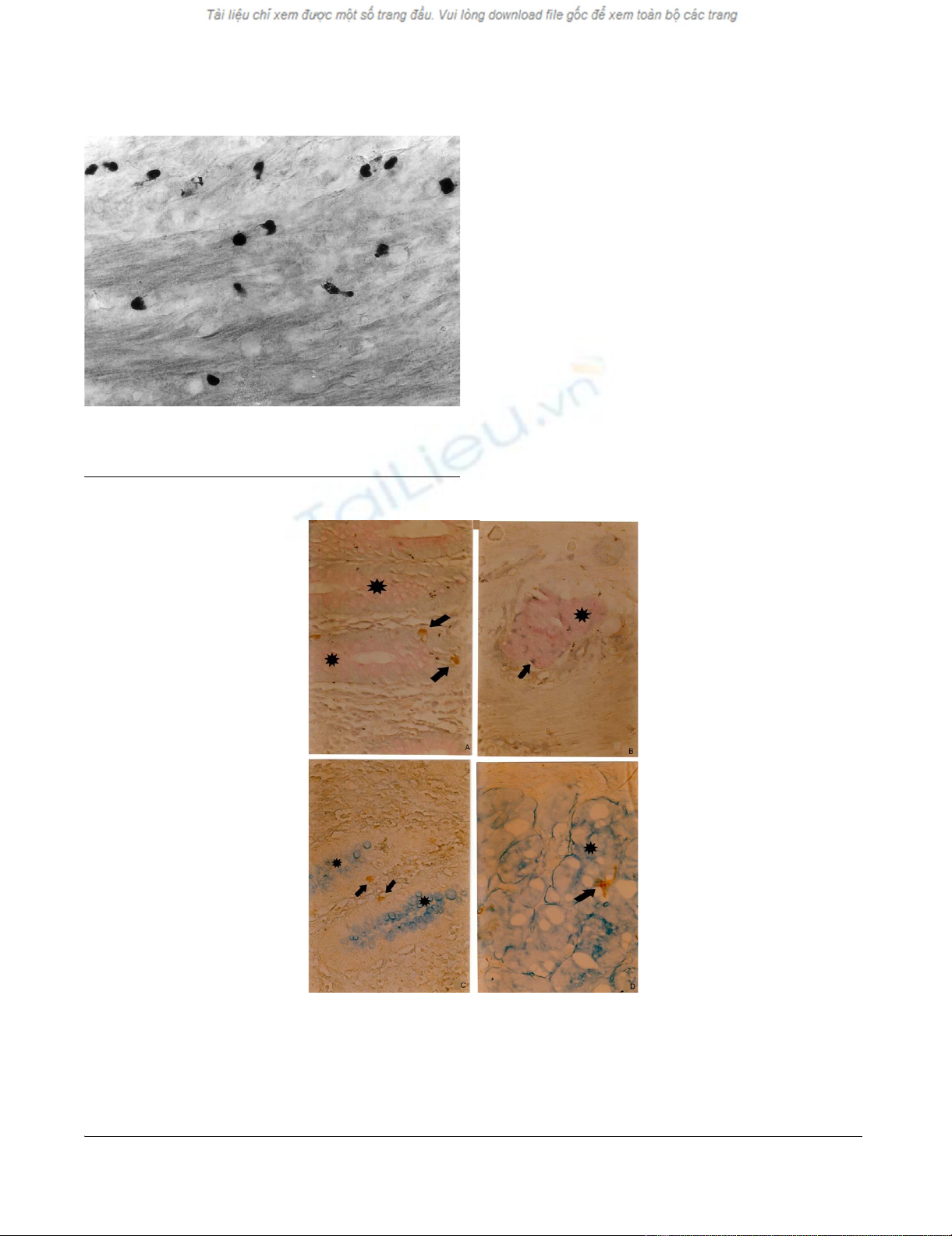
Brown serotonin-positive cells were attached to alcian
blue-positive mucous cells (Figure 4c,d).
Ultrastructural immunohistochemistry
Tumour nests resembling the normal crypts could be seen
in the submucosa of the appendix. The mucous cells
showed slight nuclear atypia. The endocrine cells were
gathered in groups of 2 or 3 and were located basally to
the mucous cells (Figure 5a). Their granules contained
chromogranin A reaction product and were from the
ovoid or discoid EC2 type. Single chromogranin A-positive
endocrine cells, likely from D type with small electron-
dense ovoid granules were found in the stroma of the sub-
mucosa (Figure 5b).
Discussion
In the appendix goblet cell carcinoids appear less fre-
quently than conventional carcinoids [3]. A review of
appendiceal tumours set their incidence at only 5% of
occurring appendiceal primary tumours [14]. A small
Serotonin-positive endocrine cells in the muscle layer of the caecumFigure 3
Serotonin-positive endocrine cells in the muscle layer of the
caecum. Magnification × 300.
a. PAS-positive mucous cells (pink, star), and brown chromogranin A-positive endocrine cells (arrow) in the normal appen-diceal mucosaFigure 4
a. PAS-positive mucous cells (pink, star), and brown chromogranin A-positive endocrine cells (arrow) in the normal appen-
diceal mucosa, b. PAS-positive mucin-secreting cells (pink, star), surrounded by brown chromogranin A-positive endocrine
cells (arrow) in a tumour gland in the submucosa, c. Alciane blue-positive mucous cells (blue, star) and brown chromogranin
A-positive endocrine cells (arrow) in the normal appendiceal mucosa, d. Alciane blue-positive mucin-secreting cells (blue,
star), and brown chromogranin A-positive endocrine cells (arrow) in a tumour gland. (A combined histochemical and immu-
nohistochemical staining). Magnifications a, b, c d × 300.


%20--%3e%3cdefs%3e%3cstyle%3e%20.st0%20{%20fill:%20%23fff;%20}%20.st1%20{%20fill:%20%237800fa;%20}%20%3c/style%3e%3c/defs%3e%3cpath%20class='st1'%20d='M117.78,12.18H43.11c2.9,3.47,4.65,7.94,4.65,12.82,0,5.6-2.3,10.66-6.01,14.29h76.02l7.22-13.56-7.22-13.56Z'/%3e%3cg%3e%3cpath%20class='st0'%20d='M53.58,26.17h-.59v-1.46h.59v-4.96h2.83c1.78,0,2.67.94,2.67,2.82v5.76c0,1.87-.89,2.81-2.67,2.81h-2.83v-4.96ZM55.36,21.37v3.34h1.1v1.46h-1.1v3.34h1.01c.61,0,.91-.37.91-1.1v-5.93c0-.74-.3-1.1-.91-1.1h-1.01Z'/%3e%3cpath%20class='st0'%20d='M65.99,31.14h-1.8l-.31-2.07h-2.19l-.31,2.07h-1.64l1.82-11.39h2.62l1.82,11.39ZM65.28,18.04c-.25.46-.51.77-.75.94-.21.15-.47.22-.79.22-.26,0-.57-.07-.92-.22l-.38-.15c-.14-.05-.26-.07-.37-.07-.3,0-.53.18-.71.54l-.91-.68c.25-.46.51-.77.75-.94.21-.14.48-.21.79-.21.26,0,.57.07.92.21l.38.15c.14.05.26.07.37.07.3,0,.53-.18.71-.54l.91.68ZM61.91,27.52h1.73l-.87-5.76-.87,5.76Z'/%3e%3cpath%20class='st0'%20d='M74.53,26.89v1.52c0,1.91-.89,2.86-2.67,2.86s-2.67-.95-2.67-2.86v-5.93c0-1.91.89-2.86,2.67-2.86s2.67.95,2.67,2.86v1.11h-1.69v-1.22c0-.75-.31-1.12-.93-1.12s-.93.37-.93,1.12v6.15c0,.74.31,1.11.93,1.11s.93-.37.93-1.11v-1.63h1.69Z'/%3e%3cpath%20class='st0'%20d='M81.4,31.14h-1.8l-.31-2.07h-2.19l-.31,2.07h-1.64l1.82-11.39h2.62l1.82,11.39ZM75.9,19.2l1.52-1.91h1.71l1.51,1.91h-1.61l-.76-.95-.75.95h-1.61ZM77.32,27.52h1.73l-.87-5.76-.87,5.76ZM83.1,15.99l-1.76,1.91h-1.26l1.17-1.91h1.86Z'/%3e%3cpath%20class='st0'%20d='M84.86,19.75c1.78,0,2.67.94,2.67,2.82v1.48c0,1.87-.89,2.81-2.67,2.81h-.85v4.28h-1.79v-11.39h2.64ZM84.01,21.37v3.86h.85c.58,0,.87-.36.87-1.08v-1.71c0-.71-.29-1.07-.87-1.07h-.85Z'/%3e%3cpath%20class='st0'%20d='M93.51,19.75c1.78,0,2.67.94,2.67,2.82v1.48c0,1.87-.89,2.81-2.67,2.81h-.85v4.28h-1.79v-11.39h2.64ZM92.66,21.37v3.86h.85c.58,0,.87-.36.87-1.08v-1.71c0-.71-.29-1.07-.87-1.07h-.85Z'/%3e%3cpath%20class='st0'%20d='M98.8,31.14h-1.79v-11.39h1.79v4.88h2.03v-4.88h1.83v11.39h-1.83v-4.88h-2.03v4.88Z'/%3e%3cpath%20class='st0'%20d='M105.36,24.55h2.46v1.62h-2.46v3.34h3.09v1.63h-4.88v-11.39h4.88v1.63h-3.09v3.18ZM108.17,17.29l-1.76,1.91h-1.26l1.17-1.91h1.86Z'/%3e%3cpath%20class='st0'%20d='M112.2,19.75c1.78,0,2.67.94,2.67,2.82v1.48c0,1.87-.89,2.81-2.67,2.81h-.85v4.28h-1.79v-11.39h2.64ZM111.35,21.37v3.86h.85c.58,0,.87-.36.87-1.08v-1.71c0-.71-.29-1.07-.87-1.07h-.85Z'/%3e%3c/g%3e%3ccircle%20class='st1'%20cx='25'%20cy='25'%20r='20'/%3e%3cpath%20class='st0'%20d='M32.78,19.27c2.92,0,4.43,2.55,5.28,5.33l.71,2.17c.14.38-.33.75-.71.75h-5.61c.19-.33.24-.71.09-1.08l-.75-2.45c-.43-1.32-.99-2.64-1.79-3.77.75-.57,1.65-.94,2.78-.94h0ZM25,18.38c3.25,0,4.9,2.78,5.89,5.89l.76,2.45c.14.42-.33.8-.8.8h-11.69c-.42,0-.94-.38-.8-.8l.75-2.45c.99-3.11,2.64-5.89,5.89-5.89h0ZM25,11.35c1.74,0,3.11,1.37,3.11,3.11s-1.37,3.11-3.11,3.11-3.11-1.41-3.11-3.11,1.41-3.11,3.11-3.11h0ZM17.27,19.27c1.08,0,1.98.38,2.73.94-.8,1.13-1.37,2.45-1.74,3.77l-.8,2.45c-.14.38-.05.75.09,1.08h-5.56c-.42,0-.9-.38-.75-.75l.71-2.17c.9-2.78,2.41-5.33,5.33-5.33h0ZM17.27,12.91c1.51,0,2.78,1.27,2.78,2.83s-1.27,2.83-2.78,2.83-2.83-1.27-2.83-2.83,1.27-2.83,2.83-2.83h0ZM32.78,12.91c1.56,0,2.78,1.27,2.78,2.83s-1.23,2.83-2.78,2.83-2.83-1.27-2.83-2.83,1.27-2.83,2.83-2.83h0ZM27.07,28.56v.09c0,.57-.24,1.08-.61,1.46h0v.05c-.38.33-.9.57-1.46.57s-1.08-.24-1.46-.61h0c-.38-.38-.61-.9-.61-1.46v-.09h1.41v.09c0,.19.05.38.19.47v.05c.09.09.28.19.47.19s.38-.09.47-.19v-.05c.14-.09.24-.28.24-.47t-.05-.09h1.41ZM30.99,28.56v.09c0,1.65-.66,3.16-1.74,4.24-1.08,1.08-2.59,1.79-4.24,1.79s-3.16-.71-4.24-1.79l-.05-.05c-1.04-1.08-1.7-2.55-1.7-4.2v-.09h1.41v.09c0,1.27.47,2.4,1.27,3.25h.05c.85.85,1.98,1.37,3.25,1.37s2.4-.52,3.25-1.37c.85-.8,1.37-1.98,1.37-3.25v-.09h1.37ZM34.99,28.56v.09c0,2.78-1.13,5.28-2.92,7.07-1.79,1.79-4.29,2.92-7.07,2.92s-5.23-1.13-7.07-2.92c-1.79-1.79-2.92-4.29-2.92-7.07v-.09h1.41v.09c0,2.4.94,4.53,2.5,6.08,1.56,1.56,3.72,2.5,6.08,2.5s4.52-.94,6.08-2.5c1.56-1.56,2.5-3.68,2.5-6.08v-.09h1.41Z'/%3e%3c/svg%3e)