
BioMed Central
Page 1 of 5
(page number not for citation purposes)
World Journal of Surgical Oncology
Open Access
Case report
Granulocyte-colony stimulating factor producing rectal cancer
Hiroki Takahashi, Akira Yasuda, Nubuo Ochi, Masaki Sakamoto,
Satoru Takayama, Takehiro Wakasugi, Hitoshi Funahashi, Hirozumi Sawai,
Mikinori Satoh, Yoshimi Akamo and Hiromitsu Takeyama*
Address: Department of Gastroenterological Surgery, Nagoya City University Graduate School of Medical Sciences, Kawasumi 1, Mizuho-cho,
Mizuho-ku, Nagoya, Japan
Email: Hiroki Takahashi - takahasi@med.nagoya-cu.ac.jp; Akira Yasuda - a-yasuda@med.nagoya-cu.ac.jp; Nubuo Ochi - nochi@med.nagoya-
cu.ac.jp; Masaki Sakamoto - m.saka@med.nagoya-cu.ac.jp; Satoru Takayama - takasato@med.nagoya-cu.ac.jp;
Takehiro Wakasugi - wakasugi@med.nagoya-cu.ac.jp; Hitoshi Funahashi - funa84@med.nagoya-cu.ac.jp; Hirozumi Sawai - sawai@med.nagoya-
cu.ac.jp; Mikinori Satoh - miki@med.nagoya-cu.ac.jp; Yoshimi Akamo - akamo@med.nagoya-cu.ac.jp;
Hiromitsu Takeyama* - takeyama@med.nagoya-cu.ac.jp
* Corresponding author
Abstract
Background: Granulocyte-colony stimulating factor (G-CSF)-producing cancer has been
reported to occur in various organs, especially the lung. However, G-CSF-producing colorectal
cancer (CRC) has never been reported in the English literature.
Case presentation: A 57-year-old man was admitted for the surgical removal of a rectal cancer.
Some hepatic tumors in the liver were revealed concurrently, and their appearance suggested
multiple liver metastases. Low anterior resection was performed. with the help of histopathological
examination and immunohistochemical studies, we diagnosed this case to be an undifferentiated
carcinoma of the rectum. After the operation, the white blood cell (WBC) count increased
gradually to 81,000 cells/μL. Modified-FOLFOX6 therapy was initiated to treat the liver metastases,
but there was no effect, and peritoneal dissemination had also occurred. The serum level of G-CSF
was elevated to 840 pg/mL (normal range, <18.1 pg/mL). Furthermore, immunohistochemistry with
a specific monoclonal antibody against G-CSF was positive; therefore, we diagnosed this tumor as
a G-CSF-producing cancer. The patient died from rapid growth of the liver metastases and
peritoneal dissemination 2 months after surgery.
Conclusion: This is the first case of G-CSF-producing rectal cancer, and its prognosis was very
poor.
Background
Granulocyte-colony stimulating factor (G-CSF)-produc-
ing cancer has been reported to occur in the lung [1],
stomach [2], esophagus [3], gall bladder [4], thyroid [5],
urinary bladder [6], liver [7,8]. However, to the best of our
knowledge, G-CSF-producing colorectal cancer (CRC) has
never been reported in the English literature. G-CSF-pro-
ducing cancers are thought to have a very poor prognosis.
Furthermore, undifferentiated CRC is very rare and this is
the first report of a G-CSF-producing undifferentiated can-
cer of the rectum. Its prognosis was very poor; therefore,
Published: 29 June 2008
World Journal of Surgical Oncology 2008, 6:70 doi:10.1186/1477-7819-6-70
Received: 31 March 2008
Accepted: 29 June 2008
This article is available from: http://www.wjso.com/content/6/1/70
© 2008 Takahashi et al; licensee BioMed Central Ltd.
This is an Open Access article distributed under the terms of the Creative Commons Attribution License (http://creativecommons.org/licenses/by/2.0),
which permits unrestricted use, distribution, and reproduction in any medium, provided the original work is properly cited.
World Journal of Surgical Oncology 2008, 6:70 http://www.wjso.com/content/6/1/70
Page 2 of 5
(page number not for citation purposes)
we would like to report this case and discuss its clinico-
pathological features.
Case presentation
A 57-year-old man was admitted to our hospital with
lower abdominal pain in June 2007. Barium enema and
colonoscopy revealed an ulcerative tumor in the rectum
(Figure 1), which, after biopsy, was diagnosed as a well
differentiated adenocarcinoma. Physical examination
showed no remarkable abnormalities. Neither hepatome-
galy nor splenomegaly was apparent. Serum was negative
for hepatitis B surface antigen and hepatitis C antibodies,
and the patient had no history of alcohol intake or blood
transfusion. Laboratory data on admission, including
liver function tests, were unremarkable. The white blood
cell (WBC) count was 8,000 cells/μL (neutrophil: 80.7%).
Levels of carcinoembryonic antigen (CEA), carbohydrate
antigen 19-9 (CA19-9), and α-fetoprotein (AFP) were
within normal ranges. Abdominal ultrasonography
showed a multiple hypoechoic, 1.5-cm diameter mass in
the liver. Computed tomography (CT) and magnetic reso-
nance imaging (MRI) with superparamagnetic iron oxide
(SPIO) were performed, and the results suggested multi-
ple liver metastases. The patient underwent low anterior
resection on 25 July 2007.
An ulcerated hard tumor was present in the rectum (Figure
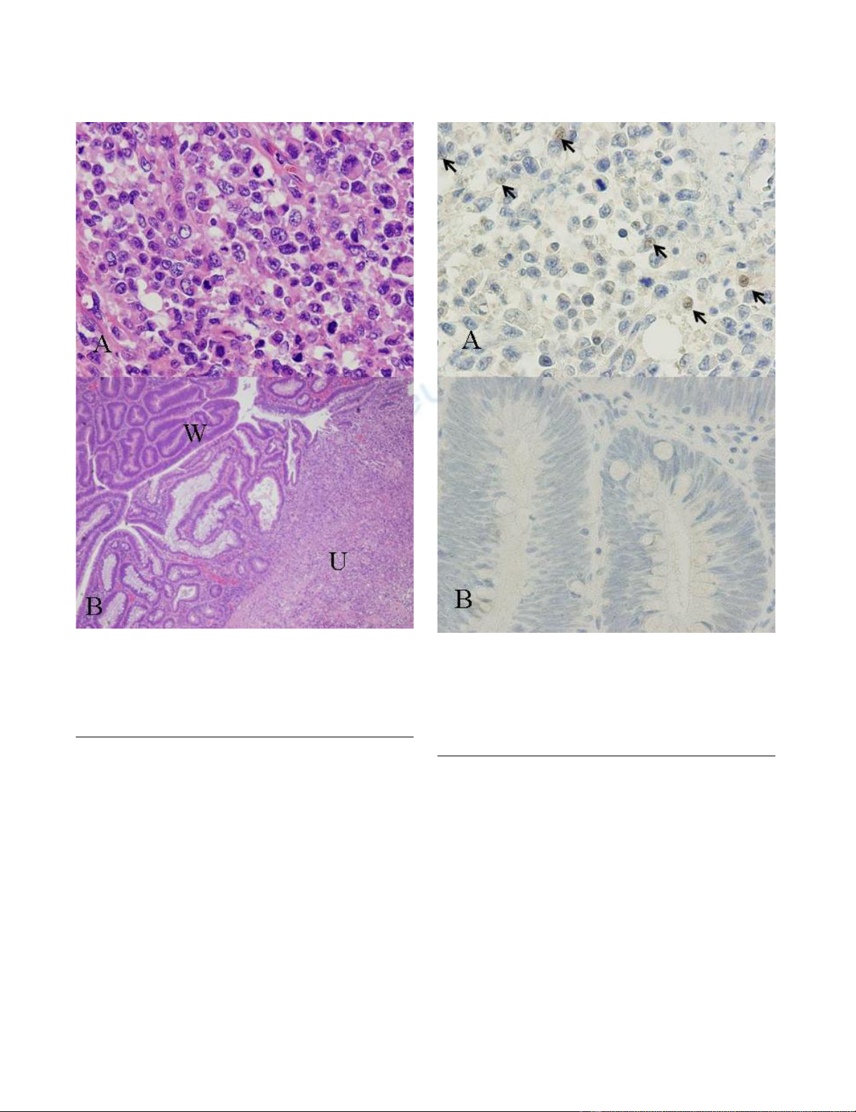
2). Histopathological examination revealed that the
tumor consisted of large abnormal cells without gland for-
mation and mucin production (Figure 3a). Immunohisto-
chemical studies were positive for cytokeratin and
vimentin; however, they were negative for CD45 and PAS.
In addition, they were negative for both neuron-specific
enolase (NSE) and synaptophysin, and histologic staining
with alcian blue was also negative. Therefore, we diag-
nosed this case to be an undifferentiated carcinoma of the
rectum. A small component of well-differentiated adeno-
carcinoma was also seen on the surface of the tumor (Fig-
ure 3b). Thus, we thought that we diagnosed this tumor as
well-differentiated adenocarcinoma at biopsy. Advanced
lymphatic vessel and venous invasion were observed.
Lymph node metastasis was also detected near the tumor,
but peritoneal dissemination was not detected. After the
operation, the WBC count gradually increased. Modified-
FOLFOX6 (mFOLFOX6) therapy was initiated to treat the
liver metastases, but it had no effect, and peritoneal dis-
semination occurred. Along with the growth of the tumor,
the WBC count increased to 81,000 cells/μL (neutrophil:
87%). On the other hand, in comparison to the grade of
leukocytosis, CRP level was not so high (6.5 mg/dl), and
there were not any obvious signs of infection, so we sus-
pected that this tumor produced G-CSF, and we measured
serum G-CSF using an enzyme-linked immunosorbent
assay (ELISA). The serum level of G-CSF was elevated to
840 pg/mL (normal range, <18.1 pg/mL). Furthermore,
immunohistochemical staining with a specific mono-
clonal antibody against human G-CSF (11041, IBL,
Gunma, Japan) was performed. G-CSF was positive in the
cytoplasm of undifferentiated carcinoma cells (Figure 4a),
but negative in the non-cancerous lesion and well-differ-
entiated adenocarcinoma cells (Figure 4b). Unfortu-
nately, we couldn't obtain biopsy specimens from the
liver tumor. But, G-CSF was positive in metastatic lymph
nodes, so we thought that G-CSF was also positive in the
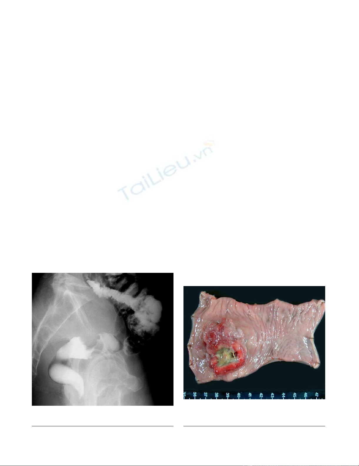
Resected specimenFigure 2
Resected specimen. An ulcerated hard tumor was present in
the rectum.
Barium enema revealed an ulcerative tumor in the rectumFigure 1
Barium enema revealed an ulcerative tumor in the rectum.
World Journal of Surgical Oncology 2008, 6:70 http://www.wjso.com/content/6/1/70
Page 3 of 5
(page number not for citation purposes)
liver tumor. Therefore, we concluded that this tumor was
a G-CSF-producing cancer. The patient died from rapid
growth of the liver metastases and peritoneal dissemina-
tion 2 months after surgery.
Discussion
G-CSF-producing cancer has been reported to occur in
various organs, especially in the lung. Histologically,
more than half of the reported cases of G-CSF-producing
lung cancer have been large cell carcinoma [1]. However,
it was reported that many cases of G-CSF-producing can-
cer of the digestive organs were poorly differentiated car-
cinoma or undifferentiated carcinoma [2,5,8,9]. Our case
was mainly an undifferentiated carcinoma, so G-CSF-pro-
ducing rectal cancer may have the same properties as other
cancers of the digestive organs. However, an area of well-
differentiated adenocarcinoma was seen on the surface of
the tumor. Yamano et al., reported a case of early-stage
gastric cancer that presented as a well-differentiated aden-
ocarcinoma that changed to a poorly differentiated aden-
ocarcinoma at the advanced stage, and acquired the
ability to produce G-CSF [2]. In our case, G-CSF immu-
nostaining was positive only in the undifferentiated cells
and negative in the well-differentiated adenocarcinoma
cells. Consequently, such histological changes might
influence the ability to produce active G-CSF. Interest-
ingly, it was reported that large cell carcinoma of the lung
is often vimentin-positive [10], and our case was also
vimentin-positive. Studies of other G-CSF-producing can-
cers have not investigated vimentin immunoreactivity, so
further examination is required.
Immunohistochemical stainingFigure 4
Immunohistochemical staining. a) Immunohistochemical
staining with a specific monoclonal antibody against recom-
binant human G-CSF was positive in the cytoplasm of undif-
ferentiated carcinoma cells (×400). b) The well-differentiated
part of the adenocarcinoma was not stained with the G-CSF
antibody (×400).
HE stainingFigure 3
HE staining. a) Large abnormal cells without gland forma-
tion and mucin production were seen in the tumor (×400). b)
A small component of well-differentiated adenocarcinoma
was also seen on the surface of the tumor (W) amidst undif-
ferentiated carcinoma (U) (×40).

World Journal of Surgical Oncology 2008, 6:70 http://www.wjso.com/content/6/1/70
Page 4 of 5
(page number not for citation purposes)
The mechanism by which certain CRCs produce G-CSF
has not been clarified. Mroczko et al., reported that
median values of G-CSF in CRC patients were significantly
higher than those in healthy subjects [11]. Furthermore, it
was reported that granulocyte-macrophage colony stimu-
lating factor (GM-CSF) secretion was also detected by
human colorectal cancer specimens and cell lines [12,13].
Tachibana et al., showed that G-CSF production by transi-
tional cell carcinoma of the bladder augments autocrine
growth, which may in part explain the poor prognoses
[14]. Savarese et al., reported that 56.5% of primary ovar-
ian carcinomas co-expressed G-CSF and the G-CSF recep-
tor (G-CSFR); potential autocrine and/or paracrine loops
involving G-CSF and its receptor occur in over 90% of pri-
mary ovarian carcinomas [15]. As mentioned earlier,
some CRCs have the ability to secrete active G-CSF. Fur-
thermore, Yang et al., reported that the G-CSFR was
expressed in 59% of CRCs [16]. Therefore, autocrine
growth is possible in CRC. Furthermore, Natori et al.,
reported that G-CSF stimulates angiogenesis and pro-
motes tumor growth [17]. In addition, Tsuruta et al.,
showed that the production of GM-CSF by squamous cell
carcinoma cell lines was closely related to their in vitro
invasiveness and MMP activity [18]. For these reasons, G-
CSF-producing cancer has a very poor prognosis, and at
the present time, there is no specific approach for G-CSF-
producing cancer. We performed mFOLFOX6 therapy to
treat the liver metastases, but it had no effect, and the
patient's general condition worsened rapidly. Usually,
preoperative WBC count of G-CSF producing tumor is
high and reduces after surgery or chemotherapy. In
present case, preoperative WBC count was not so high and
increased along with the growth of the tumor. We guess
that this is because tumor volume was not large at the
time of operation.
Conclusion
In summary, we present the first case of G-CSF-producing
rectal cancer. Its prognosis was very poor, and mFOLFOX6
therapy had no effect. But, we believe that further investi-
gation may make it clear that molecular targeted therapies
for G-CSF may become part of the treatment paradigm.
Abbreviations
CRC: colorectal cancer; G-CSF: granulocyte-colony-stimu-
lating factor; GM-CSF: granulocyte-macrophage colony
stimulating factor; ELISA: enzyme-linked immunosorbent
assay; G-CSFR: granulocyte-colony-stimulating factor
receptor; CT: Computed tomography; MRI: Magnetic res-
onance imaging; mFOLFOX: Modified-FOLFOX6.
Competing interests
The authors declare that they have no competing interests.
Authors' contributions
HT, SM, NO, ST and HT carried out the clinical examina-
tion and operation, HT, AY, HS, and YA performed the
pathological analysis, HT, HF and TW participated in the
design of the study, HT and MS conceived the study, and
participated in its design and coordination. All authors
read and approved the final manuscript.
Acknowledgements
Written consent was obtained from the patient's family for publication of
study.
References
1. Hasegawa S, Suda T, Negi K, Hattori Y: Lung large cell carcinoma
producing granulocyte-colony-stimulating factor. Ann Thorac
Surg 2007, 83:308-310.
2. Yamano T, Morii E, Ikeda J, Aozasa K: Granulocyte colony-stimu-
lating factor production and rapid progression of gastric can-
cer after histological change in the tumor. Jpn J Clin Oncol 2007,
37:793-796.
3. Ota S, Kato A, Kobayashi H, Yonezumi M, Yamaguchi J, Musashi M,
Imamura M, Asaka M: Monoclonal origin of an esophageal car-
cinosarcoma producing granulocyte-colony stimulating fac-
tor: a case report. Cancer 1998, 82:2102-2111.
4. Ikeda T, Ohgaki K, Miura M, Aishima S, Shimizu T, Maehara Y: Gran-
ulocyte-colony stimulating factor-producing gallbladder can-
cer without recurrence more than 2 years after resection:
report of a case. Surg Today 2005, 35:590-593.
5. Fujita T, Ogasawara Y, Naito M, Doihara H, Shimizu N: Anaplastic
thyroid carcinoma associated with granulocyte colony-stim-
ulating factor: report of a case. Surg Today 2006, 36:63-67.
6. Hirasawa K, Kitamura T, Oka T, Matsushita H: Bladder tumor pro-
ducing Granulocyte colony-stimulating factor and parathy-
roid hormone related protein. J Urol 2002, 167:2130.
7. Araki K, Kishihara F, Takahashi K, Matsumata T, Shimura T, Suehiro
T, Kuwano H: Hepatocellular carcinoma producing a granulo-
cyte colony-stimulating factor: report of a resected case with
a literature review. Liver Int 2007, 27:716-721.
8. Yamamoto S, Takashima S, Ogawa H, Kuroda T, Yamamoto M,
Takeda A, Nakamura H: Granulocyte-colony-stimulating-fac-
tor-producing hepatocellular carcinoma. J Gastroenterol 1999,
34:640-644.
9. Sohda T, Shiga H, Nakane H, Watanabe H, Takeshita M, Sakisaka S:
Cholangiocellular carcinoma that produced both granulo-
cyte-colony-stimulating factor and parathyroid hormone-
related protein. Int J Clin Oncol 2006, 11:246-249.
10. Kodama T, Shimosato Y, Koide T, Watanabe S, Teshima S: Large cell
carcinoma of lung: ultrastructure and immunohistochemical
studies. Jpn J Clin Oncol 1985, 15:431-441.
11. Mroczko B, Groblewska M, Wereszczynska-Siemiatkowska U, Kedra
B, Konopko M, Szmitkowski M: The diagnostic value of G-CSF
measurement in the sera of colorectal cancer and adenoma
patients. Clin Chim Acta 2006, 371:143-147.
12. Lahm H, Wyniger J, Hertig S, Yilmaz A, Fischer JR, Givel JC, Odartch-
enko N: Secretion of bioactive granulocyte-macrophage col-
ony-stimulating factor by human colorectal carcinoma cells.
Cancer Res 1994, 54:3700-3702.
13. Trutmann M, Terracciano L, Noppen C, Kloth J, Kaspar M, Peterli R,
Tondelli P, Schaeffer C, Zajac P, Heberer M, Spagnoli GC: GM-CSF
gene expression and protein production in human colorectal
cancer cell lines and clinical tumor specimens. Int J Cancer
1998, 77:378-385.
14. Tachibana M, Miyakawa A, Tazaki H, Nakamura K, Kubo A, Hata J,
Nishi T, Amano Y: Autocrine growth of transitional cell carci-
noma of the bladder induced by granulocyte-colony stimu-
lating factor. Cancer Res 1995, 55:3438-3443.
15. Savarese TM, Mitchell K, McQuain C, Campbell CL, Guardiani R,
Wuu J, Ollari C, Reale F, Nelson BE, Chen A, Quesenberry PJ: Coex-
pression of granulocyte colony stimulating factor and its
receptor in primary ovarian carcinomas. Cancer Lett 2001,
162:105-115.

Publish with BioMed Central and every
scientist can read your work free of charge
"BioMed Central will be the most significant development for
disseminating the results of biomedical research in our lifetime."
Sir Paul Nurse, Cancer Research UK
Your research papers will be:
available free of charge to the entire biomedical community
peer reviewed and published immediately upon acceptance
cited in PubMed and archived on PubMed Central
yours — you keep the copyright
Submit your manuscript here:
http://www.biomedcentral.com/info/publishing_adv.asp
BioMedcentral
World Journal of Surgical Oncology 2008, 6:70 http://www.wjso.com/content/6/1/70
Page 5 of 5
(page number not for citation purposes)
16. Yang X, Liu F, Xu Z, Chen C, Wu X, Li G, Li J: Expression of gran-
ulocyte colony stimulating factor receptor in human color-
ectal cancer. Postgrad Med J 2005, 81:333-337.
17. Natori T, Sata M, Washida M, Hirata Y, Nagai R, Makuuchi M: G-CSF
stimulates angiogenesis and promotes tumor growth: poten-
tial contribution of bone marrow-derived endothelial pro-
genitor cells. Biochem Biophys Res Commun 2002, 297:1058-1061.
18. Tsuruta N, Yatsunami J, Takayama K, Nakanishi Y, Ichinose Y, Hara
N: Granulocyte-macrophage-colony stimulating factor stim-
ulates tumor invasiveness in squamous cell lung carcinoma.
Cancer 1998, 82:2173-2183.


%20--%3e%3cdefs%3e%3cstyle%3e%20.st0%20{%20fill:%20%23fff;%20}%20.st1%20{%20fill:%20%237800fa;%20}%20%3c/style%3e%3c/defs%3e%3cpath%20class='st1'%20d='M117.78,12.18H43.11c2.9,3.47,4.65,7.94,4.65,12.82,0,5.6-2.3,10.66-6.01,14.29h76.02l7.22-13.56-7.22-13.56Z'/%3e%3cg%3e%3cpath%20class='st0'%20d='M53.58,26.17h-.59v-1.46h.59v-4.96h2.83c1.78,0,2.67.94,2.67,2.82v5.76c0,1.87-.89,2.81-2.67,2.81h-2.83v-4.96ZM55.36,21.37v3.34h1.1v1.46h-1.1v3.34h1.01c.61,0,.91-.37.91-1.1v-5.93c0-.74-.3-1.1-.91-1.1h-1.01Z'/%3e%3cpath%20class='st0'%20d='M65.99,31.14h-1.8l-.31-2.07h-2.19l-.31,2.07h-1.64l1.82-11.39h2.62l1.82,11.39ZM65.28,18.04c-.25.46-.51.77-.75.94-.21.15-.47.22-.79.22-.26,0-.57-.07-.92-.22l-.38-.15c-.14-.05-.26-.07-.37-.07-.3,0-.53.18-.71.54l-.91-.68c.25-.46.51-.77.75-.94.21-.14.48-.21.79-.21.26,0,.57.07.92.21l.38.15c.14.05.26.07.37.07.3,0,.53-.18.71-.54l.91.68ZM61.91,27.52h1.73l-.87-5.76-.87,5.76Z'/%3e%3cpath%20class='st0'%20d='M74.53,26.89v1.52c0,1.91-.89,2.86-2.67,2.86s-2.67-.95-2.67-2.86v-5.93c0-1.91.89-2.86,2.67-2.86s2.67.95,2.67,2.86v1.11h-1.69v-1.22c0-.75-.31-1.12-.93-1.12s-.93.37-.93,1.12v6.15c0,.74.31,1.11.93,1.11s.93-.37.93-1.11v-1.63h1.69Z'/%3e%3cpath%20class='st0'%20d='M81.4,31.14h-1.8l-.31-2.07h-2.19l-.31,2.07h-1.64l1.82-11.39h2.62l1.82,11.39ZM75.9,19.2l1.52-1.91h1.71l1.51,1.91h-1.61l-.76-.95-.75.95h-1.61ZM77.32,27.52h1.73l-.87-5.76-.87,5.76ZM83.1,15.99l-1.76,1.91h-1.26l1.17-1.91h1.86Z'/%3e%3cpath%20class='st0'%20d='M84.86,19.75c1.78,0,2.67.94,2.67,2.82v1.48c0,1.87-.89,2.81-2.67,2.81h-.85v4.28h-1.79v-11.39h2.64ZM84.01,21.37v3.86h.85c.58,0,.87-.36.87-1.08v-1.71c0-.71-.29-1.07-.87-1.07h-.85Z'/%3e%3cpath%20class='st0'%20d='M93.51,19.75c1.78,0,2.67.94,2.67,2.82v1.48c0,1.87-.89,2.81-2.67,2.81h-.85v4.28h-1.79v-11.39h2.64ZM92.66,21.37v3.86h.85c.58,0,.87-.36.87-1.08v-1.71c0-.71-.29-1.07-.87-1.07h-.85Z'/%3e%3cpath%20class='st0'%20d='M98.8,31.14h-1.79v-11.39h1.79v4.88h2.03v-4.88h1.83v11.39h-1.83v-4.88h-2.03v4.88Z'/%3e%3cpath%20class='st0'%20d='M105.36,24.55h2.46v1.62h-2.46v3.34h3.09v1.63h-4.88v-11.39h4.88v1.63h-3.09v3.18ZM108.17,17.29l-1.76,1.91h-1.26l1.17-1.91h1.86Z'/%3e%3cpath%20class='st0'%20d='M112.2,19.75c1.78,0,2.67.94,2.67,2.82v1.48c0,1.87-.89,2.81-2.67,2.81h-.85v4.28h-1.79v-11.39h2.64ZM111.35,21.37v3.86h.85c.58,0,.87-.36.87-1.08v-1.71c0-.71-.29-1.07-.87-1.07h-.85Z'/%3e%3c/g%3e%3ccircle%20class='st1'%20cx='25'%20cy='25'%20r='20'/%3e%3cpath%20class='st0'%20d='M32.78,19.27c2.92,0,4.43,2.55,5.28,5.33l.71,2.17c.14.38-.33.75-.71.75h-5.61c.19-.33.24-.71.09-1.08l-.75-2.45c-.43-1.32-.99-2.64-1.79-3.77.75-.57,1.65-.94,2.78-.94h0ZM25,18.38c3.25,0,4.9,2.78,5.89,5.89l.76,2.45c.14.42-.33.8-.8.8h-11.69c-.42,0-.94-.38-.8-.8l.75-2.45c.99-3.11,2.64-5.89,5.89-5.89h0ZM25,11.35c1.74,0,3.11,1.37,3.11,3.11s-1.37,3.11-3.11,3.11-3.11-1.41-3.11-3.11,1.41-3.11,3.11-3.11h0ZM17.27,19.27c1.08,0,1.98.38,2.73.94-.8,1.13-1.37,2.45-1.74,3.77l-.8,2.45c-.14.38-.05.75.09,1.08h-5.56c-.42,0-.9-.38-.75-.75l.71-2.17c.9-2.78,2.41-5.33,5.33-5.33h0ZM17.27,12.91c1.51,0,2.78,1.27,2.78,2.83s-1.27,2.83-2.78,2.83-2.83-1.27-2.83-2.83,1.27-2.83,2.83-2.83h0ZM32.78,12.91c1.56,0,2.78,1.27,2.78,2.83s-1.23,2.83-2.78,2.83-2.83-1.27-2.83-2.83,1.27-2.83,2.83-2.83h0ZM27.07,28.56v.09c0,.57-.24,1.08-.61,1.46h0v.05c-.38.33-.9.57-1.46.57s-1.08-.24-1.46-.61h0c-.38-.38-.61-.9-.61-1.46v-.09h1.41v.09c0,.19.05.38.19.47v.05c.09.09.28.19.47.19s.38-.09.47-.19v-.05c.14-.09.24-.28.24-.47t-.05-.09h1.41ZM30.99,28.56v.09c0,1.65-.66,3.16-1.74,4.24-1.08,1.08-2.59,1.79-4.24,1.79s-3.16-.71-4.24-1.79l-.05-.05c-1.04-1.08-1.7-2.55-1.7-4.2v-.09h1.41v.09c0,1.27.47,2.4,1.27,3.25h.05c.85.85,1.98,1.37,3.25,1.37s2.4-.52,3.25-1.37c.85-.8,1.37-1.98,1.37-3.25v-.09h1.37ZM34.99,28.56v.09c0,2.78-1.13,5.28-2.92,7.07-1.79,1.79-4.29,2.92-7.07,2.92s-5.23-1.13-7.07-2.92c-1.79-1.79-2.92-4.29-2.92-7.07v-.09h1.41v.09c0,2.4.94,4.53,2.5,6.08,1.56,1.56,3.72,2.5,6.08,2.5s4.52-.94,6.08-2.5c1.56-1.56,2.5-3.68,2.5-6.08v-.09h1.41Z'/%3e%3c/svg%3e)