
BioMed Central
Page 1 of 10
(page number not for citation purposes)
Virology Journal
Open Access
Research
Mechanisms of the action of povidone-iodine against human and
avian influenza A viruses: its effects on hemagglutination and
sialidase activities
Nongluk Sriwilaijaroen1,2, Prapon Wilairat3, Hiroaki Hiramatsu2,
Tadanobu Takahashi4,5, Takashi Suzuki4,5, Morihiro Ito2, Yasuhiko Ito2,
Masato Tashiro6 and Yasuo Suzuki*2,5
Address: 1Faculty of Medicine, Thammasat University (Rangsit Campus), Pathumthani 12120, Thailand, 2Health Science Hills, College of Life and
Health Sciences, Chubu University, Kasugai, Aichi 487-8501, Japan, 3Department of Biochemistry, Faculty of Science, Mahidol University,
Bangkok, Thailand, 4Department of Biochemistry, University of Shizuoka, School of Pharmaceutical Sciences, Shizuoka 422-8526, Japan, 5Global
COE Program for Innovation in Human Health Sciences, Shizuoka 422-8526, Japan and 6Department of Viral Diseases and Vaccine Control,
National Institute of Infectious Diseases, Toyama, Shinjuku-ku, Tokyo, 162-8640, Japan
Email: Nongluk Sriwilaijaroen - snongluk@hotmail.com; Prapon Wilairat - scpwl@mahidol.ac.th;
Hiroaki Hiramatsu - hiramatu@isc.chubu.ac.jp; Tadanobu Takahashi - takahasi@u-shizuoka-ken.ac.jp; Takashi Suzuki - suzukit@u-shizuoka-
ken.ac.jp; Morihiro Ito - m-ito@isc.chubu.ac.jp; Yasuhiko Ito - yito@isc.chubu.ac.jp; Masato Tashiro - mtashiro@hih.go.jp;
Yasuo Suzuki* - suzukiy@isc.chubu.ac.jp
* Corresponding author
Abstract
Background: Influenza virus infection causes significant morbidity and mortality and has marked
social and economic impacts throughout the world. The influenza surface glycoproteins,
hemagglutinin (HA) and neuraminidase (NA), act cooperatively to support efficient influenza A
virus replication and provide the most important targets for anti-influenza chemotherapy. In this
study, povidone-iodine (PVP-I), which has a broad-spectrum microbicidal property, was examined
for its inhibitory effects against influenza virus infection in MDCK cells and the mechanisms of PVP-
I action on HA and NA were revealed.
Results: Results obtained using a novel fluorescence- and chromogenic-based plaque inhibition
assay showed that 1.56 mg/ml PVP-I inhibited infections in MDCK cells of human (8 strains) and
avian (5 strains) influenza A viruses, including H1N1, H3N2, H5N3 and H9N2, from 23.0–97.5%. A
sialidase inhibition assay revealed that PVP-I inhibited N1, N2 and N3 neuraminidases with IC50
values of 9.5–212.1 μg/ml by a mixed-type inhibition mechanism. Receptor binding inhibition and
hemagglutinin inhibition assays indicated that PVP-I affected viral hemagglutinin rather than host-
specific sialic acid receptors.
Conclusion: Mechanisms of reduction of viral growth in MDCK cells by PVP-I involve blockade of
viral attachment to cellular receptors and inhibition of viral release and spread from infected cells.
Therefore, PVP-I is useful to prevent infection and limit spread of human and avian influenza viruses.
Published: 13 August 2009
Virology Journal 2009, 6:124 doi:10.1186/1743-422X-6-124
Received: 9 June 2009
Accepted: 13 August 2009
This article is available from: http://www.virologyj.com/content/6/1/124
© 2009 Sriwilaijaroen et al; licensee BioMed Central Ltd.
This is an Open Access article distributed under the terms of the Creative Commons Attribution License (http://creativecommons.org/licenses/by/2.0),
which permits unrestricted use, distribution, and reproduction in any medium, provided the original work is properly cited.

Virology Journal 2009, 6:124 http://www.virologyj.com/content/6/1/124
Page 2 of 10
(page number not for citation purposes)
Background
Among the three types (A, B and C) of influenza viruses,
A type is the most virulent, infecting various avian and
mammalian species and causing human pandemics as a
consequence of antigenic change (antigenic shift) in their
surface glycoproteins, hemagglutinin (HA) and neurami-
nidase (NA) [1]. Sixteen HA and 9 NA subtypes have been
recognized so far [2]. HA and NA interact with sialic acid
receptors on the host cell surface, the former mediating
membrane fusion that results in virus infection and the
latter possessing sialidase activity that cleaves sialyl link-
ages between viral HA and cellular receptors to release
progeny viruses and separate viruses from HA-mediated
self-aggregation, allowing the virus to infect a new host
cell for continuing virus replication [3].
Virus infection can be inhibited by the use of compounds
that bind to viral HA [4-6], inhibit NA activity [7-11] or
inhibit both HA and NA activities [12]. Two NA inhibi-
tors, sialic acid and shikimic acid analogues, have recently
been licensed for treatment of influenza A and B infec-
tions: zanamivir [13] (Relenza®), which is administered
by inhalation, and oseltamivir phosphate [14] (Tamiflu®),
which is administered orally as a prodrug and is converted
by hepatic esterase to its active form, oseltamivir carboxy-
late (OC). However, influenza A and B viruses with muta-
tions in the NA gene have developed resistance to
oseltamivir and zanamivir [15,16]. The worldwide circu-
lation of oseltamivir-resistant seasonal H1N1, highly
pathogenic avian H5N1 [17,18] and the pandemic
(H1N1) 2009 [19] have provided an impetus to develop
new antiviral and antiseptic materials.
In the nineteenth century, povidone-iodine (PVP-I), a
polyvinylpyrrolidone iodine complex, was developed and
found to have a potent broad-spectrum activity against
bacteria, mycobacteria, fungi, viruses and protozoa [20].
PVP-I has become widely used as an antiseptic and disin-
fectant. Despite long-term use, development of PVP-I
resistance in microorganisms has not been reported
[21,22].
PVP-I products have been found to be effective in inacti-
vating a variety of enveloped and nonenveloped viruses,
such as polio [23], herpes simplex, herpes zoster [24], and
human immunodeficiency viruses [25,26]. Anti-influenza
virus activity of PVP-I also has been reported recently [26-
28]. Pretreatment of avian influenza H5N1, H5N3, H7N7
and H9N2 viruses with PVP-I products, such as solution,
scrub, gargle and throat spray, in the range of 0.23–2%,
reduced viral infectious titers to undetectable values in
embryonated hen's eggs [27]. Both aqueous (Betaisod-
ona®) and liposomal PVP-I inactivated human influenza A
virus (H3N2), resulting in reduction of the virus titer by
more than 4 orders of magnitude in Madin-Darby canine
kidney (MDCK) cells [28]. However, the target sites and
mechanisms of PVP-I action on influenza A and the other
virus infections have hitherto remained unknown. In this
study, we investigated mechanisms underlying PVP-I anti-
influenza activity. The apparent reduction of influenza A
viral infectious titers after incubation with PVP-I products
within a short period of time [26-28] led us to investigate
two spike glycoproteins on the viral surface, HA and NA,
which play essential roles in viral infection, as targets of
PVP-I anti-influenza effects.
Results
Inhibition by PVP-I of influenza A virus growth in MDCK
cells
We first determined the cytotoxicity of PVP-I against
MDCK cells employed as host cells of influenza viruses in
this study by using a cell counting kit-8 assay. Half-maxi-
mum cytotoxic concentration of PVP-I after 24-h exposure
of MDCK cells to PVP-I was 2.4 ± 0.2 mg/ml. PVP-I rang-
ing from 0–1.56 mg/ml, which had no effect on MDCK
cells, reduced virus yield in MDCK cells in a dose-depend-
ent manner (Figure 1B). In comparison with virus yield in
the absence of the inhibitor, 1.56 mg/ml of PVP-I reduced
human virus yield by 59.7–97.5% and avian virus yield by
23.0–57.4%, suggesting enhanced sensitivity towards
human viruses compared to that toward avian viruses.
OC, used as control, inhibited A/Memphis/1/71 (H3N2)
infection by 62% and 73% at concentrations of 0.13 μM
and 80 μM, respectively, whereas it inhibited A/DK/HK/
313/78 (H5N3) infection by 20% and 37%, respectively,
at the same concentrations.
Binding of influenza A viruses to sialoglycopolymers and
guinea pig erythrocytes and inhibition by PVP-I
In agreement with hemagglutinins from avian and human
influenza viruses, which prefer binding to α2,3- and α2,6-
sialylated polymers, respectively [29], A/Memphis/1/71
and A/DK/HK/313/78 viruses predominately bound to
sialoglycopolymers terminated in α2,6 and α2,3 respec-
tively (Figure 2A). Binding of A/Memphis/1/71 to α2,3
and α2,6 polymers was reduced by fetuin (up to 1.25 mg/
ml) and PVP-I (up to 0.78 mg/ml), whereas that of A/DK/
HK/313/78 was inhibited by fetuin but not by PVP-I (Fig-
ure 2B).
Quantitative inhibition of viral HA binding to sialo-glyco-
conjugate receptors on the erythrocyte surface by fetuin
control and PVP-I is shown in Figure 3A and summarized
for PVP-I activity in Table 1. No erythrocyte hemolysis and
no significant change in pH (pH of each well ranging from
6.52 to 7.20) in the assay system were observed. In gen-
eral, fetuin exhibited higher inhibitory activity (ranging
from 0.02 to 1.25 mg/ml) than that of PVP-I (0.2–12.5
mg/ml).
Virology Journal 2009, 6:124 http://www.virologyj.com/content/6/1/124
Page 3 of 10
(page number not for citation purposes)
A qualitative analysis of hemagglutination inhibition
showed that hemagglutination (guinea pig erythrocyte
clumping) of human A/Memphis/1/71 (~400 hemagglu-
tination units (HAU)) and avian A/DK/HK/313/78 (~400
HAU) was completely inhibited by 2.50 mg/ml and 5.00
mg/ml of PVP-I, respectively (Figure 3B).
Effect of PVP-I on influenza A virus sialidase activity
In order to examine the effect of PVP-I on sialidase activity
of different subtypes of influenza virus strains, the enzyme
activity and Km value of each virus subtype were deter-
mined at pH 6.0 using 2'-(4-methylumbelliferyl)-α-D-N-
acetylneuraminic acid (MUNA), a sensitive fluorogenic
substrate without 2,3 and 2,6 linkages. Then an inhibition
assay was performed using 2 enzyme units of each virus
subtype and substrate concentration at its Km value. IC50
values of OC against sialidase of different virus strains
were ranged from 0.37 to 6.88 nM (data not shown).
There were marked differences in IC50 values for PVP-I,
from 9.5 to 212.1 μg/ml depending on the virus strain
(Table 1).
The kinetic mechanism by which PVP-I inhibits influenza
A virus sialidase activity was investigated by determining
kinetic parameters of human A/PR/8/34 (H1N1) sialidase
on hydrolysis of MUNA in the absence and presence of an
inhibitor. As shown in Table 2, with OC or 2-deoxy-2,3-
dehydro-N-acetylneuraminic acid (DANA), Km values
increased, but Vmax did not change. In the presence of PVP-
I, Km values increased and Vmax decreased. Vmax/Km ratio
decreased 6-fold, 6-fold and 12-fold in the presence of 4
nM OC, 75 μg/ml PVP-I and 5 μM DANA, respectively,
indicating decrease in sialidase efficiency. Lineweaver-
Burk plots showed that inhibition of A/PR/8/34 sialidase
activity by OC and DANA was of a competitive type,
whereas that by PVP-I was of a mixed type (Figure 4). The
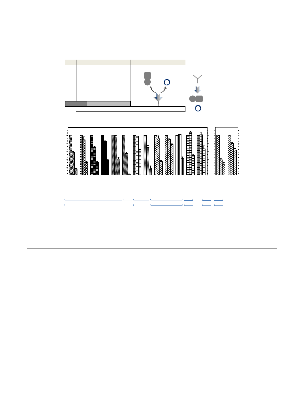
Inhibitory effect of PVP-I on influenza viral infection in MDCK cellsFigure 1
Inhibitory effect of PVP-I on influenza viral infection in MDCK cells. (A) A simplified diagram of the infection assay
used in this study. (B) Quantification of viruses in cells at Ex355/Em460 is expressed as percent virus yield (left Y axis) and per-
cent inhibition (right Y axis) of untreated infected cells.
$
$$
$
%
%%
%
0.00
0.39
1.56
0.00
0.39
1.56
0.00
0.39
1.56
0.00
0.39
1.56
0.00
0.39
1.56
0.00
0.39
1.56
0.00
0.39
1.56
0.00
0.39
1.56
0.00
0.39
1.56
0.00
0.39
1.56
0.00
0.39
1.56
0.00
0.39
1.56
0.00
0.39
1.56
0
20
40
60
80
100
5HODWLYHYLUXV\LHOG
+1
+1+1
+1ᄼᄼᄼᄼᄼᄼ
ᄼᄼᄼᄼᄼᄼᄼᄼᄼᄼᄼᄼ
ᄼᄼᄼᄼᄼᄼ +1
+1+1
+1ᄼᄼᄼᄼ
ᄼᄼᄼᄼᄼᄼᄼᄼ
ᄼᄼᄼᄼ +1
+1+1
+1ᄼᄼ
ᄼᄼᄼᄼ
ᄼᄼ +1
+1+1
+1ᄼᄼ
ᄼᄼᄼᄼ
ᄼᄼ +1
+1+1
+1ᄼ
ᄼᄼ
ᄼ+1
+1+1
+1
+XPDQᄼᄼᄼ $YLDQᄼ +XPDQᄼᄼ $YLDQᄼ $YLDQ+XPDQ $YLDQ
0.00
0.13
80.00
0.00
0.13
80.00
0
20
40
60
80
100
393
393393
393
,PJPO
,PJPO,PJPO
,PJPO
2VHOWDPLYLU
2VHOWDPLYLU2VHOWDPLYLU
2VHOWDPLYLU
FDUER[\ODWH
FDUER[\ODWHFDUER[\ODWH
FDUER[\ODWHµ
µµ
µ0
00
0
5HODWLYHLQKLELWLRQ
$%HO
$7H[DV
$8665
$35
$:웴
$:61
$'.+.
$$LFKL
$0HPSKLV
$'.+.
$'.+.
$'.+.
$'.+.
$0HPSKLV
$'.+.
웷워웪
웷워웪웷워웪
웷워웪
윉ᄼ
윉ᄼ윉ᄼ
윉ᄼ 웒
웒웒
웒K
KK
Kᄼᄼ
ᄼᄼᄼᄼ
ᄼᄼ
K
KK
Kᄼᄼᄼᄼᄼᄼᄼ
ᄼᄼᄼᄼᄼᄼᄼᄼᄼᄼᄼᄼᄼᄼ
ᄼᄼᄼᄼᄼᄼᄼ
PLQ
PLQPLQ
PLQ HDFKVWHS
HDFKVWHSHDFKVWHS
HDFKVWHSᄼ
ᄼᄼ
ᄼ
웪
웪웪
웪
3UHWUHDWPHQW
9LUDODGVRUSWLRQ
7UHDWPHQW
)OXRUHVFHQFH VWDLQLQJ
([(P
08
08
08
08
08
08
08
08ሪ
ሪ
ሪ
ሪ*$/
*$/
*$/
*$/
0J&O
0J&O
0J&O
0J&O
웮웥웤웬
웮웥웤웬웮웥웤웬
웮웥웤웬
˟JDODFWRVLGDVHODEHOHGDQWL
PRXVH,J*
$QWLQXFOHRSURWHLQPRQRFORQDO
$EFRQMXJDWHGPRXVH,J*
0HWK\OXPEHOOLIHU\OJDODFWRVLGH
0HWK\OXPEHOOLIHU\OSURGXFWV
08
0808
08
08
0808
08ሪ
ሪሪ
ሪ*$/
*$/*$/
*$/
,QKLELWRU
9LUXV
9
99
9
,
,,
,
Virology Journal 2009, 6:124 http://www.virologyj.com/content/6/1/124
Page 4 of 10
(page number not for citation purposes)
Ki values for free sialidase for OC, DANA and PVP-I were
0.66 nM, 432.60 nM and 11.74 μg/ml, respectively, and
the Ki for sialidase-MUNA complex for PVP-I was 190.63
μg/ml, whereas Km for MUNA was 14.66 μM (7.17 μg/
ml). Thus, the competitive inhibitors OC and DANA
exhibited 2.21 × 10⁴- and 34-fold higher affinities for
influenza sialidase, respectively, than that of MUNA,
whereas the mixed-type inhibitor PVP-I, with two inhibi-
tion constants, Ki for free sialidase and Kis for bound siali-
dase complex, had 1.6- and 26.6-fold lower affinities than
that of MUNA, respectively.
Discussion
Iodine is a nonmetallic essential nutrient with a potent
broad range of microbicide actions against almost all of
the important health-related microorganisms, including
bacteria, fungi, viruses and protozoa. Although a high
content of iodine species with free molecular form (I2)
and hypoiodous acid (HOI) in aqueous solution has pow-
erful microbicidal effects but can cause volatility, stinging
and cytotoxicity [30-32]. To overcome these problems,
iodine was combined with neutral carrier polymers to
increase iodine solubility and to keep low the release of
iodine as a solubilizing agent and to act as an iodine res-
ervoir [30,33]. The most popular carrier in current use is
povidone [32,33], which has no microbicidal activity
[34]. Since povidone slowly and continuously releases
free iodine into solution, these properties help to main-
tain antimicrobial capacity for a long period and to
decrease toxicity.
By using the cell counting kit-8 assay, we found that the
IC50 cytotoxicity of MDCK cells following 24-h exposure
to PVP-I was 2.4 ± 0.2 mg/ml. Based on morphological
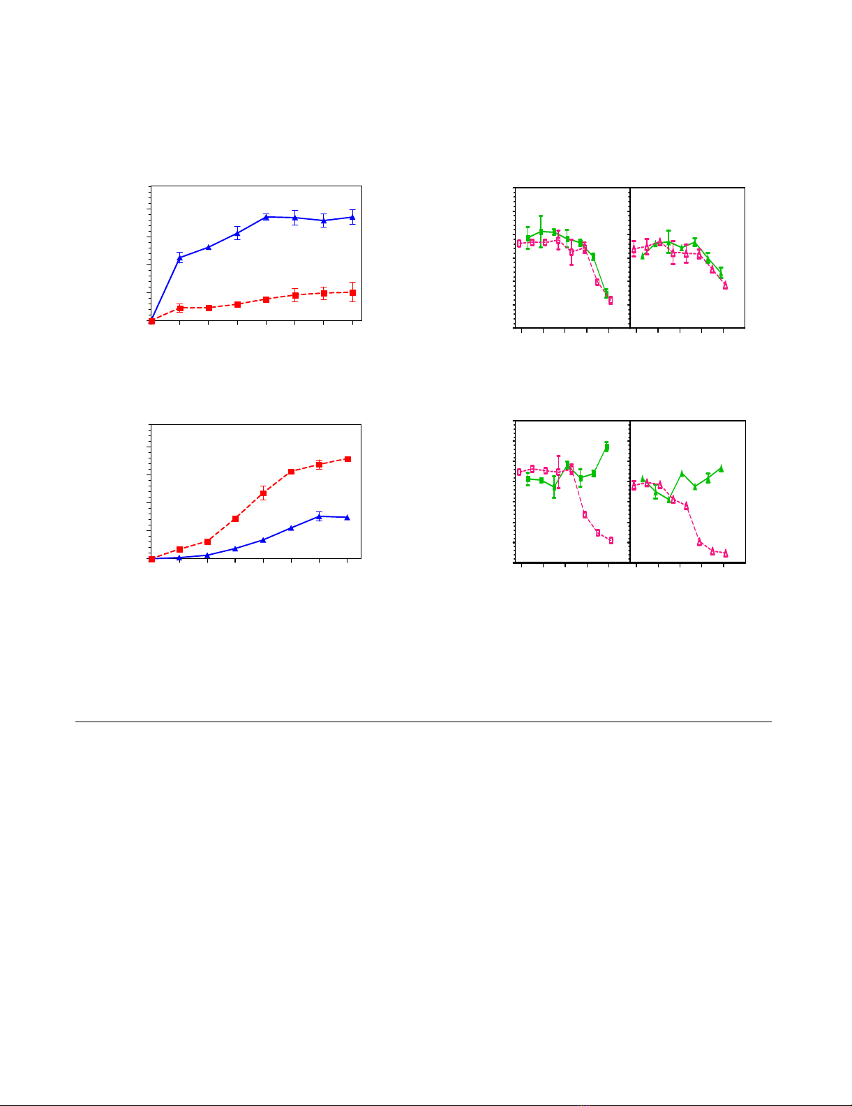
Effect of PVP-I on direct binding activity of influenza viruses to glycopolymersFigure 2
Effect of PVP-I on direct binding activity of influenza viruses to glycopolymers. (A) Virus binding activity to glyco-
polymers linked with α2,3 (filled red square) and α2,6 (filled blue triangle)-sialic acids. (B) Inhibition of virus binding to a specific
polymer. Percentage of untreated control viruses was plotted against inhibitor concentration. (filled green square) α2,3 linkage
+ PVP-I; (empty pink square) α2,3 linkage + fetuin; (filled green triangle) α2,6 linkage + PVP-I; (empty pink triangle) α2,6 linkage
+ fetuin.
0.00
0.25
0.50
0.75
1.00
0 0.25 1 3.9 15.6 62.5 250 1000
Glycopolymers, ng/ml
Binding activity at 492 nm
0.00
0.25
0.50
0.75
1.00
0 31.3 62.5 125 250 500 1000 2000
Glycopolymers, ng/ml
Binding activity at 492 nm
A/DK/HK/313/78 (H5N3)
A/Memphis/1/71 (H3N2)
AB
A/Memphis/1/71 (H3N2)
A/DK/HK/313/78 (H5N3)
0
25
50
75
100
125
150
0.1 1 10 100 1000
0.1 1 10 100 1000
Relative binding activity, %
Compounds, µg/ml
0.1 1 10 100 1000
0
25
50
75
100
125
150
175
0.1 1 10 100 1000
Compounds, µg/ml
ع
عع
ع
α2,3 polymer
ً
ًً
ً
α2,6 polymer
ع
عع
ع
α2,3 + PVP-I
غ
غغ
غ
α2,3 + fetuin
ً
ًً
ً
α2,6 + PVP-I
ٌ
ٌٌ
ٌ
α2,6 + fetuin
ع
عع
ع
α2,3 polymer
ً
ًً
ً
α2,6 polymer
Relative binding activity, %
ع
عع
ع
α2,3 + PVP-I
غ
غغ
غ
α2,3 + fetuin
ً
ًً
ً
α2,6 + PVP-I
ٌ
ٌٌ
ٌ
α2,6 + fetuin

Virology Journal 2009, 6:124 http://www.virologyj.com/content/6/1/124
Page 5 of 10
(page number not for citation purposes)
criteria [35], cell shrinkage, rounding and detachment
from the surface of the culture plate after treatment with
3.1 mg/ml of PVP-I suggested that the cells were undergo-
ing apoptosis. Therefore, we used low concentrations of
PVP-I that did not cause any toxicity to host MDCK cells
in order to investigate its anti-influenza virus activity.
Our results confirmed that PVP-I is a potent inhibitor of
influenza virus production in MDCK cells. We indicated
that PVP-I inhibits the viral replication in a dose depend-
ent manner and is more active against human viruses
(H1N1, H3N2) than avian viruses (H1N1, H5N3, H9N2).
PVP-I appeared to inhibit binding of human A/Memphis/
1/71 (H3N2) virus to specific sialoglycopolymers but not
that of avian A/DK/HK/313/78 (H5N3) virus. Hemagglu-
tination of erythrocytes induced by human viruses was
inhibited by PVP-I, while hemagglutination inhibition of
avian viruses required higher PVP-I concentrations. Dif-
ferences in hemagglutination inhibitory activity of PVP-I
against various viruses may be associated with the differ-
ent structure of HA protein of each virus type. Unlike the
α2,3 and α2,6 sialoconjugated protein fetuin [36], which
reduces HA binding activity of both avian and human
influenza viruses via competition for binding with sialylo-
ligosaccharide receptor substrates to the viruses [37],
blockage of viral HA attachment to receptor substrates by
PVP-I may result from alteration of viral HA protein struc-
ture by reaction of free iodine with basic -NH groups, phe-
nolic groups, and -SH groups of amino acid residues [30].
Although avian viruses appear to be less sensitive than
human viruses to PVP-I, based on results of the erythro-
cyte agglutination assay, which reflects viral attachment to
host cells, agglutination of avian A/DK/HK/313/78 virus
(~400 HAU) was completely inhibited after a second
exposure to 5 mg/ml of PVP-I. This is in agreement with
the finding that titers of a highly pathogenic avian virus
(H5N1) and three low pathogenic avian viruses (H5N3,
H7N7 and H9N2) cultivated in embryonated eggs
become undetectable by incubation with a commercial
PVP-I product for 10 seconds before inoculation [27].
These results suggest that gargling with PVP-I could pre-
vent human infection not only by human influenza
viruses that bind to sialyl α2,6 Gal receptors in the upper
part of human trachea but also by avian viruses that bind
to sialyl α2,3 Gal receptors that exist deep in the human
respiratory tract [38]. This could consequently minimize
the risk of avian virus mutation, either by adaptation or
reassortment, to recognize the human host predominately
carrying α2,6-linked sialic acids.
PVP-I inhibited sialidase activity as a mixed-type inhibi-
tor, indicating that free iodine is capable of binding to
either free sialidase or sialidase complexed with its sub-
strate, but iodine binding to free sialidase is more efficient
than that to sialidase-substrate complex as Ki was 16-fold
lower than Kis. This may be explained by the distribution
of lysine, arginine, histidine, cysteine and tyrosine resi-
dues throughout the sequence of the NA molecule, which
Table 1: Inhibition by PVP-I of sialidase activity, hemagglutination and infectivity activity of influenza A viruses
Virus subtype Virus strain Sialidase inhibition activity
IC50a (μg/ml)
Hemagglutination inhibition
activityb (mg/ml)
Infection inhibitory activity (%)c
H1N1 A/Bel/42 11 ± 2 1.56 84 ± 1
A/Texas/36/91 72 ± 4 0.78 68 ± 3
A/USSR/92/77 47 ± 3 0.78 68 ± 2
A/PR/8/34 9.5 ± 0.5 0.20 62 ± 2
A/WS/33 12.5 ± 0.5 0.78 60 ± 4
A/WSN/33 45 ± 1 0.39 97 ± 1
A/DK/HK/36/4 21 ± 5 12.50 40 ± 4
H3N2 A/Aichi/2/68 96 ± 2 3.13 82 ± 5
A/Memphis/1/71 61 ± 4 1.56 66 ± 3
H9N2 A/DK/HK/92/76 212 ± 9 12.50 34 ± 8
H5N3 A/DK/HK/313/78 78 ± 4 12.50 23 ± 1
A/DK/HK/23/76 124 ± 7 3.13 57 ± 4
A/DK/HK/677/1 55 ± 1 3.13 50 ± 3
aIC50values are concentrations inhibiting viral sialidase activity by 50%. Standard error means were calculated from means of two independent
experiments, each conducted in duplicate.
bMinimum concentration that inhibits hemagglutination.
cPercent viral inhibition (with 1.56 mg/ml of PVP-I) was calculated by comparison with the control without an inhibitor. Each experiment was
performed in triplicate.


%20--%3e%3cdefs%3e%3cstyle%3e%20.st0%20{%20fill:%20%23fff;%20}%20.st1%20{%20fill:%20%237800fa;%20}%20%3c/style%3e%3c/defs%3e%3cpath%20class='st1'%20d='M117.78,12.18H43.11c2.9,3.47,4.65,7.94,4.65,12.82,0,5.6-2.3,10.66-6.01,14.29h76.02l7.22-13.56-7.22-13.56Z'/%3e%3cg%3e%3cpath%20class='st0'%20d='M53.58,26.17h-.59v-1.46h.59v-4.96h2.83c1.78,0,2.67.94,2.67,2.82v5.76c0,1.87-.89,2.81-2.67,2.81h-2.83v-4.96ZM55.36,21.37v3.34h1.1v1.46h-1.1v3.34h1.01c.61,0,.91-.37.91-1.1v-5.93c0-.74-.3-1.1-.91-1.1h-1.01Z'/%3e%3cpath%20class='st0'%20d='M65.99,31.14h-1.8l-.31-2.07h-2.19l-.31,2.07h-1.64l1.82-11.39h2.62l1.82,11.39ZM65.28,18.04c-.25.46-.51.77-.75.94-.21.15-.47.22-.79.22-.26,0-.57-.07-.92-.22l-.38-.15c-.14-.05-.26-.07-.37-.07-.3,0-.53.18-.71.54l-.91-.68c.25-.46.51-.77.75-.94.21-.14.48-.21.79-.21.26,0,.57.07.92.21l.38.15c.14.05.26.07.37.07.3,0,.53-.18.71-.54l.91.68ZM61.91,27.52h1.73l-.87-5.76-.87,5.76Z'/%3e%3cpath%20class='st0'%20d='M74.53,26.89v1.52c0,1.91-.89,2.86-2.67,2.86s-2.67-.95-2.67-2.86v-5.93c0-1.91.89-2.86,2.67-2.86s2.67.95,2.67,2.86v1.11h-1.69v-1.22c0-.75-.31-1.12-.93-1.12s-.93.37-.93,1.12v6.15c0,.74.31,1.11.93,1.11s.93-.37.93-1.11v-1.63h1.69Z'/%3e%3cpath%20class='st0'%20d='M81.4,31.14h-1.8l-.31-2.07h-2.19l-.31,2.07h-1.64l1.82-11.39h2.62l1.82,11.39ZM75.9,19.2l1.52-1.91h1.71l1.51,1.91h-1.61l-.76-.95-.75.95h-1.61ZM77.32,27.52h1.73l-.87-5.76-.87,5.76ZM83.1,15.99l-1.76,1.91h-1.26l1.17-1.91h1.86Z'/%3e%3cpath%20class='st0'%20d='M84.86,19.75c1.78,0,2.67.94,2.67,2.82v1.48c0,1.87-.89,2.81-2.67,2.81h-.85v4.28h-1.79v-11.39h2.64ZM84.01,21.37v3.86h.85c.58,0,.87-.36.87-1.08v-1.71c0-.71-.29-1.07-.87-1.07h-.85Z'/%3e%3cpath%20class='st0'%20d='M93.51,19.75c1.78,0,2.67.94,2.67,2.82v1.48c0,1.87-.89,2.81-2.67,2.81h-.85v4.28h-1.79v-11.39h2.64ZM92.66,21.37v3.86h.85c.58,0,.87-.36.87-1.08v-1.71c0-.71-.29-1.07-.87-1.07h-.85Z'/%3e%3cpath%20class='st0'%20d='M98.8,31.14h-1.79v-11.39h1.79v4.88h2.03v-4.88h1.83v11.39h-1.83v-4.88h-2.03v4.88Z'/%3e%3cpath%20class='st0'%20d='M105.36,24.55h2.46v1.62h-2.46v3.34h3.09v1.63h-4.88v-11.39h4.88v1.63h-3.09v3.18ZM108.17,17.29l-1.76,1.91h-1.26l1.17-1.91h1.86Z'/%3e%3cpath%20class='st0'%20d='M112.2,19.75c1.78,0,2.67.94,2.67,2.82v1.48c0,1.87-.89,2.81-2.67,2.81h-.85v4.28h-1.79v-11.39h2.64ZM111.35,21.37v3.86h.85c.58,0,.87-.36.87-1.08v-1.71c0-.71-.29-1.07-.87-1.07h-.85Z'/%3e%3c/g%3e%3ccircle%20class='st1'%20cx='25'%20cy='25'%20r='20'/%3e%3cpath%20class='st0'%20d='M32.78,19.27c2.92,0,4.43,2.55,5.28,5.33l.71,2.17c.14.38-.33.75-.71.75h-5.61c.19-.33.24-.71.09-1.08l-.75-2.45c-.43-1.32-.99-2.64-1.79-3.77.75-.57,1.65-.94,2.78-.94h0ZM25,18.38c3.25,0,4.9,2.78,5.89,5.89l.76,2.45c.14.42-.33.8-.8.8h-11.69c-.42,0-.94-.38-.8-.8l.75-2.45c.99-3.11,2.64-5.89,5.89-5.89h0ZM25,11.35c1.74,0,3.11,1.37,3.11,3.11s-1.37,3.11-3.11,3.11-3.11-1.41-3.11-3.11,1.41-3.11,3.11-3.11h0ZM17.27,19.27c1.08,0,1.98.38,2.73.94-.8,1.13-1.37,2.45-1.74,3.77l-.8,2.45c-.14.38-.05.75.09,1.08h-5.56c-.42,0-.9-.38-.75-.75l.71-2.17c.9-2.78,2.41-5.33,5.33-5.33h0ZM17.27,12.91c1.51,0,2.78,1.27,2.78,2.83s-1.27,2.83-2.78,2.83-2.83-1.27-2.83-2.83,1.27-2.83,2.83-2.83h0ZM32.78,12.91c1.56,0,2.78,1.27,2.78,2.83s-1.23,2.83-2.78,2.83-2.83-1.27-2.83-2.83,1.27-2.83,2.83-2.83h0ZM27.07,28.56v.09c0,.57-.24,1.08-.61,1.46h0v.05c-.38.33-.9.57-1.46.57s-1.08-.24-1.46-.61h0c-.38-.38-.61-.9-.61-1.46v-.09h1.41v.09c0,.19.05.38.19.47v.05c.09.09.28.19.47.19s.38-.09.47-.19v-.05c.14-.09.24-.28.24-.47t-.05-.09h1.41ZM30.99,28.56v.09c0,1.65-.66,3.16-1.74,4.24-1.08,1.08-2.59,1.79-4.24,1.79s-3.16-.71-4.24-1.79l-.05-.05c-1.04-1.08-1.7-2.55-1.7-4.2v-.09h1.41v.09c0,1.27.47,2.4,1.27,3.25h.05c.85.85,1.98,1.37,3.25,1.37s2.4-.52,3.25-1.37c.85-.8,1.37-1.98,1.37-3.25v-.09h1.37ZM34.99,28.56v.09c0,2.78-1.13,5.28-2.92,7.07-1.79,1.79-4.29,2.92-7.07,2.92s-5.23-1.13-7.07-2.92c-1.79-1.79-2.92-4.29-2.92-7.07v-.09h1.41v.09c0,2.4.94,4.53,2.5,6.08,1.56,1.56,3.72,2.5,6.08,2.5s4.52-.94,6.08-2.5c1.56-1.56,2.5-3.68,2.5-6.08v-.09h1.41Z'/%3e%3c/svg%3e)