
BioMed Central
Page 1 of 4
(page number not for citation purposes)
World Journal of Surgical Oncology
Open Access
Case report
Transsacral colon fistula: late complication after resection,
irradiation and free flap transfer of sacral chondrosarcoma
Lars Steinstraesser*1, Michael Sand1, Stefan Langer1, Gert Muhr2,
Thomas A Schildhauer2 and Hans-Ulrich Steinau1
Address: 1Department of Plastic Surgery, Burn Center, Hand Center, Sarcoma Reference Center Ruhr-University Bochum, Bergmannsheil, Bürkle-
de-la-Camp Platz 1, 44791 Bochum, Germany and 2Department of Surgery, Ruhr-University Bochum, Bergmannsheil, Bürkle-de-la-Camp Platz
1, 44791 Bochum, Germany
Email: Lars Steinstraesser* - lars.steinstraesser@rub.de; Michael Sand - michael.sand@rub.de; Stefan Langer - stefan.langer@rub.de;
Gert Muhr - gert.muhr@rub.de; Thomas ASchildhauer-Thomas.A.Schildhauer@rub.de; Hans-Ulrich Steinau - hans-
ulrich.steinau@bergmannsheil.de
* Corresponding author
Abstract
Background: Primary sacral tumors are rare and experience related to accompanying effects of
these tumors is therefore limited to observations on a small number of patients.
Case presentation: In this case report we present a patient with a history of primary sacral
chondrosarcoma, an infection of an implanted spinal stabilization device and discuss the challenges
that resulted from a colonic fistula associated with large, life threatening abscesses as late
complications of radiotherapy.
Conclusion: In patients with sacral tumors enterocutaneous fistulas after free musculotaneous
free flaps transfer are rare and can occur in the setting of surgical damage followed by radiotherapy
or advanced disease. They are associated with prolonged morbidity and high mortality.
Identification of high-risk patients and management of fistulas at an early stage may delay the need
for subsequent therapy and decrease morbidity.
Background
Primary sacral tumors are rare and experience related to
accompanying effects of these tumors is therefore limited
to observations on a small number of patients [1,2]. These
include individuals with benign neoplasms such as osteo-
chondroma, giant cell tumors and osteoid osteomas and,
more commonly chordoma, myeloma, osteosarcoma and
chondrosarcoma [3-6].
Sacral neoplasms cause mild but noticeable symptoms at
an early stage. In these cases it is essential to achieve the
right diagnosis in time for wide excision margins. A radi-
cal surgical approach with partial or total sacrectomy,
including sacrifice of sacral roots and spinal-pelvic fixa-
tion, is technically challenging and may jeopardize axial
stability. Surgical approaches are therefore often limited
by the size of the tumor and additionally dictated by the
proximity to vital structures. As a consequence a resection
in sano is feasible only up to a certain size of the tumor.
By the time of diagnosis sacral tumors are often too large
for achieving adequate margins. Although chondrosarco-
mas are reported to have low radio-sensitivity, local con-
trol is sometimes achieved through radiation in patients
Published: 11 November 2008
World Journal of Surgical Oncology 2008, 6:121 doi:10.1186/1477-7819-6-121
Received: 22 August 2008
Accepted: 11 November 2008
This article is available from: http://www.wjso.com/content/6/1/121
© 2008 Steinstraesser et al; licensee BioMed Central Ltd.
This is an Open Access article distributed under the terms of the Creative Commons Attribution License (http://creativecommons.org/licenses/by/2.0),
which permits unrestricted use, distribution, and reproduction in any medium, provided the original work is properly cited.
World Journal of Surgical Oncology 2008, 6:121 http://www.wjso.com/content/6/1/121
Page 2 of 4
(page number not for citation purposes)
who have not been radically resected [7-9]. Nonetheless
radiation-induced damage can cause major early and late
post-radiation side effects, requiring management by the
plastic surgeon [10,11]. Spinal stabilization devices,
which are commonly used after resection of large sacral
tumors, can become infected. After control of sepsis,
wound drainage and debridement myocutaneous flaps
enable long-term spinal stabilization and in some cases
salvage of the implanted stabilization devices can be
achieved [12].
In this case report we present a patient with a history of
primary sacral chondrosarcoma, an infection of the
implanted spinal stabilization device and discuss the chal-
lenges that resulted from a colonic fistula associated with
large, life threatening abscesses as late complications of
radiotherapy.
Case presentation
A 57-year-old man with a history of chondrosarcoma of
the Os sacrum was treated 1985 by a R1 resection at
another institution. To support stabilization, implanta-
tion of an Universal Spine System (USS, Synthes, Inc.,
West Chester, PA) was followed by osteosynthesis of L4/
pelvis and additional spongiosaplasty with fibula chips.
Post-operatively neutron irradiation was started due to
intralesional surgical margins. Ten years later (1995), the
patient developed a fulminant osteomyelitis ending up
with soft tissue defect of 38 × 26 cm. Following radical
debridement with removal of both iliac crests, the fibula
chips and the USS system. After extended wound treat-
ment a new USS system with fixation at the arcus root of
L4, L5 and the lateral mass of the sacrum was implanted
for stabilization of the sacrum. The large lumbal soft tis-
sue defect with exposed vertebrae and hardware was cov-
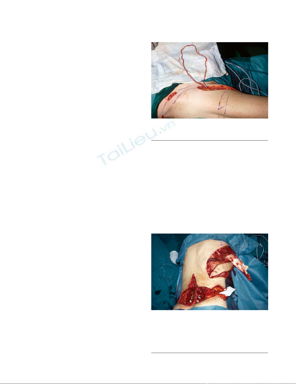
ered with a free flap. For vascular supply the complete
saphenous vein graft (76 cm long) was served as an arte-
rio-venous loop and was anastomosed end-to-side to the
superficial femoral artery as previously described (Fig 1)
[13]. The large wound was covered with a latissimus dorsi
free-flap and anastomosed to the AV-loop (Fig 2). After
uneventful postoperative period the patient could be dis-
charged and the wound conditions have remained stable
for over 10 years with a small fluid drainage from a
cephalic sinus.
In 2004 the patient presented with a recurrent inguinal
hernia on the left side outside of our sarcoma center. The
Shouldice repair of his hernia was uneventful. Three
weeks after the operation hematological laboratory find-
ings and blood chemistry values showed signs of infec-
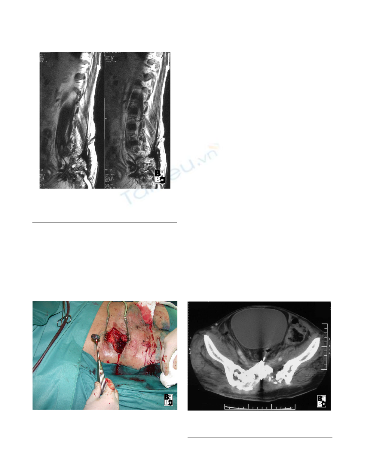
tion. A CT-scan of the abdomen and pelvis showed a
massive inflammatory infiltrate with air trapping contigu-
ous to a large abscess in the right iliacal muscle (7 × 5 cm)
and a collateral infiltrate of the psoas muscle. Addition-
ally extended osseous destructions of the sacrum were
documented. To differentiate between postoperative
defects, the previously diagnosed sequestrating chronic
osteomyelitis or a possible relapse of his chondrosarcoma
was hardly possible (Fig 3). After antibiotic treatment of a
urinary tract infection the patient developed an antiobi-
otic-associated diarrhea. The patient was then referred to
us for further therapy of his life threatening hematoge-
nous dissemination of bacteria from his multiple
abscesses. By the time of referral in addition to the previ-
ously described ilacal and psoas abscesses he had devel-
oped an active discharging praesternal/mediastinal
abscess and bilateral intracarpal infections. The abscesses
were surgically drained and a bacterial smear revealed
Saphenous vein graft anastomosed end-to-side to the superfi-cial femoral artery (arterio-venous loop)Figure 1
Saphenous vein graft anastomosed end-to-side to the
superficial femoral artery (arterio-venous loop).
Latissimus dorsi free flap with the superficial femoral artery (arterio-venous loop) at the bottom right corner and an implanted Universal Spine System with fixation at the arcus root of L4, L5 and the lateral mass of the sacrum for stabili-zation at the bottom left cornerFigure 2
Latissimus dorsi free flap with the superficial femoral
artery (arterio-venous loop) at the bottom right cor-
ner and an implanted Universal Spine System with
fixation at the arcus root of L4, L5 and the lateral
mass of the sacrum for stabilization at the bottom
left corner.
World Journal of Surgical Oncology 2008, 6:121 http://www.wjso.com/content/6/1/121
Page 3 of 4
(page number not for citation purposes)
Escherichia coli in massive numbers. Antibiotic therapy
was initiated with Imipenem and Metronidazol. Methyl-
ene blue dye was injected into the sacral fistulas, and mul-
tiple fistulectomys and a sequestrectomy were performed
(Fig 4).
After ensuring sufficient dorsal drainage a gastrographin
enema was performed to determine the site of a possible
gastrointestinal perforation which was suspected as a pos-
sible source of infection. The contrast medium was shown
to be leaking from the ascending colon and the caecum
into the iliacal and psoas muscle, reaching to the sacral
lacunae (Fig 5). A right hemicolectomy was performed.
During the operation a perforation of the dorsal wall and
the basis of the caecum were found. After clearing out a
fecal abscess drainage was established and covered by split
omentum-plasty followed by multiple rinses, using poly-
meric biguanide-hydrochloride (Lavasept®, Fresenius Kabi
AG, Bad Homburg, Germany). Additionally the sacral and
praesternal abscesses were once more debrided in the
operating room. The right ureter was adherent to the
abscess formation and was mobilized. Postoperatively the
patient was referred to an intensive care unit. The bilateral
septic inflammations of the carpal joints were successfully
treated with high dose antibiotics after surgical excision
and drainage (Imipenem and Metronidazol). After multi-
ple irrigations of the large wounds and decreasing inflam-
mation, granulation tissue developed. In order to further
minimize the dead space of the large wounds, microdefor-
mational wound therapy by means of Vacuum Assisted
Closure (V.A.C.-Therapy®, KCI Medizinprodukte GmbH,
Wiesbaden, Germany) was applied to the bilateral sacral
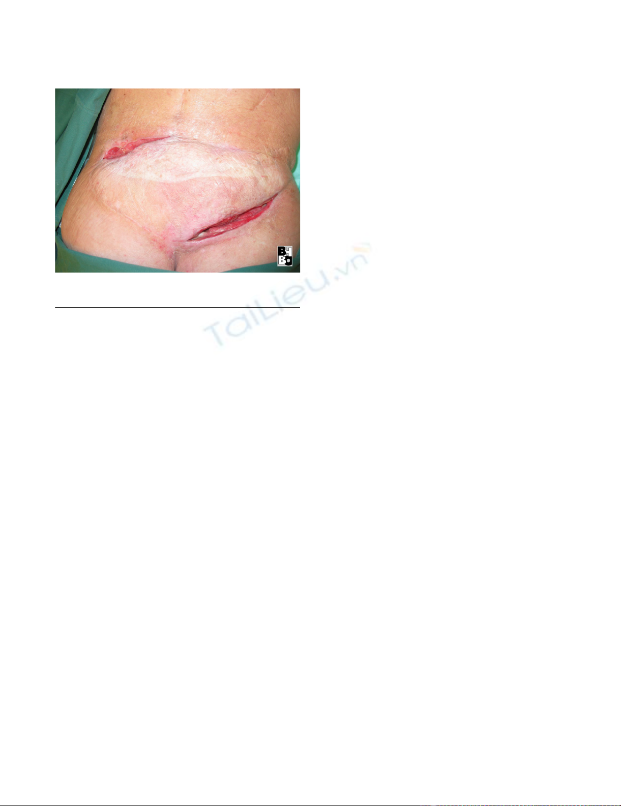
wounds. Despite four weeks of intensive care his latis-
simus dorsi free flap was saved and the patient was dis-
charged with wounds showing no signs of infection and a
tendency towards good granulation (Fig 6). A control MRI
in 2008 showed a stable fistula with no signs of recur-
rence.
List of used products
Universal Spine System (USS, Synthes, Inc., West Chester,
PA)
Gastrographin enema showing a gastrointestinal perforation reaching to the sacral lacunaeFigure 5
Gastrographin enema showing a gastrointestinal per-
foration reaching to the sacral lacunae.
Sagital sections (MRI) of the lumbal and sacral spine showing extended osseous destructions of the sacrumFigure 3
Sagital sections (MRI) of the lumbal and sacral spine
showing extended osseous destructions of the sac-
rum.
Sequestrectomy after methylene blue dye injection into the sacral fistulasFigure 4
Sequestrectomy after methylene blue dye injection
into the sacral fistulas.
World Journal of Surgical Oncology 2008, 6:121 http://www.wjso.com/content/6/1/121
Page 4 of 4
(page number not for citation purposes)
Polymeric biguanide-hydrochloride (Lavasept®, Fresenius
Kabi AG, Bad Homburg, Germany)
Vacuum Assisted Closure (V.A.C.-Therapy®, KCI Medizin-
produkte GmbH, Wiesbaden, Germany)
Discussion
In patients with sacral tumors enterocutaneous fistulas
after free musculotaneous free flaps transfer are rare. They
occur in the setting of surgical damage followed by radio-
therapy or advanced disease and are associated with pro-
longed morbidity and high mortality. Identification of
high-risk patients and management of fistulas at an early
stage may delay the need for subsequent therapy and
decrease morbidity [14].
As in our case, ulceration of the gut and development of a
fistula is based on changes in the collagen tissues and par-
ticularly in vascular tissue of the gut [15]. The bowel
mucosal lining cells divide roughly every 22 days which is
very fast compared to the other types of human tissue. Red
and white cell precursors are the only cells which divide
faster. Therefore radiation poisoning affects these two sys-
tems more than others which can ultimately, even several
years after radiation therapy, result in enterocutaneous fis-
tulas with all the possible side effects discussed in this case
report.
Conclusion
In summary, we have described a 57-yr-old sacral chond-
rosarcoma patient with a transsacral colon fistula compli-
cated by E. coli bacteremia and multiple extra-intestinal
manifestations.
Consent
Written informed consent was obtained from the patient
for publication of this case report and any accompanying
images. A copy of the written consent is available for
review by the Editor-in-Chief of this journal.
Competing interests
All authors hereby disclose any commercial associations
which might pose or create a conflict of interest with
information presented in this manuscript. All authors
declare that they have no competing interests.
Authors' contributions
LS documented and prepared most of the draft. MS docu-
mented and prepared most of the draft. SL Literature
research, revision of bibliography. GM Edited the manu-
script and helped in preparing the draft. TAS Documented
and prepared part of the draft. HUS Edited the manu-
script, revision of bibliography and helped in preparing
the draft. All authors read and approved final manuscript.
References
1. Randall RL, Bruckner J, Lloyd C, Pohlman TH, Conrad EU 3rd: Sacral
resection and reconstruction for tumors and tumor-like con-
ditions. Orthopedics 2005, 28:307-313.
2. Randall RL: Giant cell tumor of the sacrum. Neurosurg Focus
2003, 15:E13.
3. Deutsch H, Mummaneni PV, Haid RW, Rodts GE, Ondra SL: Benign
sacral tumors. Neurosurg Focus 2003, 15:E14.
4. Biagini R, Orsini U, Demitri S, Bibiloni J, Ruggieri P, Mercuri M,
Capanna R, Majorana B, Bertoni F, Bacchini P, Briccoli A: Osteoid
osteoma and osteoblastoma of the sacrum. Orthopedics 2001,
24:1061-1064.
5. Bergh P, Gunterberg B, Meis-Kindblom JM, Kindblom LG: Prognos-
tic factors and outcome of pelvic, sacral, and spinal chondro-
sarcomas: a center-based study of 69 cases. Cancer 2001,
91:1201-1212.
6. Leone A, Costantini A, Guglielmi G, Settecasi C, Priolo F: Primary
bone tumors and pseudotumors of the lumbosacral spine.
Rays 2000, 25:89-103.
7. Rhomberg W, Eiter H, Böhler F, Dertinger S: Combined radio-
therapy and razoxane in the treatment of chondrosarcomas
and chordomas. Anticancer Res 2006, 26:2407-2411.
8. Nedea EA, DeLaney TF: Sarcoma and skin radiation oncology.
Hematol Oncol Clin North Am 2006, 20:401-429.
9. Pritchard DJ, Lunke RJ, Taylor WF, Dahlin DC, Medley BE: Chond-
rosarcoma: a clinicopathologic and statistical analysis. Cancer
1980, 45:149-157.
10. Novak JM, Collins JT, Donowitz M, Farman J, Sheahan DG, Spiro HM:
Effects of radiation on the human gastrointestinal tract. J Clin
Gastroenterol 1979, 1:9-39.
11. Albu E, Gerst PH, Ene C, Carvajal S, Rao SK: Jejunal-rectal fistula
as a complication of postoperative radiotherapy. Am Surg
1990, 56:697-699.
12. Hultman CS, Jones GE, Losken A, Seify H, Schaefer TG, Zapiach LA,
Carlson GW: Salvage of infected spinal hardware with parasp-
inous muscle flaps: anatomic considerations with clinical cor-
relation. Ann Plast Surg 2006, 57:521-528.
13. Germann G, Steinau HU: The clinical reliability of vein grafts in
free-flap transfer. J Reconstr Microsurg 1996, 12:11-7.
14. Chamberlain RS, Kaufman HL, Danforth DN: Enterocutaneous fis-
tula in cancer patients: etiology, management, outcome, and
impact on further treatment. Am Surg 1998, 64:1204-1211.
15. Novak JM, Collins JT, Donowitz M, Farman J, Sheahan DG, Spiro HM:
Effects of radiation on the human gastrointestinal tract. J Clin
Gastroenterol 1979, 1:9-39.
Latissimus dorsi free flap with fistulaFigure 6
Latissimus dorsi free flap with fistula.


%20--%3e%3cdefs%3e%3cstyle%3e%20.st0%20{%20fill:%20%23fff;%20}%20.st1%20{%20fill:%20%237800fa;%20}%20%3c/style%3e%3c/defs%3e%3cpath%20class='st1'%20d='M117.78,12.18H43.11c2.9,3.47,4.65,7.94,4.65,12.82,0,5.6-2.3,10.66-6.01,14.29h76.02l7.22-13.56-7.22-13.56Z'/%3e%3cg%3e%3cpath%20class='st0'%20d='M53.58,26.17h-.59v-1.46h.59v-4.96h2.83c1.78,0,2.67.94,2.67,2.82v5.76c0,1.87-.89,2.81-2.67,2.81h-2.83v-4.96ZM55.36,21.37v3.34h1.1v1.46h-1.1v3.34h1.01c.61,0,.91-.37.91-1.1v-5.93c0-.74-.3-1.1-.91-1.1h-1.01Z'/%3e%3cpath%20class='st0'%20d='M65.99,31.14h-1.8l-.31-2.07h-2.19l-.31,2.07h-1.64l1.82-11.39h2.62l1.82,11.39ZM65.28,18.04c-.25.46-.51.77-.75.94-.21.15-.47.22-.79.22-.26,0-.57-.07-.92-.22l-.38-.15c-.14-.05-.26-.07-.37-.07-.3,0-.53.18-.71.54l-.91-.68c.25-.46.51-.77.75-.94.21-.14.48-.21.79-.21.26,0,.57.07.92.21l.38.15c.14.05.26.07.37.07.3,0,.53-.18.71-.54l.91.68ZM61.91,27.52h1.73l-.87-5.76-.87,5.76Z'/%3e%3cpath%20class='st0'%20d='M74.53,26.89v1.52c0,1.91-.89,2.86-2.67,2.86s-2.67-.95-2.67-2.86v-5.93c0-1.91.89-2.86,2.67-2.86s2.67.95,2.67,2.86v1.11h-1.69v-1.22c0-.75-.31-1.12-.93-1.12s-.93.37-.93,1.12v6.15c0,.74.31,1.11.93,1.11s.93-.37.93-1.11v-1.63h1.69Z'/%3e%3cpath%20class='st0'%20d='M81.4,31.14h-1.8l-.31-2.07h-2.19l-.31,2.07h-1.64l1.82-11.39h2.62l1.82,11.39ZM75.9,19.2l1.52-1.91h1.71l1.51,1.91h-1.61l-.76-.95-.75.95h-1.61ZM77.32,27.52h1.73l-.87-5.76-.87,5.76ZM83.1,15.99l-1.76,1.91h-1.26l1.17-1.91h1.86Z'/%3e%3cpath%20class='st0'%20d='M84.86,19.75c1.78,0,2.67.94,2.67,2.82v1.48c0,1.87-.89,2.81-2.67,2.81h-.85v4.28h-1.79v-11.39h2.64ZM84.01,21.37v3.86h.85c.58,0,.87-.36.87-1.08v-1.71c0-.71-.29-1.07-.87-1.07h-.85Z'/%3e%3cpath%20class='st0'%20d='M93.51,19.75c1.78,0,2.67.94,2.67,2.82v1.48c0,1.87-.89,2.81-2.67,2.81h-.85v4.28h-1.79v-11.39h2.64ZM92.66,21.37v3.86h.85c.58,0,.87-.36.87-1.08v-1.71c0-.71-.29-1.07-.87-1.07h-.85Z'/%3e%3cpath%20class='st0'%20d='M98.8,31.14h-1.79v-11.39h1.79v4.88h2.03v-4.88h1.83v11.39h-1.83v-4.88h-2.03v4.88Z'/%3e%3cpath%20class='st0'%20d='M105.36,24.55h2.46v1.62h-2.46v3.34h3.09v1.63h-4.88v-11.39h4.88v1.63h-3.09v3.18ZM108.17,17.29l-1.76,1.91h-1.26l1.17-1.91h1.86Z'/%3e%3cpath%20class='st0'%20d='M112.2,19.75c1.78,0,2.67.94,2.67,2.82v1.48c0,1.87-.89,2.81-2.67,2.81h-.85v4.28h-1.79v-11.39h2.64ZM111.35,21.37v3.86h.85c.58,0,.87-.36.87-1.08v-1.71c0-.71-.29-1.07-.87-1.07h-.85Z'/%3e%3c/g%3e%3ccircle%20class='st1'%20cx='25'%20cy='25'%20r='20'/%3e%3cpath%20class='st0'%20d='M32.78,19.27c2.92,0,4.43,2.55,5.28,5.33l.71,2.17c.14.38-.33.75-.71.75h-5.61c.19-.33.24-.71.09-1.08l-.75-2.45c-.43-1.32-.99-2.64-1.79-3.77.75-.57,1.65-.94,2.78-.94h0ZM25,18.38c3.25,0,4.9,2.78,5.89,5.89l.76,2.45c.14.42-.33.8-.8.8h-11.69c-.42,0-.94-.38-.8-.8l.75-2.45c.99-3.11,2.64-5.89,5.89-5.89h0ZM25,11.35c1.74,0,3.11,1.37,3.11,3.11s-1.37,3.11-3.11,3.11-3.11-1.41-3.11-3.11,1.41-3.11,3.11-3.11h0ZM17.27,19.27c1.08,0,1.98.38,2.73.94-.8,1.13-1.37,2.45-1.74,3.77l-.8,2.45c-.14.38-.05.75.09,1.08h-5.56c-.42,0-.9-.38-.75-.75l.71-2.17c.9-2.78,2.41-5.33,5.33-5.33h0ZM17.27,12.91c1.51,0,2.78,1.27,2.78,2.83s-1.27,2.83-2.78,2.83-2.83-1.27-2.83-2.83,1.27-2.83,2.83-2.83h0ZM32.78,12.91c1.56,0,2.78,1.27,2.78,2.83s-1.23,2.83-2.78,2.83-2.83-1.27-2.83-2.83,1.27-2.83,2.83-2.83h0ZM27.07,28.56v.09c0,.57-.24,1.08-.61,1.46h0v.05c-.38.33-.9.57-1.46.57s-1.08-.24-1.46-.61h0c-.38-.38-.61-.9-.61-1.46v-.09h1.41v.09c0,.19.05.38.19.47v.05c.09.09.28.19.47.19s.38-.09.47-.19v-.05c.14-.09.24-.28.24-.47t-.05-.09h1.41ZM30.99,28.56v.09c0,1.65-.66,3.16-1.74,4.24-1.08,1.08-2.59,1.79-4.24,1.79s-3.16-.71-4.24-1.79l-.05-.05c-1.04-1.08-1.7-2.55-1.7-4.2v-.09h1.41v.09c0,1.27.47,2.4,1.27,3.25h.05c.85.85,1.98,1.37,3.25,1.37s2.4-.52,3.25-1.37c.85-.8,1.37-1.98,1.37-3.25v-.09h1.37ZM34.99,28.56v.09c0,2.78-1.13,5.28-2.92,7.07-1.79,1.79-4.29,2.92-7.07,2.92s-5.23-1.13-7.07-2.92c-1.79-1.79-2.92-4.29-2.92-7.07v-.09h1.41v.09c0,2.4.94,4.53,2.5,6.08,1.56,1.56,3.72,2.5,6.08,2.5s4.52-.94,6.08-2.5c1.56-1.56,2.5-3.68,2.5-6.08v-.09h1.41Z'/%3e%3c/svg%3e)