
BioMed Central
Page 1 of 4
(page number not for citation purposes)
Journal of Medical Case Reports
Open Access
Case report
Left-sided appendicitis in a patient with congenital gastrointestinal
malrotation: a case report
Frank J Welte* and Mario Grosso
Address: Department of Radiology, Baystate Medical Center, Tufts University School of Medicine, Springfield, Massachusetts, USA
Email: Frank J Welte* - fjwelte@gmail.com; Mario Grosso - Mario.GrossoMD@bhs.org
* Corresponding author
Abstract
Background: While appendicitis is the most common abdominal disease requiring surgical
intervention seen in the emergency room setting, intestinal malrotation is relatively uncommon.
When patients with asymptomatic undiagnosed gastrointestinal malrotation clinically present with
abdominal pain, accurate diagnosis and definitive therapy may be delayed, possibly increasing the
risk of morbidity and mortality. We present a case where CT was crucial diagnostically and helpful
for pre-surgical planning in a patient presenting with an acute abdomen superimposed on complete
congenital gastrointestinal malrotation.
Case presentation: A 46-year-old previously healthy male with four days of primarily left-sided
abdominal pain, low-grade fevers, nausea and anorexia presented to the Emergency Department.
His medical history was significant for poorly controlled diabetes and dyslipidemia. His white blood
count at that time was elevated. Initial abdominal plain films suggested small bowel obstruction. A
CT scan of the abdomen and pelvis was performed with oral and IV contrast to exclude
diverticulitis, revealing acute appendicitis superimposed on congenital intestinal malrotation.
Following consultation with the surgical team for surgical planning, the patient went on to
laparoscopic appendectomy and did well postoperatively.
Conclusion: Atypical presentations of acute abdominal conditions superimposed on
asymptomatic gastrointestinal malrotation can result in delays in delivery of definitive therapy and
potentially increase morbidity and mortality if not diagnosed in a timely manner. Appropriate
imaging can be helpful in hastening diagnosis and guiding intervention.
Background
Appendicitis is the most common surgical disease diag-
nosed in the emergency room setting. Gastrointestinal
malrotation is, by comparison, relatively uncommon.
Depending upon the location of the cecum and appendix,
patients with acute appendicitis and malrotation may
present atypically with left-sided abdominal pain. Left-
sided abdominal pain most commonly raises the diagnos-
tic question of possible diverticulitis, creating a diagnostic
dilemma in these patients. Furthermore, a trend may be
developing where diverticulitis, once a disease primarily
of older adults, may be becoming more prevalent in
younger adults [1] and may exhibit a somewhat different
demographic and clinical course [2]. This phenomenon
may further bias a clinician confronted with a middle-
aged patient with an acute abdomen and left-sided symp-
toms.
Published: 19 September 2007
Journal of Medical Case Reports 2007, 1:92 doi:10.1186/1752-1947-1-92
Received: 2 July 2007
Accepted: 19 September 2007
This article is available from: http://www.jmedicalcasereports.com/content/1/1/92
© 2007 Welte and Grosso; licensee BioMed Central Ltd.
This is an Open Access article distributed under the terms of the Creative Commons Attribution License (http://creativecommons.org/licenses/by/2.0),
which permits unrestricted use, distribution, and reproduction in any medium, provided the original work is properly cited.

Journal of Medical Case Reports 2007, 1:92 http://www.jmedicalcasereports.com/content/1/1/92
Page 2 of 4
(page number not for citation purposes)
We present a case where the relatively common entity of
appendicitis was in no way suspected prior to cross sec-
tional imaging, which incidentally revealed gastrointesti-
nal malrotation, significantly changing clinical
management.
Case presentation
A 46-year-old previously healthy male presented to Emer-
gency Department with four days of primarily left-sided
abdominal pain, low-grade fevers, nausea, and anorexia.
His medical history was significant for poorly controlled
diabetes and dyslipidemia. No history of abdominal sur-
gery was reported. Two months prior to admission, the
patient's serum hemoglobin A1C was markedly elevated
at 10.7 and his serum lipid profile was quite abnormal
(total cholesterol: 313, triglycerides: 287, HDL: 39, LDL:
217). On admission, the patient's home medications
included atorvastatin (Lipitor®; 40 mg orally per day) and
subcutaneous isophane/regular insulin (Insulin Novolin®
70/30) twice per day.
The patient's physical exam at presentation was significant
for left lower quadrant pain and voluntary guarding. The
patient's white blood count at that time was 18.1 × 10⁹
cells/L (absolute neutrophil count: 13.6; 75.1%), hemat-
ocrit: 45.2%, serum glucose: 279, blood urea nitrogen
(BUN): 31, and serum creatinine: 1.5. Serum electrolytes
were as follows: sodium: 133, potassium: 4.2, chloride:
95, and CO2: 23 (anion gap 15). Urinalysis was positive
for 2+ albumin, 1+glucose, 1+ ketones, 1+ bilirubin, and
hyaline casts. Plain films of the abdomen were obtained
in the Emergency Department. A spiral CT scan of the
abdomen and pelvis with oral and IV contrast was subse-
quently performed to exclude colonic diverticulitis.
Imaging findings
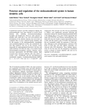
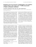
Abdominal plain films demonstrated multiple dilated
loops of small bowel with air/fluid levels in the right
abdomen (arrows in Figure 1), consistent with small
bowel obstruction, but also noted, unusually, to herald
acute appendicitis. Intestinal malrotation was not consid-
ered in the differential diagnosis at that point. No pneu-
moperitoneum was evident.
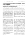
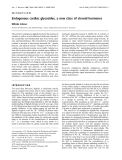
Abdominal CT with oral and IV contrast was then per-
formed, which demonstrated the majority of the small
bowel positioned in the right abdomen, the cecum
located in the left mid abdomen, and absence of the liga-
ment of Treitz. The orientation of the superior mesenteric
vessels was abnormal. These findings together were con-
sistent with complete intestinal malrotation. No associ-
ated situs, caval, or other congenital anomalies were
present and no evidence of volvulus was identified. A
dilated, tubular, blind-ending structure was identified
arising from the cecumin the left mid-abdomen (arrows
in Figures 2A,B), with significant stranding in the adjacent
fat, indicative of acute appendicitis.
Management
Fluid resuscitation was initiated in the Emergency Depart-
ment with Lactated Ringer's solution (125 cc/hr). Based
on plain film findings of possible small bowel obstruction
and absence of history of any abdominal surgery, an
abdominal CT was performed, as discussed above.
After extensive review of the CT data for presurgical plan-
ning, the patient was taken to the operating room for
laparoscopic exploration and appendectomy, where the
imaging findings were confirmed and the appendix was
found to be perforated. No surgical intervention to
address the malrotation, such as Ladd's procedure, was
performed. Pathologic examination of the excised appen-
dix verified the diagnosis of acute gangrenous appendici-
tis with perforation. The patient's post-operative course
was uneventful. The patient was treated perioperatively
with a 5 day regimen of intravenous cefoxitin (1,000 mg
every 6 hours) and as an outpatient with a 14 day course
of oral levofloxacin (500 mg per day; Levaquin®).
Conclusion
Intestinal malrotation represents errors of rotation of the
midgut about the superior mesenteric artery during weeks
5–10 of fetal life and subsequent abnormal fixation to the
peritoneal wall [3]. Intestinal malrotation, while often
associated with other congenital anomalies, is an isolated
finding in the majority of adult cases. Approximately 60%
of patients with intestinal malrotation present in the first
month of life and 20% present between 1 and 12 months,
classically with bilious vomiting secondary to duodenal
obstruction distal to the Ampulla of Vater. The estimated
incidence of intestinal malrotation is 1 in every 500 live
births (0.2%; range 0.03 – 0.5%) [3,4] based on autopsy
series, retrospective reviews [5], and prospective barium
enema studies [6]. The true incidence of adults with
asymptomatic malrotation remains difficult to accurately
determine.
More typically, symptomatic adults with bowel malrota-
tion present with acute bowel ischemia or bowel obstruc-
tion secondary to midgut orcecal volvulus, or with
chronic abdominal pain. Treatment of incidentally dis-
covered malrotation in asymptomatic patients older than
1–2 years of age remains somewhat controversial [5,7].
Even in patients in whom a Ladd's procedure is per-
formed, the appendix will be positioned in an abnormal
location, unless appendectomy is performed contempora-
neously. In the present case, malrotation was incidentally
discovered in a previously asymptomatic adult patient
presenting with an acute abdomen, confounding the diag-
nosis.

Journal of Medical Case Reports 2007, 1:92 http://www.jmedicalcasereports.com/content/1/1/92
Page 3 of 4
(page number not for citation purposes)
Abdominal plain filmsFigure 1
Abdominal plain films. Supine (A) and upright (B) abdominal plain films demonstrate multiple loops of dilated small bowel
(arrows in A) with air/fluid levels (arrows in B) in the right abdomen, suggestive of small bowel obstruction; this finding can also
be seen as an unusual sign of acute appendicitis. Intestinal malrotation was not considered at this time.
Abdominal CT: Atypical acute appendicitisFigure 2
Abdominal CT: Atypical acute appendicitis. Axial spiral CT with oral and IV contrast (A) and coronal multiplanar recon-
struction (B) demonstrate dilated appendix in the left mid-abdomen (arrows) with adjacent fat stranding.

Publish with BioMed Central and every
scientist can read your work free of charge
"BioMed Central will be the most significant development for
disseminating the results of biomedical research in our lifetime."
Sir Paul Nurse, Cancer Research UK
Your research papers will be:
available free of charge to the entire biomedical community
peer reviewed and published immediately upon acceptance
cited in PubMed and archived on PubMed Central
yours — you keep the copyright
Submit your manuscript here:
http://www.biomedcentral.com/info/publishing_adv.asp
BioMedcentral
Journal of Medical Case Reports 2007, 1:92 http://www.jmedicalcasereports.com/content/1/1/92
Page 4 of 4
(page number not for citation purposes)
Other cases of appendicitis in adults or adolescents in the
context of intestinal malrotation have been described pre-
viously, (see, for example, [4,5,8-15]) frequently with
delayed or incorrect initial diagnoses. Several of these
prior reports describe diagnosis of acute appendicitis and
gastrointestinal malrotation based on abdominal CT, but
go on to describe additional imaging such as upper GI or
barium enema to verify or further characterize the malro-
tation prior to surgical intervention. An important point is
that if CT is diagnostic of malrotation and of a superim-
posed acute condition requiring urgent intervention such
as acute appendicitis, further immediate dynamic imaging
of the intestinal malrotation is often unnecessary, pro-
vided the CT is sufficient for surgical planning, as in this
case. Furthermore, the sensitivity and specificity of upper
GI, fluoroscopic barium enema or abdominal ultrasound
in adolescent or adult patients for detection of causes of
an acute abdomen are limited compared to CT.
Complete intestinal malrotation can result in common
acute clinical entities such as appendicitis presenting atyp-
ically due to the a priori unexpected location of the cecum
and appendix, causing a diagnostic dilemma. In this case,
a patient whose clinical presentation was initially most
suggestive of diverticulitis was found to actually have
acute appendicitis in the context of congenital bowel mal-
rotation.
CT was critical in this case in redirecting the primary clin-
ical team, hastening the administration of definitive ther-
apy, and for presurgical planning. The atypical
presentation of acute appendicitis in patients with intesti-
nal malrotation presents a diagnostic challenge. Appropri-
ate imaging can be diagnostically decisive in identifying
patients who present with such an atypical presentation of
a common emergent clinical entity.
Competing interests
The author(s) declare that they have no competing inter-
ests.
Authors' contributions
FJW: Primary author; wrote and approved the final manu-
script. MG: Provided clinical background and commen-
tary. All authors read and approved the final manuscript.
Acknowledgements
We thank the Baystate Medical Center Department of Radiology and the
Tufts University School of Medicine for financial support for the publication
of this report. We thank Sharon D. Wayne for editorial comments on the
manuscript. Written consent was obtained from the patient for publication
of this case report.
References
1. Zaidi E, Daly B: CT and clinical features of acute diverticulitis
in an urban U.S. population: rising frequency in young, obese
adults. AJR Am J Roentgenol 2006, 187:689-694.
2. Lahat A, Menachem Y, Avidan B, Yanai H, Sakhnini E, Bardan E, Bar-
Meir S: Diverticulitis in the young patient – is it different?
World J Gastroenterol 2006, 12:2932-2935.
3. Anjali P, Hatley R: Intestinal Malrotation. [http://www.emedi
cine.com/ped/topic1200.htm]. WebMD. Accessed: February 25, 2007
4. Keith JCTJ, Buday SJ, Price PD, Smear J: Asymptomatic Midgut
Rotational Anomalies in Adults: 2 Case Reports and Review
of the Literature. Contemporary Surgery 2003, 59:322-325.
5. Malek MM, Burd RS: Surgical treatment of malrotation after
infancy: a population-based study. J Pediatr Surg 2005,
40:285-289.
6. Kantor JL: Anomalies of the colon. Radiology 1934, 23:651-662.
7. Dilley AV, Pereira J, Shi EC, Adams S, Kern IB, Currie B, Henry GM:
The radiologist says malrotation: does the surgeon operate?
Pediatr Surg Int 2000, 16:45-49.
8. Hollander SC, Springer SA: The diagnosis of acute left-sided
appendicitis with computed tomography. Pediatr Radiol 2003,
33:70-71.
9. Kamiyama T, Fujiyoshi F, Hamada H, Nakajo M, Harada O, Haraguchi
Y: Left-sided acute appendicitis with intestinal malrotation.
Radiat Med 2005, 23:125-127.
10. Lin CJ, Tiu CM, Chou YH, Chen JD, Liang WY, Chang CY: CT pres-
entation of ruptured appendicitis in an adult with incom-
plete intestinal malrotation. Emerg Radiol 2004, 10:210-212.
11. Lee MR, Kim JH, Hwang Y, Kim YK: A left-sided periappendiceal
abscess in an adult with intestinal malrotation. World J Gastro-
enterol 2006, 12:5399-5400.
12. Hou SK, Chern CH, How CK, Kao WF, Chen JD, Wang LM, Huang
CI: Diagnosis of appendicitis with left lower quadrant pain. J
Chin Med Assoc 2005, 68:599-603.
13. Tsumura H, Ichikawa T, Kagawa T, Nishihara M: Successful laparo-
scopic Ladd's procedure and appendectomy for intestinal
malrotation with appendicitis. Surg Endosc 2003, 17:657-658.
14. Pinto A, Di Raimondo D, Tuttolomondo A, Fernandez P, Caronia A,
Lagalla R, Arnao V, Law RL, Licata G: An atypical clinical presen-
tation of acute appendicitis in a young man with midgut mal-
rotation. Radiography 2007, 13:164-168.
15. de Roo RA, van Breda Vriesman AC, Steenvoorde P: [Diagnostic
image (186) A man with abdominal pain in the left upper
quadrant. Acute appendicitis with malrotation of the colon].
Ned Tijdschr Geneeskd 2004, 148:825.


%20--%3e%3cdefs%3e%3cstyle%3e%20.st0%20{%20fill:%20%23fff;%20}%20.st1%20{%20fill:%20%237800fa;%20}%20%3c/style%3e%3c/defs%3e%3cpath%20class='st1'%20d='M117.78,12.18H43.11c2.9,3.47,4.65,7.94,4.65,12.82,0,5.6-2.3,10.66-6.01,14.29h76.02l7.22-13.56-7.22-13.56Z'/%3e%3cg%3e%3cpath%20class='st0'%20d='M53.58,26.17h-.59v-1.46h.59v-4.96h2.83c1.78,0,2.67.94,2.67,2.82v5.76c0,1.87-.89,2.81-2.67,2.81h-2.83v-4.96ZM55.36,21.37v3.34h1.1v1.46h-1.1v3.34h1.01c.61,0,.91-.37.91-1.1v-5.93c0-.74-.3-1.1-.91-1.1h-1.01Z'/%3e%3cpath%20class='st0'%20d='M65.99,31.14h-1.8l-.31-2.07h-2.19l-.31,2.07h-1.64l1.82-11.39h2.62l1.82,11.39ZM65.28,18.04c-.25.46-.51.77-.75.94-.21.15-.47.22-.79.22-.26,0-.57-.07-.92-.22l-.38-.15c-.14-.05-.26-.07-.37-.07-.3,0-.53.18-.71.54l-.91-.68c.25-.46.51-.77.75-.94.21-.14.48-.21.79-.21.26,0,.57.07.92.21l.38.15c.14.05.26.07.37.07.3,0,.53-.18.71-.54l.91.68ZM61.91,27.52h1.73l-.87-5.76-.87,5.76Z'/%3e%3cpath%20class='st0'%20d='M74.53,26.89v1.52c0,1.91-.89,2.86-2.67,2.86s-2.67-.95-2.67-2.86v-5.93c0-1.91.89-2.86,2.67-2.86s2.67.95,2.67,2.86v1.11h-1.69v-1.22c0-.75-.31-1.12-.93-1.12s-.93.37-.93,1.12v6.15c0,.74.31,1.11.93,1.11s.93-.37.93-1.11v-1.63h1.69Z'/%3e%3cpath%20class='st0'%20d='M81.4,31.14h-1.8l-.31-2.07h-2.19l-.31,2.07h-1.64l1.82-11.39h2.62l1.82,11.39ZM75.9,19.2l1.52-1.91h1.71l1.51,1.91h-1.61l-.76-.95-.75.95h-1.61ZM77.32,27.52h1.73l-.87-5.76-.87,5.76ZM83.1,15.99l-1.76,1.91h-1.26l1.17-1.91h1.86Z'/%3e%3cpath%20class='st0'%20d='M84.86,19.75c1.78,0,2.67.94,2.67,2.82v1.48c0,1.87-.89,2.81-2.67,2.81h-.85v4.28h-1.79v-11.39h2.64ZM84.01,21.37v3.86h.85c.58,0,.87-.36.87-1.08v-1.71c0-.71-.29-1.07-.87-1.07h-.85Z'/%3e%3cpath%20class='st0'%20d='M93.51,19.75c1.78,0,2.67.94,2.67,2.82v1.48c0,1.87-.89,2.81-2.67,2.81h-.85v4.28h-1.79v-11.39h2.64ZM92.66,21.37v3.86h.85c.58,0,.87-.36.87-1.08v-1.71c0-.71-.29-1.07-.87-1.07h-.85Z'/%3e%3cpath%20class='st0'%20d='M98.8,31.14h-1.79v-11.39h1.79v4.88h2.03v-4.88h1.83v11.39h-1.83v-4.88h-2.03v4.88Z'/%3e%3cpath%20class='st0'%20d='M105.36,24.55h2.46v1.62h-2.46v3.34h3.09v1.63h-4.88v-11.39h4.88v1.63h-3.09v3.18ZM108.17,17.29l-1.76,1.91h-1.26l1.17-1.91h1.86Z'/%3e%3cpath%20class='st0'%20d='M112.2,19.75c1.78,0,2.67.94,2.67,2.82v1.48c0,1.87-.89,2.81-2.67,2.81h-.85v4.28h-1.79v-11.39h2.64ZM111.35,21.37v3.86h.85c.58,0,.87-.36.87-1.08v-1.71c0-.71-.29-1.07-.87-1.07h-.85Z'/%3e%3c/g%3e%3ccircle%20class='st1'%20cx='25'%20cy='25'%20r='20'/%3e%3cpath%20class='st0'%20d='M32.78,19.27c2.92,0,4.43,2.55,5.28,5.33l.71,2.17c.14.38-.33.75-.71.75h-5.61c.19-.33.24-.71.09-1.08l-.75-2.45c-.43-1.32-.99-2.64-1.79-3.77.75-.57,1.65-.94,2.78-.94h0ZM25,18.38c3.25,0,4.9,2.78,5.89,5.89l.76,2.45c.14.42-.33.8-.8.8h-11.69c-.42,0-.94-.38-.8-.8l.75-2.45c.99-3.11,2.64-5.89,5.89-5.89h0ZM25,11.35c1.74,0,3.11,1.37,3.11,3.11s-1.37,3.11-3.11,3.11-3.11-1.41-3.11-3.11,1.41-3.11,3.11-3.11h0ZM17.27,19.27c1.08,0,1.98.38,2.73.94-.8,1.13-1.37,2.45-1.74,3.77l-.8,2.45c-.14.38-.05.75.09,1.08h-5.56c-.42,0-.9-.38-.75-.75l.71-2.17c.9-2.78,2.41-5.33,5.33-5.33h0ZM17.27,12.91c1.51,0,2.78,1.27,2.78,2.83s-1.27,2.83-2.78,2.83-2.83-1.27-2.83-2.83,1.27-2.83,2.83-2.83h0ZM32.78,12.91c1.56,0,2.78,1.27,2.78,2.83s-1.23,2.83-2.78,2.83-2.83-1.27-2.83-2.83,1.27-2.83,2.83-2.83h0ZM27.07,28.56v.09c0,.57-.24,1.08-.61,1.46h0v.05c-.38.33-.9.57-1.46.57s-1.08-.24-1.46-.61h0c-.38-.38-.61-.9-.61-1.46v-.09h1.41v.09c0,.19.05.38.19.47v.05c.09.09.28.19.47.19s.38-.09.47-.19v-.05c.14-.09.24-.28.24-.47t-.05-.09h1.41ZM30.99,28.56v.09c0,1.65-.66,3.16-1.74,4.24-1.08,1.08-2.59,1.79-4.24,1.79s-3.16-.71-4.24-1.79l-.05-.05c-1.04-1.08-1.7-2.55-1.7-4.2v-.09h1.41v.09c0,1.27.47,2.4,1.27,3.25h.05c.85.85,1.98,1.37,3.25,1.37s2.4-.52,3.25-1.37c.85-.8,1.37-1.98,1.37-3.25v-.09h1.37ZM34.99,28.56v.09c0,2.78-1.13,5.28-2.92,7.07-1.79,1.79-4.29,2.92-7.07,2.92s-5.23-1.13-7.07-2.92c-1.79-1.79-2.92-4.29-2.92-7.07v-.09h1.41v.09c0,2.4.94,4.53,2.5,6.08,1.56,1.56,3.72,2.5,6.08,2.5s4.52-.94,6.08-2.5c1.56-1.56,2.5-3.68,2.5-6.08v-.09h1.41Z'/%3e%3c/svg%3e)