
CAS E REP O R T Open Access
Lung adenocarcinoma presenting as a solitary
gingival metastasis: a case report
Armando Orlandi
1*
, Michele Basso
1
, Mariantonietta Di Salvatore
1
, Francesco Federico
2
, Alessandra Cassano
1
and
Carlo Barone
1
Abstract
Introduction: Gingival metastases are very rare and generally occur in disseminated tumors. We report a case of
solitary gingival metastasis of lung cancer.
Case presentation: We report the case of a 74-year-old asymptomatic Caucasian woman affected by a rapidly
growing, painless gingival swelling. Histopathologic examination of the excisional biopsy showed metastasis of
poorly differentiated thyroid transcription factor 1-positive adenocarcinoma. A total-body computed tomographic
scan revealed a tumor of the right lung lower lobe with ipsilateral, mediastinal lymph node swelling. Moreover,
bone scintigraphy revealed no bone metastases. No other metastases were found, so we planned a multi-modal
therapeutic approach with a curative intent. However, the tumor proved to be intrinsically resistant and highly
aggressive.
Conclusion: The presentation of solitary gingival metastasis is exceptional. In view of its rapid clinical evolution,
our case confirms that gingival metastasis is an important prognostic factor. This behavior raises the question
whether the poor prognosis for patients with tumors with oral metastases depends on its diffuse spread or on its
highly malignant nature.
Introduction
Oral metastatic tumors are rare, comprising approxi-
mately 1% of all oral tumors [1]. The jawbones are
affected in 90% of the cases, whereas metastases to the
soft tissues of the oral cavity occur very rarely and
mostly involve the gingiva (54% of soft tissue metas-
tases), followed by the alveolar mucosa or the tongue
[2,3]. Metastases may reach the oral cavity hematogen-
ously, mainly through inversion of the venous flow in
the cervical Batson’s plexus [4]. Alternatively, exfoliating
cancer cells might be implanted in the oral mucosa by
retrograde spreading along the respiratory tract or by
cough [5]. The hyper-vascularization in inflamed period-
ontaltissuesmaybeacausative factor [6]. In 30% of
cases, oral metastasis is the first manifestation of cancer,
but it is often a sign of advanced disease with
multi-metastatic involvement [7]. In fact, survival after
recognition of gingival metastasis ranges from a few
weeks to less than six months, with five-year survival
lower than 5% [7-10]. The poor prognosis related to this
condition points out the importance of differentiating
oral metastases from benign lesions, which often is
achievable only by surgical excision. The case that we
report here shows that a gingival metastasis may be the
only presenting sign of lung adenocarcinoma, but it
remains associated with a dismal outcome.
Case presentation
An apparently healthy, 74-year-old Caucasian woman who
was a non-smoker and had no history of alcohol addiction
presented with swelling of the vestibular gingival mucosa
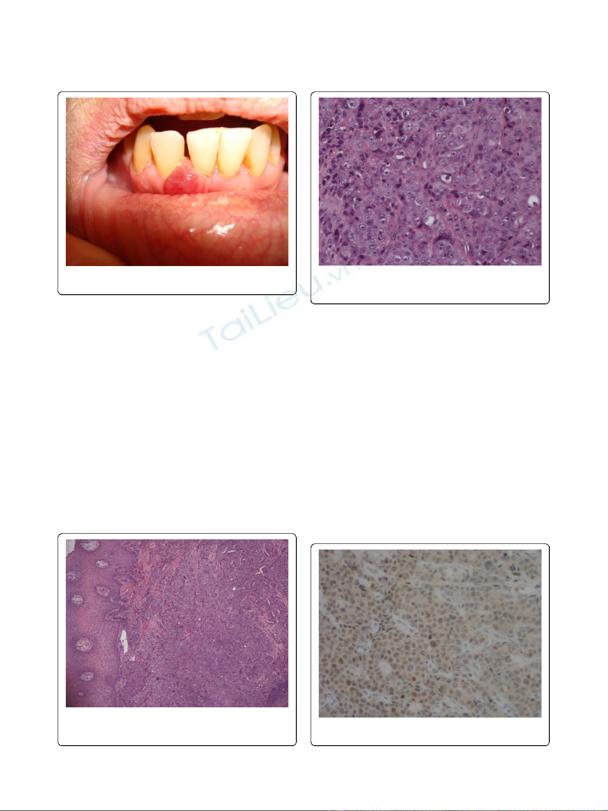
at the level of the lower right incisors (Figure 1). No other
pathologic finding was noticed during the physical exami-
nation. She underwent an excisional biopsy of the lesion,
and histopathologic immunohistochemistry showed a
poorly differentiated adenocarcinoma expressing cytokera-
tin 7 and thyroid transcription factor 1, whereas cytokera-
tins 5, 6 and 20 were absent. The pattern suggested a
metastasis of lung cancer (Figures 2, 3, 4). The total-body
computed tomographic (CT) scan with contrast-
* Correspondence: armando.orlandi@edu.rm.unicatt.it
1
Division of Medical Oncology, Catholic University of Sacred Heart, Rome,
Italy
Full list of author information is available at the end of the article
Orlandi et al.Journal of Medical Case Reports 2011, 5:202
http://www.jmedicalcasereports.com/content/5/1/202 JOURNAL OF MEDICAL
CASE REPORTS
© 2011 Orlandi et al; licensee BioMed Central Ltd. This is an Open Access article distributed under the terms of the Creative Commons
Attribution License (http://creativecommons.org/licenses/by/2.0), which permits unrestricted use, distribution, and reproduction in
any medium, provided the original work is properly cited.
enhancing medium revealed a 7.4 cm-sized tumor of the
lower lobe of the right lung with metastases to the ipsilat-
eral mediastinal lymph nodes (cT3N2). No other metas-
tases were detected, and her bone scan was also negative.
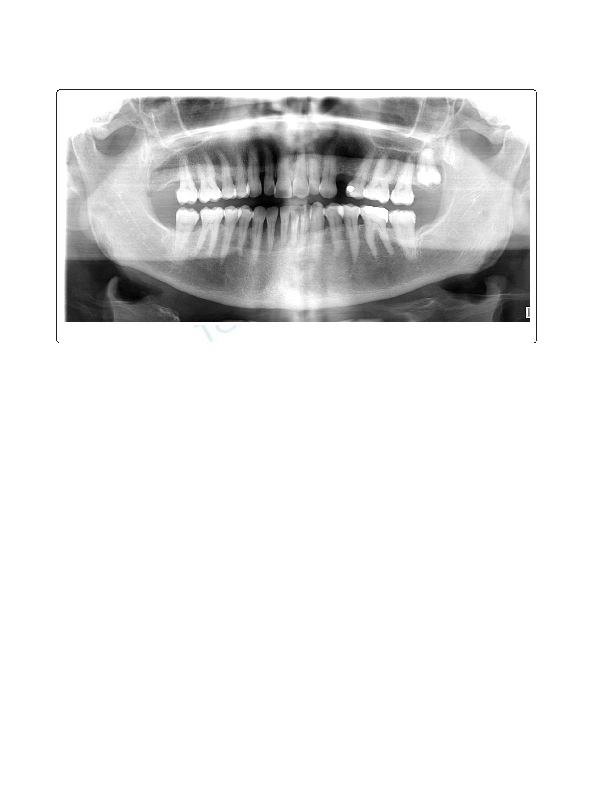
An orthopantomogram of thedentalarchesexcluded
metastases to the jawbones (Figure 5). After multi-disci-
plinary clinical evaluation, sequential treatment was
planned, including neoadjuvant chemotherapy (ChT) fol-
lowed by concomitant chemoradiation and surgery. Plati-
num-based combination therapy was selected, but
cisplatinum was excluded because the patient had low-
grade renal insufficiency with a serum creatinine level of
1.8 mg/dL. Therefore, carboplatin area under the curve 6
on day one and gemcitabine (1000 mg/mq on days one
and eight) every three weeks were started. Two months
later, after she had undergone three cycles of ChT, her CT
scan showed clear expansion of the primary tumor with
diffuse infiltration of the right lung. A second-line treat-
ment with docetaxel was attempted, but the tumor rapidly
progressed and the patient died six weeks later as a result
of respiratory failure.
Discussion
Metastatic tumors to the oral region are rare and mostly
produced by breast, lung and kidney cancer, but other
tumors may be also included [6]. Bone involvement is
much more frequent than soft tissue involvement, and
in the latter case lung cancer is the most common pri-
mary source. Hirshberg et al. [6] reviewed cases of oral
metastases reported from 1916 to 1991 and found 157
cases of oral soft tissue metastases, 86 of which had gin-
gival localization. The primary tumors were located in
the lung (25.5%), kidney (15.1%), bone (10.4%), breast
(9.3%) and liver (8.1%). Yoshii et al. [11] estimated that
Figure 1 Intra-oral view of the lesion developing in front of
right jaw incisors.
Figure 2 Histopathologic study showing proliferation of
adenocarcinoma cells below the gingival epithelium
(hematoxylin and eosin stain; original magnification, × 4).
Figure 3 Histopathologic study showing proliferation of
adenocarcinoma cells below the gingival epithelium
(hematoxylin and eosin stain; original magnification, × 20).
Figure 4 The tumor cells were immunoreactive for thyroid
transcription factor 1 (original magnification, × 20).
Orlandi et al.Journal of Medical Case Reports 2011, 5:202
http://www.jmedicalcasereports.com/content/5/1/202
Page 2 of 4
the probability of lung cancer involving a diagnosis of
gingival metastasis is about 10% to 20%. Other authors
have emphasized that the prognosis of patients with oral
metastases is very poor, with a median survival of less
than six months, mainly because of the fact that oral
metastases are an expression of a multi-metastatic dis-
ease [12]. A recent review of 39 patients with oral
metastases confirmed a median survival of 5.2 months
without significant differences according to oral localiza-
tion or to the site of the primary cancer [13]. In our
patient, oral localization was the only metastasis detect-
able at presentation. To the best of our knowledge, no
other similar cases have been described in the literature,
and this calls attention to the importance of recognition
of metastases to oral soft tissues. Most gingival lesions
in patients with prior or current non-oral malignancies
are not metastases [14]. Generally, gingival or oral
mucosal metastases extend from mandibular or maxil-
lary lesions and spread beyond the peri-osteum to cause
visiblegingivalororalmucosal masses [14]. Therefore,
gingival metastases are polypoid or exophytic and highly
vascularized, and bleeding is very common [8-10,15-17].
The same characteristics are also displayed by a number
of benign lesions, such as pyogenic granuloma (or vas-
cular epulis), peripheral giant cell granuloma (giant cell
epulis) or fibrous epulis [18]. From a clinical point of
view, the aspects suggestive of malignancy are only the
rapid growth and the propensity for either necrosis or
hemorrhage. In these cases, the possibility of metastasis
should be kept in mind, and biopsy is mandatory.
In our patient, no other metastases were found; there-
fore, we planned a multi-modal therapeutic approach
with a curative intent. However, the tumor proved
intrinsically resistant and highly aggressive. This beha-
vior raises the question whether the poor prognosis of
patients with tumors with oral metastases depends on
their diffuse spread or on their highly malignant nature.
Early detection might be important in metastases from
chemosensitive tumors, whereas chemoresistant tumors,
such as lung cancer, the present therapeutic strategies
are largely ineffective, and oral metastases should be
considered as only a negative prognostic factor.
Conclusion
In view of the rapid clinical evolution, in spite of the
fact that this is a single case report and no clear diag-
nostic recommendations can be made on the basis of a
single report, the present case of our patient supports
the fact that gingival metastasis is an important prog-
nostic factor. Thus, given the malignant potential and
the diagnostic value of a gingival metastasis, it is essen-
tial to carry out the excision of any presumed benign
tumor within healthy boundaries and to ask for a sys-
tematic histopathological examination.
Consent
Written informed consent was obtained from the patient
for publication of this case report and any accompany-
ing images. A copy of the written consent is available
for review by the Editor-in-Chief of this journal.
Figure 5 Panoramic radiography showing generalized alveolysis.
Orlandi et al.Journal of Medical Case Reports 2011, 5:202
http://www.jmedicalcasereports.com/content/5/1/202
Page 3 of 4

Author details
1
Division of Medical Oncology, Catholic University of Sacred Heart, Rome,
Italy.
2
Department of Pathology, Catholic University of Sacred Heart, Rome,
Italy.
Authors’contributions
OA collected the data and was involved in drafting the manuscript. DM and
FF participated in the acquisition of data. BM, CA and BC were involved in
drafting the manuscript or revising it for important intellectual content. All
authors read and approved the final manuscript.
Competing interests
The authors declare that they have no competing interests.
Received: 23 June 2010 Accepted: 25 May 2011 Published: 25 May 2011
References
1. Meyer I, Shklar G: Malignant tumors metastatic to mouth and jaws. Oral
Surg Oral Med Oral Pathol 1965, 20:350-362.
2. Sánchez Aniceto G, García Peñín A, de la Mata Pages R, Montalvo
Moreno JJ: Tumors metastatic to the mandible: analysis of nine cases
and review of the literature. J Oral Maxillofac Surg 1990, 48:246-251.
3. Hirshberg A, Leibovich P, Buchner A: Metastases to the oral mucosa:
analysis of 157 cases. J Oral Pathol Med 1993, 22:385-390.
4. Batson OV: The function of the vertebral veins and their role in the
spread of metastases. Ann Surg 1940, 112:138-149.
5. Chossegros C, Blanc JL, Cheynet F, Bataille JF, Tessier H: [Metastatic
localization in the buccal cavity: case report and literature review] [in
French]. Rev Stomatol Chir Maxillofac 1991, 92:160-164.
6. Lamster IB, Karabin SD: Periodontal disease activity. Curr Opin Dent 1992,
2:39-52.
7. Hirshberg A, Leibovitch P, Buchner A: Metastatic tumours to the jaw
bones: analysis of 390 cases. J Oral Pathol 1994, 23:337-341.
8. McDaniel RK, Luna MA, Stimson PG: Metastatic tumors in the jaws. Oral
Surg Oral Med Oral Pathol 1971, 31:380-386.
9. Morishita M, Fukud J: Hepatocellular carcinoma metastatic to the
maxillary incisal gingiva. J Oral Maxillofac Surg 1984, 42:812-815.
10. Maiorano E, Piatelli A, Favia G: Hepatocellular carcinoma metastatic to the
oral mucosa: report of a case with multiple gingival localizations.
J Periodontol 2000, 71:641-645.
11. Yoshii T, Muraoka S, Sano N, Furudoi S, Takahide K: Large cell carcinoma of
the lung metastatic to the mandibular gingiva. J Periodontol 2002,
73:571-574.
12. Van der Waal RI, Buter J, van der Waal I: Oral metastases: report of 24
cases. Br J Oral Maxillofac Surg 2003, 41:3-6.
13. Seoane J, Van der Waal I, Van der Waal RI, Cameselle-Teijeiro J, Antón I,
Tardio A, Alcázar-Otero JJ, Varela-Centelles P, Diz P: Metastatic tumours to
the oral cavity: a survival study with a special focus on gingival
metastases. J Clin Periodontol 2009, 36:488-492.
14. Kadokura M, Yamamoto S, Kataoka D, Nonaka M, Tanio N, Kunimura T,
Kushima M, Kushihashi T, Kawada T, Takaba T: Pulmonary adenocarcinoma
metastatic to the gingiva. Int J Clin Oncol 1999, 4:253-255.
15. Wegwood D, Rusen D, Balks S: Gingival metastasis from primary
hepatocellular carcinoma: report of a case. Oral Surg Oral Med Oral Pathol
1979, 47:263-266.
16. Nishimura Y, Yakata H, Kawasaki T, Nakajima T: Metastatic tumours of the
mouth and jaws: a review of the Japanese literature. J Oral Maxillofac
Surg 1982, 10:253-258.
17. Kanazawa H, Sato K: Gingival metastasis from primary hepatocellular
carcinoma: report of a case and review of literature. J Oral Maxillofac Surg
1989, 47:987-990.
18. Hirshberg A, Buchner A: Metastatic tumours in the oral region: an
overview. Eur J Cancer B Oral Oncol 1995, 31B:355-360.
doi:10.1186/1752-1947-5-202
Cite this article as: Orlandi et al.: Lung adenocarcinoma presenting as a
solitary gingival metastasis: a case report. Journal of Medical Case Reports
2011 5:202.
Submit your next manuscript to BioMed Central
and take full advantage of:
• Convenient online submission
• Thorough peer review
• No space constraints or color figure charges
• Immediate publication on acceptance
• Inclusion in PubMed, CAS, Scopus and Google Scholar
• Research which is freely available for redistribution
Submit your manuscript at
www.biomedcentral.com/submit
Orlandi et al.Journal of Medical Case Reports 2011, 5:202
http://www.jmedicalcasereports.com/content/5/1/202
Page 4 of 4


![PET/CT trong ung thư phổi: Báo cáo [Năm]](https://cdn.tailieu.vn/images/document/thumbnail/2024/20240705/sanhobien01/135x160/8121720150427.jpg)


%20--%3e%3cdefs%3e%3cstyle%3e%20.st0%20{%20fill:%20%23fff;%20}%20.st1%20{%20fill:%20%237800fa;%20}%20%3c/style%3e%3c/defs%3e%3cpath%20class='st1'%20d='M117.78,12.18H43.11c2.9,3.47,4.65,7.94,4.65,12.82,0,5.6-2.3,10.66-6.01,14.29h76.02l7.22-13.56-7.22-13.56Z'/%3e%3cg%3e%3cpath%20class='st0'%20d='M53.58,26.17h-.59v-1.46h.59v-4.96h2.83c1.78,0,2.67.94,2.67,2.82v5.76c0,1.87-.89,2.81-2.67,2.81h-2.83v-4.96ZM55.36,21.37v3.34h1.1v1.46h-1.1v3.34h1.01c.61,0,.91-.37.91-1.1v-5.93c0-.74-.3-1.1-.91-1.1h-1.01Z'/%3e%3cpath%20class='st0'%20d='M65.99,31.14h-1.8l-.31-2.07h-2.19l-.31,2.07h-1.64l1.82-11.39h2.62l1.82,11.39ZM65.28,18.04c-.25.46-.51.77-.75.94-.21.15-.47.22-.79.22-.26,0-.57-.07-.92-.22l-.38-.15c-.14-.05-.26-.07-.37-.07-.3,0-.53.18-.71.54l-.91-.68c.25-.46.51-.77.75-.94.21-.14.48-.21.79-.21.26,0,.57.07.92.21l.38.15c.14.05.26.07.37.07.3,0,.53-.18.71-.54l.91.68ZM61.91,27.52h1.73l-.87-5.76-.87,5.76Z'/%3e%3cpath%20class='st0'%20d='M74.53,26.89v1.52c0,1.91-.89,2.86-2.67,2.86s-2.67-.95-2.67-2.86v-5.93c0-1.91.89-2.86,2.67-2.86s2.67.95,2.67,2.86v1.11h-1.69v-1.22c0-.75-.31-1.12-.93-1.12s-.93.37-.93,1.12v6.15c0,.74.31,1.11.93,1.11s.93-.37.93-1.11v-1.63h1.69Z'/%3e%3cpath%20class='st0'%20d='M81.4,31.14h-1.8l-.31-2.07h-2.19l-.31,2.07h-1.64l1.82-11.39h2.62l1.82,11.39ZM75.9,19.2l1.52-1.91h1.71l1.51,1.91h-1.61l-.76-.95-.75.95h-1.61ZM77.32,27.52h1.73l-.87-5.76-.87,5.76ZM83.1,15.99l-1.76,1.91h-1.26l1.17-1.91h1.86Z'/%3e%3cpath%20class='st0'%20d='M84.86,19.75c1.78,0,2.67.94,2.67,2.82v1.48c0,1.87-.89,2.81-2.67,2.81h-.85v4.28h-1.79v-11.39h2.64ZM84.01,21.37v3.86h.85c.58,0,.87-.36.87-1.08v-1.71c0-.71-.29-1.07-.87-1.07h-.85Z'/%3e%3cpath%20class='st0'%20d='M93.51,19.75c1.78,0,2.67.94,2.67,2.82v1.48c0,1.87-.89,2.81-2.67,2.81h-.85v4.28h-1.79v-11.39h2.64ZM92.66,21.37v3.86h.85c.58,0,.87-.36.87-1.08v-1.71c0-.71-.29-1.07-.87-1.07h-.85Z'/%3e%3cpath%20class='st0'%20d='M98.8,31.14h-1.79v-11.39h1.79v4.88h2.03v-4.88h1.83v11.39h-1.83v-4.88h-2.03v4.88Z'/%3e%3cpath%20class='st0'%20d='M105.36,24.55h2.46v1.62h-2.46v3.34h3.09v1.63h-4.88v-11.39h4.88v1.63h-3.09v3.18ZM108.17,17.29l-1.76,1.91h-1.26l1.17-1.91h1.86Z'/%3e%3cpath%20class='st0'%20d='M112.2,19.75c1.78,0,2.67.94,2.67,2.82v1.48c0,1.87-.89,2.81-2.67,2.81h-.85v4.28h-1.79v-11.39h2.64ZM111.35,21.37v3.86h.85c.58,0,.87-.36.87-1.08v-1.71c0-.71-.29-1.07-.87-1.07h-.85Z'/%3e%3c/g%3e%3ccircle%20class='st1'%20cx='25'%20cy='25'%20r='20'/%3e%3cpath%20class='st0'%20d='M32.78,19.27c2.92,0,4.43,2.55,5.28,5.33l.71,2.17c.14.38-.33.75-.71.75h-5.61c.19-.33.24-.71.09-1.08l-.75-2.45c-.43-1.32-.99-2.64-1.79-3.77.75-.57,1.65-.94,2.78-.94h0ZM25,18.38c3.25,0,4.9,2.78,5.89,5.89l.76,2.45c.14.42-.33.8-.8.8h-11.69c-.42,0-.94-.38-.8-.8l.75-2.45c.99-3.11,2.64-5.89,5.89-5.89h0ZM25,11.35c1.74,0,3.11,1.37,3.11,3.11s-1.37,3.11-3.11,3.11-3.11-1.41-3.11-3.11,1.41-3.11,3.11-3.11h0ZM17.27,19.27c1.08,0,1.98.38,2.73.94-.8,1.13-1.37,2.45-1.74,3.77l-.8,2.45c-.14.38-.05.75.09,1.08h-5.56c-.42,0-.9-.38-.75-.75l.71-2.17c.9-2.78,2.41-5.33,5.33-5.33h0ZM17.27,12.91c1.51,0,2.78,1.27,2.78,2.83s-1.27,2.83-2.78,2.83-2.83-1.27-2.83-2.83,1.27-2.83,2.83-2.83h0ZM32.78,12.91c1.56,0,2.78,1.27,2.78,2.83s-1.23,2.83-2.78,2.83-2.83-1.27-2.83-2.83,1.27-2.83,2.83-2.83h0ZM27.07,28.56v.09c0,.57-.24,1.08-.61,1.46h0v.05c-.38.33-.9.57-1.46.57s-1.08-.24-1.46-.61h0c-.38-.38-.61-.9-.61-1.46v-.09h1.41v.09c0,.19.05.38.19.47v.05c.09.09.28.19.47.19s.38-.09.47-.19v-.05c.14-.09.24-.28.24-.47t-.05-.09h1.41ZM30.99,28.56v.09c0,1.65-.66,3.16-1.74,4.24-1.08,1.08-2.59,1.79-4.24,1.79s-3.16-.71-4.24-1.79l-.05-.05c-1.04-1.08-1.7-2.55-1.7-4.2v-.09h1.41v.09c0,1.27.47,2.4,1.27,3.25h.05c.85.85,1.98,1.37,3.25,1.37s2.4-.52,3.25-1.37c.85-.8,1.37-1.98,1.37-3.25v-.09h1.37ZM34.99,28.56v.09c0,2.78-1.13,5.28-2.92,7.07-1.79,1.79-4.29,2.92-7.07,2.92s-5.23-1.13-7.07-2.92c-1.79-1.79-2.92-4.29-2.92-7.07v-.09h1.41v.09c0,2.4.94,4.53,2.5,6.08,1.56,1.56,3.72,2.5,6.08,2.5s4.52-.94,6.08-2.5c1.56-1.56,2.5-3.68,2.5-6.08v-.09h1.41Z'/%3e%3c/svg%3e)