J. Vet. Sci. (2003),(cid:1)4(3), 209–212
(cid:1) (cid:2) (cid:3) (cid:4) (cid:5) (cid:6) (cid:7) (cid:8) (cid:2) (cid:9) (cid:10)(cid:11)(cid:12)(cid:11)(cid:13)(cid:14)(cid:15)(cid:16)(cid:13)(cid:17)(cid:8) (cid:18)(cid:19)(cid:14)(cid:11)(cid:15)(cid:19)(cid:11)
Glutamate and GABA concentrations in the cerebellum of novel ataxic mutant Pogo mice
Ki-Hyung Kim, Jeoung-Hee Ha1, Seung-Hyuk Chung, Chul-Tae Kim, Sun-Kyung Kim, Byung-Hwa Hyun2, Kazuhiko Sawada3, Yoshihiro Fukui3, Il-Kwon Park4, Geun-jwa Lee5, Bum-Kyeong Kim, Nam-Seob Lee and Young-Gil Jeong*
Department of Anatomy & Pathology, College of Medicine, Konyang Univiversity, Nonsan 320-711, Korea 1Department of Pharmacology, College of Medicine, Yeungnam University, Gyeongsan 712-749, Korea 2Genetic Resource Center, KRIBB, Daejon 305-333, Korea 3Department of Anatomy, University of Tokushima School of Medicine, Tokushima, Japan 4Angio-Lab., Paichai University RRC, Daejeon 302-161, Korea 5Chungnam Livestock & Veterinary Service institute, Hongsung 350-821, Korea
the most [6], whereas
is synthesized
The Pogo mouse is an autosomal recessive ataxic mutant that arose spontaneously in the inbred KJR/MsKist strain derived originally from Korean wild mice. The ataxic phenotype is characterized by difficulty in maintaining posture and side to side stability, faulty coordination between limbs and trunk, and the consequent inability to walk straight. In the present study, the cerebellar concentrations of glutamate and GABA were analyzed, since glutamate is a most prevalent excitatory neurotransmitter whereas γ-aminobutyric acid (GABA) is one of the most abundant inhibitory neurotransmitters, which may be the main neurotransmitters related with the ataxia and epilepsy. The concentration of glutamate of cerebellum decreased significantly in ataxic mutant Pogo mouse compared to those of control mouse. However, GABA concentration was not decrease. These results suggested that the decrease in glutamate concentration may contribute to ataxia in mutant Pogo mouse.
Key words: Pogo, glutamate, GABA, cerebellum
for
walk straight [16,18]. The Pogo mutation is inherited as a trait on chromosome 8 as well as the tottering, leaner, and rolling mutations. prevalent excitatory Glutamate is γ-aminobutyric acid neurotransmitter (GABA) is the most abundant inhibitory neurotransmitter [20]. Glutamate is the main excitatory neurotransmitter in the brain [10] and all glutamate is formed from glucose within the central nervous system because glutamate dose not readily cross the blood-brain barrier [11,15,21,25]. from 2-oxoglutarate by Glutamate transmination either with alanine, aspartate or one of the branched chain amino acids leucine, isoleucine and valine, and can also be formed from glutamine by phosphate- activated glutaminase [28]. Glutamate is accumulated into vesicles to a high concentration and released to the synapses by calcium-dependent exocytosis upon the arrival of an action potential. As a high extracellular concentration of glutamate is also neurotoxic, high-affinity glutamate terminating synaptic transporters are essential transmission and for maintaining a low extracellular glutamate concentration.
Introduction
the action of
in
inhibitory neurotransmitter
The Pogo mouse is an autosomal recessive ataxic mutant that arose spontaneously in the inbred KJR/MsKist strain derived originally from Korean wild mice. The ataxic phenotype is characterized by difficulty in maintaining posture and side to side stability, faulty coordination between limbs and trunk, and the consequent inability to
to be released from
the
*Corresponding author Phone: +82-41-730-5115; Fax: +82-41-736-5318 E-mail: ygjeong@konyang.ac.kr
GABA is the primary inhibitory neurotransmitter known the excitatory to counterbalance neurotransmitter glutamate. The importance of GABA as an the mammalian cerebellum is well documented [23,27]. The excitatory granule cells, by far the most numerous neuronal type in the cerebellum [9], receive input from GABAergic cells; thus it has been suggested that the majority of GABA receptors are located on granule cells [24]. GABA is interneurons by thought feedforward inhibition from the granule cells or by feedback inhibition from the pyramidal cells controlled by
210 Ki-Hyung Kim et al.
Table 1. Condition of HPLC for the determination of brain glutamate and GABA concentration in mice
glutamatergic nerve endings [12]. The present study examined that the concentrations of glutamate and GABA in the cerebellum of Pogo mouse.
Parameter Conditions
Materials and Methods
Column Flow rate
Mobile phase
Animals
ex: 340 nm, λ
em: 450 nm)
Mice were generated from a breeding colony of Pogo mice developed from original breeding pairs obtained from Korea Rearch Institute Bioscience and Biotechnology (KRIBB). 30-day-old ataxic Pogo (pogo/pogo) and normal wild mice (+/+, control) were used in all experiments. All experimental procedures were carried out in accordance with the NIH Guidelines for the Care and Use of Laboratory Animals.
alcohols and mounted in DPX (BDH Chemicals Inc., Toronto, Canada).
Gradient Attenuation Detector RP-C18 (150 × 4.0 mm I.D., 10 µm) 0.6 ml/min 10 mM potassium acetate buffer (pH 6.5)-methanol Methanol 20-70%/40 min 10 Fluorescence detector (λ
Immunohistochemistry
Determination of glutamate/ GABA levels
in
Concentarations of glutamate and GABA
the cerebellums were measured using a modified method of Allen et al. [1]. Tissues were homogenized in 0.3 M triethanolamine buffer, pH 6.8, containing of 1 mM aminoetylisothiouronium bromide and 2 mM pyridoxal 5'- phosphate, then centrifuged (Hanil Supra 22K, ROK) at 15,000 g for 20 mins. Postmitochondrial fraction from each extract was resuspened in 20 mM potassium phosphate buffer, deproteinizied, and then centrifuged. Supernatants were filtered by membrane filter (0.2 µm: 13 mm), and then o-phtalaldhyde derivatives were used for the detection of fluorescence in the HPLC measurement (fluorescence detector, SHIMADZU, Japan, Table 1). The amounts of glutamate and GABA in cerebellums were represented as nmole per mg protein.
temperature
Results
A. Immunohistochemistry
Calbindin is expressed in the cerebellum exclusively by Purkinje cells [8,22]. Anti-CaBP immunohistochemistry deposited peroxidase reaction product throughout all Purkinje cells, including the somata, dendrites, dendritic spines and axons, in both normal wild type and pogo/pogo
All mice were deeply anaesthetized with sodium pentobarbital (60 mg/kg body weight) and transcardially perfused with 0.9% NaCl in 0.1 M phosphate buffer saline (PBS, pH 7.4) followed by 150 ml of 4% paraformaldehyde in 0.1 M PBS (pH 7.4). These brains were the removed from the skull and placed immediately in the same fixative at 4oC for 24 hours. The post-fixed brains were transferred to 0.1 M PBS, and after the brains were cryoprotected in 10%, 20% and 30% sucrose in 0.1 M PBS and cryostat sectioned in the frontal plane 20 µm thickness. After several rinses in 0.1 M PBS (pH 7.4) the sections were quenched for 10 min in 1% H2O2, and rinsed in 0.1 M Tris phosphate-buffered saline (TPBS; 8.5 mM Na2HPO47H2O, 3 mM KH2PO4, 125 mM NaCl, 30 mM Tris-HCl, 0.03 mM NaN3, pH 7.7). Sections were incubated overnight at room in rabbit polyclonal anti-calbindin-D (anti-CaBP, Sigma Inc., St. Louis MO). They were then washed three times for 5 min in 0.1 M TPBS, and incubated in 1 : 100 peroxidase- conjugated anti-rabbit IgG (Dakopatts Inc., Mississauga, Canada) for 2 hours at room temperature. After three additional rinses in TPBS, antibody-binding sites were revealed by a 15 min incubation in 0.2% diaminobenzidine in TPBS. Sections were then dehydrated though graded
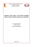
Fig. 1. Anti-calbindin immunoreaction were showed in frontal sections through the vermis of the cerebellum of a +/+ normal wild type mouse [control] (A) and ataxic Pogo mouse [pogo/pogo homozygote] (B) in lobule VIII and IX. The loss of Purkinje cells (arrow) in ataxic Pogo mouse is seen when compared with corresponding lobule of the +/+ normal wild type mouse. Scale bar = 100 µm.
Glutamate and GABA concentrations in the cerebellum of novel ataxic mutant Pogo mice 211
their parallel fibers and,
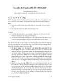
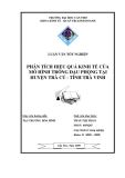
Table 2. Concentrations of glutamate and GABA in cerebellums
Control Pogo Glutamate (µmol/g) GABA (µmol/g) 1.353 ± 0.055 1.360 ± 0.074
11.327 ± 1.561 9.147 ± 1.457* *Data are represented as Mean ± S.D. of 6-9 animals. *p<0.05: Significantly different from control
of granule cells and in consequence, to the formation of ‘heterologous’ mossy fiber contacts on Purkinje cells and a persistent multi- innervation of Purkinje cells by olivary climbing fibers [7,26]. The phase relationships to sinusoidal vestibular stimulation of floccular Purkinje cells are greatly distorted and irregular under these conditions making a precise time matching of convergent input onto target neurons in the vestibular nuclei highly uncertain [13,14]. In addition, sprouting and enlargement of GABAergic synaptic boutons in the dorsal part of the lateral vestibular nuclei was observerd recently in this mutant [2,3]. In Purkinje cell degeneration mutants, where cell loss affects the mature cerebellum, a clear increase in somatal parvalbumin- immunoreactivity in the vestibular nuclei and deep cerebellar nuclei suggests an enhanced activity of mainly inhibitory neurons. However, GABAergic reinnervation was not found [2,4,5]. In leaner, reinnervative reactions of both Purkinje cell GABAergic and extracerebellar GABAergic sources, that would substitute for the lost Purkinje cell-input, are not indicated by the present findings using GABA-immunohistochemistry. GABAergic innervation density is diminished to one-half in the dorsal part of the lateral vestibular nuclei of leaner, which is only slightly higher than in Purkinje cell degeneration mutants [2]. This reduction corresponds well with the massive Purkinje cell loss in the anterior lobe and shows that, in contrast to weaver, GABAgergic reinnervation does not occur under these conditions. In addition, GABAergic terminals in leaner are reduced in size to such a degree, comparatively only with that found after experimental removal of the cerebellum or in Purkinje cell degeneration mutants [2].
mutant mice (Fig. 1). The Purkinje cells appeared normal in pogo/pogo mutant mice with respect to their size and arrangement as a monolayer in the Purkinje cell layer (Fig. 1). Purkinje cell ectopia was rare. Individual Purkinje cells in the vermis of pogo/pogo mutant mice are grossly normal with parasagittaly oriented dendritic arbors extending to the surface of the molecular layer (Fig 1A). However, in pogo/pogo mutant mice there was a loss of Purkinje cells throughout the cerebellar vermis (Fig. 1B). The loss of somata and dendrites of Purkinje cells was clearly demonstrated by using anti-CaBP immunostaining (Fig. 1B).
Fig. 2. Concentrations of glutamate and GABA in the cerebellums of control and ataxic Pogo (pogo/pogo) mice.
B. Concentration of glutamate and GABA of cerebellum As illustrated in Table 2 and Fig. 2 the concentration of glutamate of cerebellum decreased significantly in ataxic mutant Pogo/Pogo mouse compared to those of control mouse. But GABA concentration was not decrease.
Discussion
In our present experiment,
to be central
Then, there is a report of the glutamate concentration in E1 mouse. The E1 mouse is a genetically susceptible model of complex-partial epilepsy with secondary generalization of seizures. This model shows elevated GABA (40-50%) and lowered glutamine and glutamate (30%) in its most epileptic state E1 (+) compared with control or E1 (−) mice (i.e. same genetic type but not multiply stimulated to become responded to handling by having seizures). However, there was a great increase in glutamate level during the pre-convulsive state, and the themselves were blocked by AP5 given seizures intraventricularly 30 mins before seizure induction, in which GABA levels increased transitorily immediately [19]. Thus both glutamatergic and after seizures the to GABAergic systems appear mechanisms generating seizures in the E1 mouse [17].
to
In this study, we have provided that the concentration of glutamate in pogo/pogo mouse cerebellum decreased control mouse. However, GABA compared concentration was not changed.
the concentration of glutamate decreased in pogo/pogo mouse. There have been several reports of glutamate and GABA concentration in weaver, Purkinje cell degeneration (PCD), leaner and E1 mouse cerebellum. In weaver mice cerebellum, neuronal loss occurs during postnatal development and leads to a partial Purkinje cell degeneration, an almost complete loss
These results suggested that the reduction of glutamate
212 Ki-Hyung Kim et al.
concentration may related with disarrangement in synapse and contribute to motor ataxia in Pogo mouse.
Acknowledgments
This work was supported by grant No. R05-2002-000- 00710-0 from Basic Research Program of the Korea Science & Engineering Foundation.
D-aspartate receptor channels in hippocampal neurons maintained in culture. Mol. Pharmacol. 1990, 37, 477-481. 13. Grüsser-Cornehls, U. Responses of vestibular neurons from the nucleus vestibularis and the flocculus in wild type mice and mutants. Soc. Neurosci. Abstr. 1983, 9, 524.
Referneces
14. Grüsser-Cornehls, U. (ed), Compensatory mechanisms at the level of the vestibular nuclei following post-natal degeneration of specific cerebellar cell classes and ablation of the cerebellum in mutant mice. In H. Flohr, Post-lesion Neural Plasticity, pp. 431-442, Springer, Berlin/Heidelberg. 1988.
chromatographic method liquid 15. Hawkins, R. A., DeJosepf, M. R. and Hawkins, P. A. Regional brain glutamate transport in rats at normal and raised concentrations of circulating glutamate. Cell Tissue Res. 1995, 281, 207-214.
1. Allen, I. C. and Griffthis, R. Reversed-phase high performance for determination of brain glutamate decarboxylase suitable for use in kinetic studies. J. Chromatography 1984, 336, 385- 391.
16. Hyun, B. H., Kim, M. S., Choi, Y. K., Yoon, W. K., Suh, J. G., Jeong, Y. G., Park, S. K. and Lee, C. H. Mapping of the Pogo gene, a new ataxic mutant from Korean wild mice, on central mouse chromosome 8, Mamm. Genome 2001, 12, 250-252. 2. Bäurle, J., Grover, B. G. and Grüsser-Cornehls, U. Plasticity of GABAergic terminals in Deiters nucleus of weaver mutant and normal mice: a quantitative light microscopics study. Brain Res. 1992, 591, 305-318. in
17. Janjua, N., Hiramatsu. M., Kabuto, H. and Mori, A. Glutamic acid and gama-aminobutyric acid the biochemical and genetic mechanisms of E1 mouse epilepsy. Neurosciences 1992, 18 (Suppl. 2), 43-47. 3. Bäurle, J., and Grüsser-Cornehls, U. Calbindin D-28K in the lateral vestibular nucleus of mutant mice as a tool to reveal Purkinje cell plasticity, Neurosci. Lett. 1994, 167, 85- 88.
18. Jeong, Y. G., Hyun, B. H. and Hawkes, R. Abnormalities in cerebellar Purkinje cells in the novel ataxic mutant mouse, Pogo. Dev. Brain Res. 2000, 125, 61-67. 19. Mori, A. E1 Mice: Neurochemical approach to the seizure mechanism. Neuroscience 1998, 14, 275-285.
4. Bäurle, J. and Grüsser-Cornehls, U. (ed), A possible mechanism to compensate for the loss of cerebellar inhibition in Purkinje cell degeneration mutant mice. In N. Flsner and H. Breer (Dds.), Proceeding of 22nd Gottingen Neurobiology Conference. Vol. 2, p. 409, Georg Thieme, Stuttgart/New York. 1994.
20. Olsen, R. W. and DeLorey, T. M. (ed), GABA and glycine. In: Siegel, G.J., Agranoff, B. W., Albers, R.W., Fisher, S.K., Uhler, M.D., Basic Neurochemisty. pp. 335-346, Lipincott Williams and Wilkins, Philadelphia. 1999. (GAP-43) in
21. Oldendorf, W. H. Brain uptake of radiolabeled amino acids, amines and hexoses after arterial injection. Am. J. Physiol. 1971, 221, 1629-1635. 5. Bäurle, J., Oestreicher, A. B., Gispen, W. H. and Grüsser- immuno- Cornehls, U. Lesion-specific pattern of the cytochemical distribution of B-50 cerebellum of weaver and pcd mutant mice: Lack of B-50 involvment in neuroplasticity of Purkinje cell terminals, J. Neurosci. Res.1994, 38, 327-335.
22. Ozol, K., Hayden, J. M., Oberdick, J. and Hawkes, R. Transverse zones in the vermis of the mouse cerebellum, J. Comp. Neurol. 1999, 412, 95-111. 6. Cotman, C. W., Foster, A. C. and Lanhorn, T. An overview of glutmate as a neurotransmitter. Adv. Biochem. Psychopharmacol. 1981, 27, 1-27.
7. Crepel, F. and Mariani, J. Multiple innervation of Purkinje cells by climbing fibers in the cerebellum of the weaver mutant mouse. J. Neurobiol. 1976, 7, 579-582.
8. De Camilli, P., Miller, P. E., Levitt, P., Walter, U. and Greengard, P. Anatomy of cerebellar Purkinje cells in the rat determined by a specific immunohistochemical marker, Neuroscience 1984, 11, 761-817.
9. Eccles, J. C., Ito, M. and Sentagothai, J. (ed), The cerebellum as a neuronal machine, pp. 11-16, Springer, New York. 1967. 10. Fonnum F. Glutamate: a neurotransmitter in mammalian 23. Roberts, F., Chase, T. N. and Tower, D. B. (ed), GABA in nervous system function, pp. 9-13, Raven, New york. 1976. 24. Simantov, R., Oster-Granite, M. L., Nerndon, R. M. and Snyder, S. H. Gamma-aminobutyric acid (GABA) receptor binding selectively depleted by viral induced granule cell loss in hamster cerebellum. Brain Res. 1976, 105, 365-371. 25. Smith, Q. R., Momma, S., Aoyagi, M. and Rapoport, S. I. Kinetics of neutral amino acid transport across the across the blood-brain barrier. J. Neurochem. 1987, 49, 1651-1658. 26. Sotelo, C. Dendritic abnormalities of Purkinje cells in cerebellum of neurological mutant mice (weaver and staggerer). Adv. Neurol. 1975, 12, 335-351. brain. J. Neurochem. 1984, 42, 1-11. 27. Tebecis, A. K. Transmitters and identified neurons in the 86-115, nervous system. central pp. mammalian Scientichnica. Bristol. 1974. 28. Yudkoff, M. Brain metabolism of branched-chain amino acids. Glia 1997, 21, 92-98. 11. Gruetter, R., Novotny, E. J., Boulware, S. D., Mason, G. F., Rothman, D. L., Shulman, G. I., Prichard, J. W. and Shulman, R. G. Localized 13C NMR spectroscopy in the human brain of amino acid labeling from D-[1 13C]glucose. J. Neurochem. 1994, 63, 1377-1385. 12. Grudt, T. J. and Jahr, C. E. Quisqualate activates N-methyl-