84
Tạp chí Y Dược học - Trường Đại học Y Dược Huế - Tập 9, số 2 - tháng 4/2019
Địa chỉ liên hệ: Phan Anh Chi, email: anchitogether@gmail.com
Ngày nhận bài: 9/2/2019, Ngày đồng ý đăng: 16/3/2019; Ngày xuất bản: 25/4/2019
NGHIÊN CỨU VỊ TRÍ LỒI CẦU Ở TƯƠNG QUAN TRUNG TÂM VÀ Ở
LỒNG MÚI TỐI ĐA TRÊN HÌNH ẢNH CẮT LỚP VI TÍNH HÌNH NÓN
Phan Anh chi, Hồ Xuân Anh Ngọc
Khoa răng Hàm Mặt, Trường Đại học Y Dược, Đại học Huế
Tóm tắt
Đặt vấn đề: Sự khác biệt về vị trí lồi cầu giữa vị trí tương quan trung tâm và lồng múi tối đa luôn là vấn đề
gây bàn cãi và có tính thời sự cao. Mục tiêu: So sánh vị trí lồi cầu giữa vị trí tương quan trung tâm và lồng múi
tối đa ở những người không có dấu chứng rối loạn thái dương hàm trên hình ảnh cắt lớp vi tính hình nón. Đối
tượng và phương pháp: 40 sinh viên lớp RHM5 và RHM6 Trường Đại học Y Dược Huế, không có dấu chứng
rối loạn thái dương hàm được đánh giá vị trí lồi cầu tại vị trí tương quan trung tâm và lồng múi tối đa trên
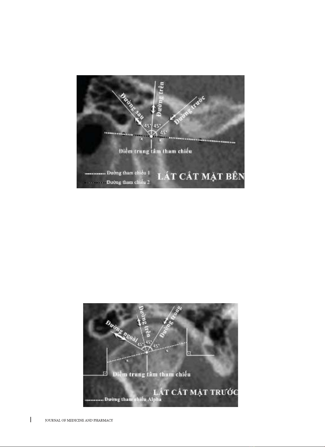
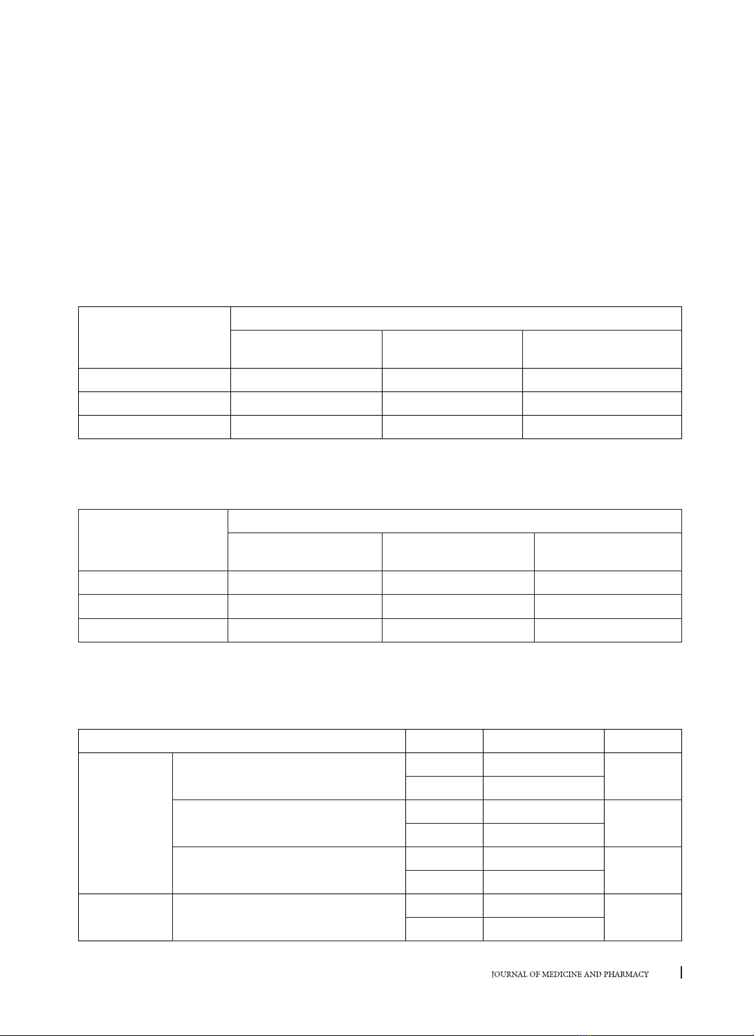
phim chụp cắt lớp vi tính hình nón. Vị trí lồi cầu được đánh giá theo phân loại của Sener (2009) và được so
sánh giá trị trung bình tại vị trí tương quan trung tâm và lồng múi tối đa bằng phép kiểm định Paired-Samples
t-test và Mann-Whitney U-test. Kết quả: Trong tổng số 480 cặp khoảng gian khớp xác định vị trí lồi cầu có
91,2% cặp có sự khác nhau giữa hai vị trí tham chiếu. Nhưng sự khác biệt không mang ý nghĩa thống kê. Vị trí
lồi cầu nằm ở trung tâm hõm khớp có tỷ lệ cao nhất ở cả tương quan trung tâm và lồng múi tối đa (43,8% ở
tương quan trung tâm và 51,2% ở lồng múi tối đa). Sau đó là tỷ lệ lồi cầu nằm ở vị trí lui sau (32,5% ở tương
quan trung tâm và 36,3% ở lồng múi tối đa). Cuối cùng thấp nhất là tỷ lệ lồi cầu nằm ở vị trí ra trước (23,7%
ở tương quan trung tâm và 12,5% ở lồng múi tối đa). Kết luận: Không có sự khác biệt có ý nghĩa thống kê về
vị trí lồi cầu giữa vị trí tương quan trung tâm và lồng múi tối đa ở những người không có dấu chứng rối loạn
thái dương hàm.
Từ khóa: Vị trí lồi cầu, tương quan trung tâm, lồng múi tối đa, cắt lớp vi tính hình nón
Abstract
A STUDY OF CONDYLAR POSITION IN CENTRIC RELATION AND
MAXIMAL INTERCUSPATION USING CONE-BEAM TOMOGRAPHY
Phan Anh Chi, Ho Xuan Anh Ngoc
Faculty of Odonto-Stomatology, Hue University of Medicine and Pharmacy, Hue University
Background: The condylar position discrepancy between centric relation and maximal intercuspation has
been still a controversial issue. Aims: To compare the condylar position between centric relation and maximal
intercuspation using cone-beam tomography in patients without temporomandibular joints disorder.
Materials and methods: To assess the condylar position in centric relation and maximal intercuspation using
cone-beam tomography on 40 fifth-year and sixth-year dental students of Hue University of Medicine and
Pharmacy without temporomandibular joints disorder. The condylar positions are assessed following Sener
classification (2009) and are compared between centric relation and maximal intercuspation using paired-
samples t-test and Mann-Whitney U-test. Results: Among 480 condye-to-fossa measurement pairs, there
are 91.2% pairs having difference between two reference position but there is no significant difference. The
condylar position at the superior of mandibular fossa has the greatest percentage in both centric relation
and maximal intercuspation (43.8% in centric relation and 51.2% in maximal intercuspation). This greatest
percentage is followed by the condylar position at posterior of mandibular fossa (32.5% in centric relation
and 36.3% in maximal intercuspation). Lastly, the condylar position at the anterior of mandibular fossa has
the fewest percentage (23.7% in centric relation and 12.5% in maximal intercuspation). Conclusion: There is
no significant difference of condylar position between centric relation and maximal intercuspation in patients
without temporomandibular joints disorder.
Keywords: Condylar position, centric relation, maximal intercuspation, cone-beam tomography
DOI: 10.34071/jmp.2019.2.14