
Review Article
Theme: Heterotrimeric G Protein-based Drug Development: Beyond Simple Receptor Ligands
Guest Editor: Shelley Hooks
Regulator of G-protein Signaling (RGS)1 and RGS10 Proteins as Potential Drug
Targets for Neuroinflammatory and Neurodegenerative Diseases
Jae-Kyung Lee
1,2
and Josephine Bou Dagher
1
Received 3 December 2015; accepted 2 February 2016; published online 22 February 2016
Abstract. Regulator of G-protein signaling (RGS) proteins were originally identified as negative
regulators of G-protein-coupled receptor (GPCR) signaling via their GTPase-accelerating protein (GAP)
activity. All RGS proteins contain evolutionarily conserved RGS domain; however, they differ in their
size and regulatory domains. RGS1 and RGS10 are smaller than other RGS proteins, and their functions
involve various inflammatory responses including autoimmune responses in both the periphery and the
central nervous system (CNS). Neuroinflammation is the chronic inflammatory response in the CNS.
Acute inflammatory response in the CNS is believed to be beneficial by involving the neuroprotective
actions of immune cells in the brain, particularly microglia, to limit tissue damage and to aid in neuronal
repair. However, chronically elevated levels of cytokines serve to maintain activation of abundant
numbers of immune cells potentiating prolonged inflammatory responses and creating an environment of
oxidative stress, which further hastens oxidative damage of neurons. In this review, we describe the
implications and features of RGS proteins (specifically RGS1 and RGS10) in neuroinflammation and
neurodegenerative diseases. We will discuss the experimental and epidemiological evidence on the
benefits of anti-inflammatory interventions by targeting RGS1 and/or RGS10 protein function or
expression in order to delay or attenuate the progression of neurodegeneration, particularly in multiple
sclerosis (MS) and Parkinson’s disease (PD).
KEY WORDS: G-protein-coupled receptor (GPCR); multiple sclerosis; neuroinflammation; Parkinson’s
disease; regulator of G-protein signaling.
INTRODUCTION
G-protein-coupled receptors (GPCRs) signal through
heterotrimeric G-proteins that consist of an αsubunit and a
βγ heterodimer (1). Regulator of G-protein signaling (RGS)
proteins play a role in turning off GPCR signaling. All of the
RGS proteins contain a conserved RGS domain that interacts
with a Gαi, Gαq/11, or Gα12/13 subunit with variable
selectivity, which accelerates the GTPase-activating activity
of the Gαsubunit (2–4). Since the mid-1990s, more than 30
functional RGS genes have been identified and subdivided
into eight subfamilies that are expressed in eukaryotic
organisms, from fungi to animals such as mice and humans
(3,5,6). RGS proteins differ widely in their size and contain a
variety of structural domains in addition to the RGS domain
and motifs that regulate their activity and determine regula-
tory binding partners (3,5; also reviewed in 6–9). Early
evidence suggested that RGS proteins acted primarily as
negative regulators of G-protein signaling. Today, it is well
documented that these proteins act as tightly regulated
modulators and multifunctional interactors of G-protein
signaling (reviewed in 8). In addition, it has been recently
appreciated that the non-RGS regions of RGS proteins can
provide non-canonical functions distinct from the inactivation
of Gαsubunits or even from G-protein signaling entirely
(reviewed in 9,10).
RGS proteins are highly conserved from yeast to mammals
and are abundant in the retina, brain, heart, and immune organs
(11; see review in 12). Tissue-specific patterns of RGS protein
expression in the human peripheral tissues and brain were
reported by Larminie and his group (13). They showed that the
major RGS10 proteins in human lymphocytes are RGS1, RGS2,
RGS10, RGS13, RGS14, RGS16, and RGS18 (13). The RGS
proteins may acquire functional diversity in immune cells by a
fine-tuned and dynamic regulation of the expression of multiple
RGS proteins.
RGS protein profiling in human lymphocytes displays a
similar expression profile to rodent lymphocytes except for
RGS18 (14), suggesting that observations in rodent lympho-
cytes may be translated into what would be occurring in
human lymphocytes. In this review, we will discuss how RGS
proteins, and more precisely RGS1 and RGS10, play
1
Department of Physiology and Pharmacology, College of Veterinary
Medicine, University of Georgia, 501 D. W. Brooks Dr., Athens,
Georgia 30602, USA.
2
To whom correspondence should be addressed. (e-mail:
jamlee@uga.edu)
The AAPS Journal, Vol. 18, No. 3, May 2016 ( #2016)
DOI: 10.1208/s12248-016-9883-4
545 1550-7416/16/0300-0545/0 #2016 American Association of Pharmaceutical Scientists
important roles in inflammatory and neurodegenerative
diseases and act as therapeutic potentials mainly in
Parkinson’s disease (PD) and multiple sclerosis (MS).
RGS PROTEIN FAMILY AND MS
MS is a chronic inflammatory disease of the central
nervous system (CNS) associated with demyelination that is
thought to have an underlying autoimmune etiology. Cur-
rently in the USA, around 250,000–300,000 people have been
diagnosed with MS and there are 200 new cases diagnosed
every week. Although there are varieties of immune-based
therapeutic drugs available for the treatment of MS, it is
difficult for clinicians to predict which drugs would work best
for an individual patient due to a lack of mechanistic
information of the disease (15). Also, major MS drugs target
broad ranges of immune cells, which significantly affect
leukocyte trafficking and function. Therefore, it is important
to identify biomarkers and/or cellular regulators specifically
modulating function of autoimmune-reactive leukocytes.
Although the etiology of MS has not been identified,
increasing evidence indicates that disease onset involves the
combined influence of environmental factors and genetic suscep-
tibility (16). GPCR signaling plays an important role in various
aspects of MS pathogenesis including : antigen presentation,
cytokine/chemokine production, and T-cell differentiation, prolif-
eration, and invasion (see review in (17)). RGS family proteins
that are important modulators of GPCR signaling pathways are
recently implicated in the development of MS and other
autoimmune diseases. Multiple points of genetic evidences have
shown the following: (1) single nucleotide polymorphisms (SNPs)
of RGS1, RGS7, RGS9, and RGS14 are reported to be of high
correlation with the diagnosis of MS, Crohn’sdisease,and
ulcerative colitis (18–22), and (2) the messenger RNA (mRNA)
level of RGS10 and RGS1 is higher in peripheral blood
mononuclear cells (PBMCs) from patients with MS according to
the Gene Expression Omnibus (GEO) profile database (23,24).
However, the role of RGS proteins in the context of the onset or
progression of autoimmune diseases is yet to be explored.
RGS1 AND MS
RGS1 is a novel MS susceptibility locus as recently
identified by the International Multiple Sclerosis Genetics
Consortium (IMSGC) (18). A search of the GEO profile
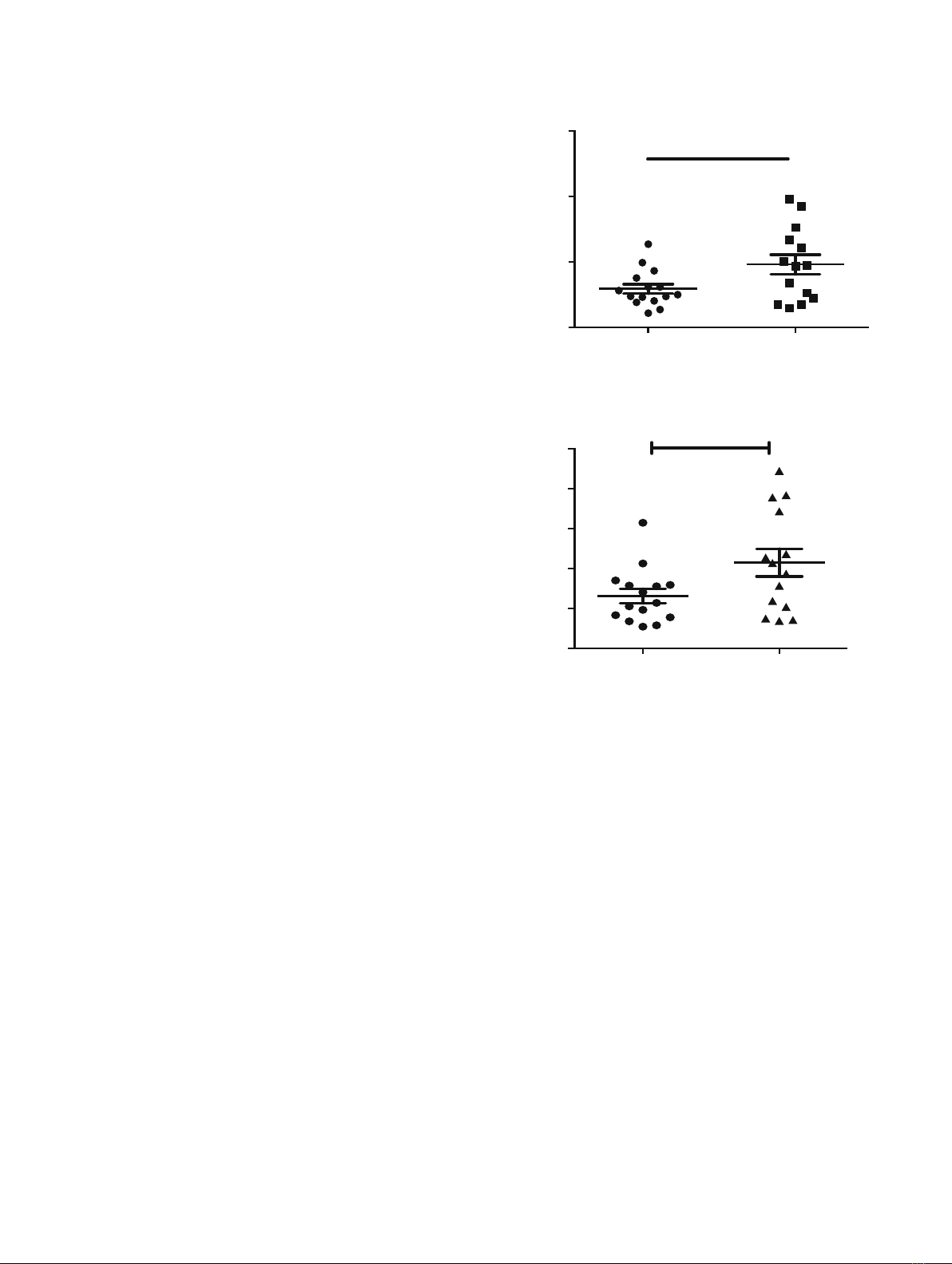
database (25) revealed that levels of RGS1 gene expression
are higher in MS patients (Fig. 1) and are induced in response
to interferon (IFN)-γtherapy in early treatment on day 1
(NCBI GEO database, accession number GDS2419). Tran et
al. reported that IFN-γinduced RGS1 mRNA and protein
expression in PBMC from human MS patients as early as 4 h
after treatment (26). However, the role of RGS1 in the onset
or progression of MS has never been explored.
RGS1 is expressed in lymphocytes, dendritic cells, mono-
cytes (27), and microglia (28). In B lymphocytes, RGS1 impairs
Gαi signaling responses (29) and its silencing enhances respon-
siveness to chemokines such as chemokine (C-X-C motif) ligand
(CXCL)12 and CXCL13 and impairs desensitization (30).
RGS1 overexpression inhibits T cell migration in response to
chemokines that control lymphoid homing, while depletion of
RGS1 selectively enhances such chemotaxis. These chemokines
include chemokine (C-C motif) ligand 19 (CCL19) and CXCL12
in gut T cells (31). CCL19 is mediated by chemokine (C-C motif)
receptor 7 (CCR7), and CXCL12 is mediated by chemokine (C-
X-C motif) receptor 4 (CXCR4) (31) which both promote T cell
egress from tissues to lymph nodes. The high level of RGS1 in
human T cells in the gut especially in inflammatory bowel
disease (IBD) suggests that RGS1 may contribute to the
colitogenic potential of T cells via limiting gut T cell response
to chemokines (31).
It has been shown that chemokine receptor signaling
plays an important role in disease progression in mouse
experimental autoimmune encephalitis (EAE), the most
commonly used murine model of MS. The key chemokines
that are overproduced at the site of active MS lesions are
CXCL10, CCL3, CCL4, CCL2, CCL8, and CCL5 (32) all of
which are important for lymphocyte recruitment to the CNS.
It will be particularly interesting to explore the role of the
microglial response to chemokines that become elevated in
Control MS patient
0
100
200
300
400
500
*
RGS1 (NM_002922)
Control MS patient
0
50
100
150 *
GC-RMA normalized signal intensity
(Affimatrix array)
GC-RMA normalized signal intensity
(Affimatrix array)
RGS1 (S59049)
Fig. 1. RGS1 mRNA expression level is higher in peripheral blood
mononuclear cells (PBMCs) of MS patients. Microarray experiments
identify genes and pathways involved in MS pathogenesis (data
accessible at the NCBI GEO database, accession number GSE21942).
PBMCs were isolated from the whole blood from 12 MS patients and 15
controls, and total RNA was extracted (22). . GC-RMA refers Guanine
Cytosine Robust Multi-Array Analysis. *p<0.05, Student's t test.
546 Lee and Bou Dagher

MS. Microglia are the myeloid-derived brain-resident macro-
phages of the CNS that are responsible for immune surveil-
lance and have been proposed to have regulatory and effector
functions in disease progression during MS (33). Microglia
activation has been shown to correlate with demyelination
and occurs in early pre-demyelinating lesions (34–36). Abla-
tion of microglia suppresses onset and severity of EAE (37–
39). However, it has been difficult to determine the role of
microglia in the onset and progression of MS in part because
they are also likely to play a beneficial role in the termination
of the inflammatory response (40). Microglia express various
chemokine receptors including those present in active human
MS lesions such as CCR1, CCR2, CCR3, CCR5, and CCR8
(41). Chemokines regulate leukocyte trafficking during in-
flammatory responses. In addition to their role in immune cell
trafficking, chemokines are known to have other functional
roles including induction of phagocytosis, modulation of cell
adhesion, cytokine activation, cell activation, and apoptosis
(42). It has been shown that chemokine receptor signaling
plays an important role in mouse EAE progression. For
example, it has been shown that blockade or genetic deletion
of CCR2 (which is involved in monocyte and T cell
trafficking) prevents severe disease and can lead to faster
remission in the mouse EAE model (43). Many of the RGS
proteins have been shown to desensitize (limit the signal of) a
variety of chemotactic receptors that are G-protein-coupled
receptors. Given that RGS1 is the most abundant RGS
protein in microglia, it may also play a role in microglia
chemotaxis which is yet to be addressed.
RGS10 AND MS
RGS10 is a 20-kDa protein that selectively accelerates
the GTPase activity of Gαi3, Gαq, and Gαz(
44). RGS10
belongs to the R12 subfamily and is highly expressed in the
brain, thymus, and lymph nodes (3,44–46). Burgon et al.
demonstrated that RGS10 can be phosphorylated at Ser168
by the cAMP-dependent protein kinase A (PKA), which is
required for its nuclear translocation (47), implicating a non-
canonical role of RGS10. RGS10 is also one of the most
highly expressed RGS proteins in mouse lymphocytes (14).
Garcia-Bernal et al. reported that RGS10 modulates
CXCL12-induced T cell adhesion through integrin α4β1
(48). They showed that RGS10 could function as an inhibitor
of CXCL12-induced T cell adhesion, which is mediated by
α4β1 and αLβ2(
48). Integrin α4 is known as a critical
molecule for the homing of lymphocytes to the CNS (49).
Currently, a humanized antibody that blocks integrin α4,
natalizumab, has been approved for treatment of MS (50).
These suggest a potential role of RGS10 in lymphocyte
adhesion and/or migration. Interestingly, the mRNA level of
RGS10 is higher in PBMC from patients with MS (NCBI
GEO database, accession number GDS2419) (23), Crohn’s
disease, and ulcerative colitis (NCBI GEO database, acces-
sion number GDS1615) (24).
The pathogenesis of MS is far more complicated than it
had been predicted from early studies of the EAE model
system. However, it is now well documented from studies of
human MS patients as well as animal models that autoim-
mune T cells mediate the initial stages of MS lesions similar to
the EAE model (51–53). From our arrays of experiments, we
showed that RGS10-null mice immunized with myelin
oligodendrocyte glycoprotein peptide fragment 35–55
(MOG
35–55
) displayed significantly milder clinical EAE in
the acute phase and remained mild all throughout the
progressive phases of the disease (54). Moreover, they
displayed significantly fewer incidences of EAE with a
delayed onset and a lower mean maximum score (54). These
data suggest the role for RGS10 in augmenting instead of
attenuating autoimmune response through autoreactive Th1
response. In-depth investigations are needed to establish how
RGS10 mediates lymphocyte effector function to augment
autoimmune responses. If RGS10 plays a role in specific
subsets of autoreactive lymphocytes during EAE, this may
enable the targeting of RGS10 in disease-modifying
strategies.
RGS10 IN NEUROINFLAMMATION AND
PARKINSON’S DISEASE
In brain-resident microglia, RGS10 has been shown to
play a critical role in inflammatory microglial activation via
negative regulation of NF-kB signaling. In dopaminergic
neuroblastoma cells, RGS10 modulates sensitivity to
inflammation-induced cell survival by interaction with the
PKA/CREB pathway (55–57). Chronic peripheral adminis-
tration of lipopolysaccharide (LPS) in RGS10-null mice
results in chronic microgliosis and loss of dopaminergic
(DA) neurons (57). RGS10-null mice displayed increased
microglial burden in the CNS and dysregulated
inflammation-related gene expression in microglia as well
as nigral DA neuron degeneration with repeated systemic
administration of low-dose LPS injection. Moreover, RGS10-
null microglia produced significantly higher levels of proin-
flammatory cytokines including TNF, IL-1β, and IL-6 than
wild-type (WT) microglia upon LPS treatment. Another
study showed that RGS10-null mice exhibited impaired
osteoclast differentiation due to the absence of RGS10-
dependent calcium current oscillations and the loss of
nuclear factor of activated T cell c1 (NFATc1) expression
(58). This suggests that RGS10 may regulate intracellular
calcium oscillations in microglia. The primary function of
RGS proteins is believed to be the regulation of
heterotrimeric G-protein signaling at the plasma membrane.
However, our findings as well as those of others (47,59,60)
reveal that RGS proteins translocate to the nucleus and are
found at high abundance at other intracellular sites. Given
that the cellular distribution of RGS10 is both nuclear and
cytoplasmic (59), it would be interesting to study the possible
function of RGS10 in regulating microglial stress responses
through mechanisms that include changes in gene transcrip-
tion and cellular calcium regulation in addition to its
GTPase-accelerating protein (GAP) activity at the plasma
membrane. A better understanding of the normal function of
RGS10 in the CNS along with proof-of-concept studies to
demonstrate that manipulation of RGS10 levels and/or its
activity in the ventral midbrain has positive effects on DA
neuron survival without exerting unwanted effects on other
cell types may reveal its potential as a therapeutic target for
blocking or delaying the progressive loss of nigrostriatal DA
neurons in PD.
547Drug Targets for Neuroinflammatory and Neurodegenerative Diseases

In conclusion, it is possible that an increase in the RGS
activity, especially RGS10, would be beneficial in PD-
associated neuroinflammation but detrimental in the treat-
ment of MS. However, identifying novel modulators and their
mechanism will open up the possibility for the development
of new mechanism-based therapeutic interventions for the
treatment of neurodegenerative diseases.
REFERENCES
1. Neves SR, Ram PT, Iyengar R. G protein pathways. Science.
2002;296(5573):1636–9.
2. Berman DM, Wilkie TM, Gilman AG. GAIP and RGS4 are
GTPase-activating proteins for the Gi subfamily of G protein
alpha subunits. Cell. 1996;86(3):445–52.
3. Ross EM, Wilkie TM. GTPase-activating proteins for
heterotrimeric G proteins: regulators of G protein signaling
(RGS) and RGS-like proteins. Annu Rev Biochem.
2000;69:795–827.
4. Siderovski DP, Diverse-Pierluissi M, De Vries L. The GoLoco
motif: a Galphai/o binding motif and potential guanine-
nucleotide exchange factor. Trends Biochem Sci.
1999;24(9):340–1.
5. Zheng B, De Vries L, Gist Farquhar M. Divergence of RGS
proteins: evidence for the existence of six mammalian RGS
subfamilies. Trends Biochem Sci. 1999;24(11):411–4.
6. Willars GB. Mammalian RGS, proteins: multifunctional regulators of
cellular signalling. Semin Cell Dev Biol. 2006;17(3):363–76.
7. Abramow-Newerly M, Roy AA, Nunn C, Chidiac P. RGS
proteins have a signalling complex: interactions between RGS
proteins and GPCRs, effectors, and auxiliary proteins. Cell
Signal. 2006;18(5):579–91.
8. Hollinger S, Hepler JR. Cellular regulation of RGS proteins:
modulators and integrators of G protein signaling. Pharmacol
Rev. 2002;54(3):527–59.
9. Kach J, Sethakorn N, Dulin NO. A finer tuning of G-protein
signaling through regulated control of RGS proteins. Am J
Physiol Heart Circ Physiol. 2012;303(1):H19–35.
10. Sethakorn N, Yau DM, Dulin NO. Non-canonical functions of
RGS proteins. Cell Signal. 2010;22(9):1274–81.
11. Dohlman HG, Thorner J. RGS proteins and signaling by
heterotrimeric G proteins. J Biol Chem. 1997;272(7):3871–4.
12. Koelle MR. A new family of G-protein regulators—the RGS
proteins. Curr Opin Cell Biol. 1997;9(2):143–7.
13. Larminie C, Murdock P, Walhin JP, Duckworth M, Blumer KJ,
Scheideler MA, et al. Selective expression of regulators of G-
protein signaling (RGS) in the human central nervous system.
Brain Res Mol Brain Res. 2004;122(1):24–34.
14. Moratz C, Harrison K, Kehrl JH. Regulation of chemokine-
induced lymphocyte migration by RGS proteins. Methods
Enzymol. 2004;389:15–32.
15. Steinman L, Merrill JT, McInnes IB, Peakman M. Optimization
of current and future therapy for autoimmune diseases. Nat
Med. 2012;18(1):59–65.
16. Handel AE, Handunnetthi L, Giovannoni G, Ebers GC,
Ramagopalan SV. Genetic and environmental factors and the
distribution of multiple sclerosis in Europe. Eur J Neurol.
2010;17(9):1210–4.
17. Du C, Xie X. G protein-coupled receptors as therapeutic targets
for multiple sclerosis. Cell Res. 2012;22(7):1108–28.
18. International Multiple Sclerosis Genetics Consortium (IMSGC).
IL12A, MPHOSPH9/CDK2AP1 and RGS1 are novel multiple
sclerosis susceptibility loci. Genes Immun. 2010;11(5):397–405.
19. Gourraud PA. When is the absence of evidence, evidence of
absence? Use of equivalence-based analyses in genetic epidemi-
ology and a conclusion for the KIF1B rs10492972*C allelic
associationinmultiplesclerosis. Genet Epidemiol.
2011;35(6):568–71.
20. Hunt KA, Zhernakova A, Turner G, Heap GA, Franke L,
Bruinenberg M, et al. Newly identified genetic risk variants for
celiac disease related to the immune response. Nat Genet.
2008;40(4):395–402.
21. Smyth DJ, Plagnol V, Walker NM, Cooper JD, Downes K, Yang
JH, et al. Shared and distinct genetic variants in type 1 diabetes
and celiac disease. N Engl J Med. 2008;359(26):2767–77.
22. Johnson BA, Wang J, Taylor EM, Caillier SJ, Herbert J, Khan
OA, et al. Multiple sclerosis susceptibility alleles in African
Americans. Genes Immun. 2010;11(4):343–50.
23. Kemppinen AK, Kaprio J, Palotie A, Saarela J. Systematic
review of genome-wide expression studies in multiple sclerosis.
BMJ Open. 2011;1(1), e000053.
24. Burczynski ME, Peterson RL, Twine NC, Zuberek KA, Brodeur
BJ, Casciotti L, et al. Molecular classification of Crohn’s disease
and ulcerative colitis patients using transcriptional profiles in
peripheral blood mononuclear cells. J Mol Diagn. 2006;8(1):51–
61.
25. Singh MK, Scott TF, LaFramboise WA, Hu FZ, Post JC, Ehrlich
GD. Gene expression changes in peripheral blood mononuclear
cells from multiple sclerosis patients undergoing beta-interferon
therapy. J Neurol Sci. 2007;258(1–2):52–9.
26. Tran T, Paz P, Velichko S, Cifrese J, Belur P, Yamaguchi KD, et
al. Interferonβ-1b induces the expression of RGS1 a negative
regulator of G-protein signaling. Int J Cell Biol.
2010;2010:529376.
27. Bansal G, Druey KM, Xie Z. R4 RGS proteins: regulation of G-
protein signaling and beyond. Pharmacol Ther. 2007;116(3):473–95.
28. Atwood BK, Lopez J, Wager-Miller J, Mackie K, Straiker A.
Expression of G protein-coupled receptors and related proteins
in HEK293, AtT20, BV2, and N18 cell lines as revealed by
microarray analysis. BMC Genomics. 2011;12:14.
29. Moratz C, Kang VH, Druey KM, Shi CS, Scheschonka A,
Murphy PM, et al. Regulator of G protein signaling 1 (RGS1)
markedly impairs Gi alpha signaling responses of B lymphocytes.
J Immunol. 2000;164(4):1829–38.
30. Han JI, Huang NN, Kim DU, Kehrl JH. RGS1 and RGS13
mRNA silencing in a human B lymphoma line enhances
responsiveness to chemoattractants and impairs desensitization.
J Leukoc Biol. 2006;79(6):1357–68.
31. Gibbons DL, Abeler-Dorner L, Raine T, Hwang IY, Jandke A,
Wencker M, et al. Cutting edge: regulator of G protein signaling-
1 selectively regulates gut T cell trafficking and colitic potential. J
Immunol. 2011;187(5):2067–71.
32. Cartier L, Hartley O, Dubois-Dauphin M, Krause KH. Chemo-
kine receptors in the central nervous system: role in brain
inflammation and neurodegenerative diseases. Brain Res Brain
Res Rev. 2005;48(1):16–42.
33. Sanders P, De Keyser J. Janus faces of microglia in multiple
sclerosis. Brain Res Rev. 2007;54(2):274–85.
34. Marik C, Felts PA, Bauer J, Lassmann H, Smith KJ. Lesion
genesis in a subset of patients with multiple sclerosis: a role for
innate immunity? Brain. 2007;130(Pt 11):2800–15.
35. Barnett MH, Prineas JW. Relapsing and remitting multiple
sclerosis: pathology of the newly forming lesion. Ann Neurol.
2004;55(4):458–68.
36. Vos CM, Geurts JJ, Montagne L, van Haastert ES, Bo L, van der
Valk P, et al. Blood-brain barrier alterations in both focal and
diffuse abnormalities on postmortem MRI in multiple sclerosis.
Neurobiol Dis. 2005;20(3):953–60.
37. Heppner FL, Greter M, Marino D, Falsig J, Raivich G, Hovelmeyer
N, et al. Experimental autoimmune encephalomyelitis repressed by
microglial paralysis. Nat Med. 2005;11(2):146–52.
38. Greter M, Heppner FL, Lemos MP, Odermatt BM, Goebels N,
Laufer T, et al. Dendritic cells permit immune invasion of the
CNS in an animal model of multiple sclerosis. Nat Med.
2005;11(3):328–34.
39. McMahon EJ, Bailey SL, Castenada CV, Waldner H, Miller SD.
Epitope spreading initiates in the CNS in two mouse models of
multiple sclerosis. Nat Med. 2005;11(3):335–9.
40. Chan A, Seguin R, Magnus T, Papadimitriou C, Toyka KV,
Antel JP, et al. Phagocytosis of apoptotic inflammatory cells by
microglia and its therapeutic implications: termination of CNS
autoimmune inflammation and modulation by interferon-beta.
Glia. 2003;43(3):231–42.
41. Balashov KE, Rottman JB, Weiner HL, Hancock WW. CCR5(+)
and CXCR3(+) T cells are increased in multiple sclerosis and
548 Lee and Bou Dagher

their ligands MIP-1alpha and IP-10 are expressed in demyelin-
ating brain lesions. Proc Natl Acad Sci U S A. 1999;96(12):6873–
8.
42. Matejuk A, Dwyer J, Ito A, Bruender Z, Vandenbark AA,
Offner H. Effects of cytokine deficiency on chemokine expres-
sion in CNS of mice with EAE. J Neurosci Res. 2002;67(5):680–
8.
43. Gaupp S, Pitt D, Kuziel WA, Cannella B, Raine CS. Experi-
mental autoimmune encephalomyelitis (EAE) in CCR2(−/−)
mice: susceptibility in multiple strains. Am J Pathol.
2003;162(1):139–50.
44. Hunt TW, Fields TA, Casey PJ, Peralta EG. RGS10 is a selective
activator of G alpha i GTPase activity. Nature. 1996;383(6596):
175–7.
45. Gold SJ, Ni YG, Dohlman HG, Nestler EJ. Regulators of G-
protein signaling (RGS) proteins: region-specific expression of
nine subtypes in rat brain. J Neurosci. 1997;17(20):8024–37.
46. Sierra DA, Gilbert DJ, Householder D, Grishin NV, Yu K,
Ukidwe P, et al. Evolution of the regulators of G-protein
signaling multigene family in mouse and human. Genomics.
2002;79(2):177–85.
47. Burgon PG, Lee WL, Nixon AB, Peralta EG, Casey PJ.
Phosphorylation and nuclear translocation of a regulator of G
protein signaling (RGS10). J Biol Chem. 2001;276(35):32828–34.
48. Garcia-Bernal D, Dios-Esponera A, Sotillo-Mallo E, Garcia-
Verdugo R, Arellano-Sanchez N, Teixido J. RGS10 restricts
upregulation by chemokines of T cell adhesion mediated by
α4β1 and αLβ2 integrins. J Immunol. 2011;187(3):1264–72.
49. Yednock TA, Butcher EC, Stoolman LM, Rosen SD. Receptors
involved in lymphocyte homing: relationship between a
carbohydrate-binding receptor and the MEL-14 antigen. J Cell
Biol. 1987;104(3):725–31.
50. Polman CH, O’Connor PW, Havrdova E, Hutchinson M, Kappos
L, Miller DH, et al. A randomized, placebo-controlled trial of
natalizumab for relapsing multiple sclerosis. N Engl J Med.
2006;354(9):899–910.
51. Hartung HP, Gonsette R, Konig N, Kwiecinski H, Guseo A,
Morrissey SP, et al. Mitoxantrone in progressive multiple
sclerosis: a placebo-controlled, double-blind, randomised,
multicentre trial. Lancet. 2002;360(9350):2018–25.
52. McFarland HF, Martin R. Multiple sclerosis: a complicated
picture of autoimmunity. Nat Immunol. 2007;8(9):913–9.
53. Coles AJ, Wing MG, Molyneux P, Paolillo A, Davie CM, Hale
G, et al. Monoclonal antibody treatment exposes three mecha-
nisms underlying the clinical course of multiple sclerosis. Ann
Neurol. 1999;46(3):296–304.
54. Lee JK, Kannarkat GT, Chung J, Lee HJ, Graham KL, Tansey
MG. RGS10 deficiency ameliorates the severity of disease in
experimental autoimmune encephalomyelitis. J Neuroinflamma-
tion. 2016;13(1):24.
55. Lee JK, Chung J, Druey KM, Tansey MG. RGS10 exerts a
neuroprotective role through the PKA/c-AMP response-element
(CREB) pathway in dopaminergic neuron-like cells. J
Neurochem. 2012;122(2):333–43.
56. Lee JK, Chung J, McAlpine FE, Tansey MG. Regulator of G-
protein signaling-10 negatively regulates NF-{kappa}B in microg-
lia and neuroprotects dopaminergic neurons in
hemiparkinsonian rats. J Neurosci. 2011;31(33):11879–88.
57. Lee JK, McCoy MK, Harms AS, Ruhn KA, Gold SJ, Tansey
MG. Regulator of G-protein signaling 10 promotes dopaminergic
neuron survival via regulation of the microglial inflammatory
response. J Neurosci. 2008;28(34):8517–28.
58. Yang S, Li YP. RGS10-null mutation impairs osteoclast differ-
entiation resulting from the loss of [Ca2+]i oscillation regulation.
Genes Dev. 2007;21(14):1803–16.
59. Chatterjee TK, Fisher RA. Cytoplasmic, nuclear, and Golgi
localization of RGS proteins. Evidence for N-terminal and RGS
domain sequences as intracellular targeting motifs. J Biol Chem.
2000;275(31):24013–21.
60. Waugh JL, Lou AC, Eisch AJ, Monteggia LM, Muly EC, Gold
SJ. Regional, cellular, and subcellular localization of RGS10 in
rodent brain. J Comp Neurol. 2005;481(3):299–313.
549Drug Targets for Neuroinflammatory and Neurodegenerative Diseases


![Bài giảng Độc học môi trường Phần 2: [Thêm nội dung cụ thể của bài giảng để tối ưu SEO]](https://cdn.tailieu.vn/images/document/thumbnail/2026/20260424/vispacex_27/135x160/16741777445751.jpg)


