
Review Article
Theme: Heterotrimeric G Protein-based Drug Development: Beyond Simple Receptor Ligands
Guest Editor: Shelley Hooks
RGS6 as a Novel Therapeutic Target in CNS Diseases and Cancer
Katelin E. Ahlers,
1
Bandana Chakravarti,
1
and Rory A. Fisher
1,2,3
Received 13 November 2015; accepted 25 February 2016; published online 22 March 2016
Abstract. Regulator of G protein signaling (RGS) proteins are gatekeepers regulating the cellular
responses induced by G protein-coupled receptor (GPCR)-mediated activation of heterotrimeric G
proteins. Specifically, RGS proteins determine the magnitude and duration of GPCR signaling by acting
as a GTPase-activating protein for Gαsubunits, an activity facilitated by their semiconserved RGS
domain. The R7 subfamily of RGS proteins is distinguished by two unique domains, DEP/DHEX and
GGL, which mediate membrane targeting and stability of these proteins. RGS6, a member of the R7
subfamily, has been shown to specifically modulate Gα
i/o
protein activity which is critically important in
the central nervous system (CNS) for neuronal responses to a wide array of neurotransmitters. As such,
RGS6 has been implicated in several CNS pathologies associated with altered neurotransmission,
including the following: alcoholism, anxiety/depression, and Parkinson’s disease. In addition, unlike other
members of the R7 subfamily, RGS6 has been shown to regulate G protein-independent signaling
mechanisms which appear to promote both apoptotic and growth-suppressive pathways that are
important in its tumor suppressor function in breast and possibly other tissues. Further highlighting the
importance of RGS6 as a target in cancer, RGS6 mediates the chemotherapeutic actions of doxorubicin
and blocks reticular activating system (Ras)-induced cellular transformation by promoting degradation of
DNA (cytosine-5)-methyltransferase 1 (DNMT1) to prevent its silencing of pro-apoptotic and tumor
suppressor genes. Together, these findings demonstrate the critical role of RGS6 in regulating both G
protein-dependent CNS pathology and G protein-independent cancer pathology implicating RGS6 as a
novel therapeutic target.
KEY WORDS: alcoholism; depression; doxorubicin; Parkinson’s disease; RGS protein.
INTRODUCTION
G protein-coupled receptors (GPCRs) are involved in
virtually every known physiological process, and dysfunction
in their signaling is linked to many human diseases. GPCRs
become active in response to extracellular agonist binding
which induces conformational changes in the receptor pro-
moting its association with heterotrimeric G proteins (1),
consisting of three functional subunits: the GDP/GTP-binding
αsubunit, and the βand γsubunits. Agonist-activated
GPCRs function as GTP exchange factors (GEFs) for Gα
subunits, promoting exchange of GDP for GTP and resulting
in Gαsubunit activation and dissociation from Gβγ subunits,
with both Gα-GTP and Gβγ activating downstream signaling
pathways (2). Four families of Gαsubunits, Gα
i
,Gα
s
,Gα
q
,
and Gα
12
, that exhibit selectivity in terms of their coupling to
GPCRs and their downstream signaling actions, contribute in
part to the signaling specificity of different GPCRs. The
intrinsic GTPase activity of Gαsubunits is responsible for
hydrolysis of GTP, reformation of inactive Gα-GDP subunits
and their reassociation with Gβγ, effectively terminating both
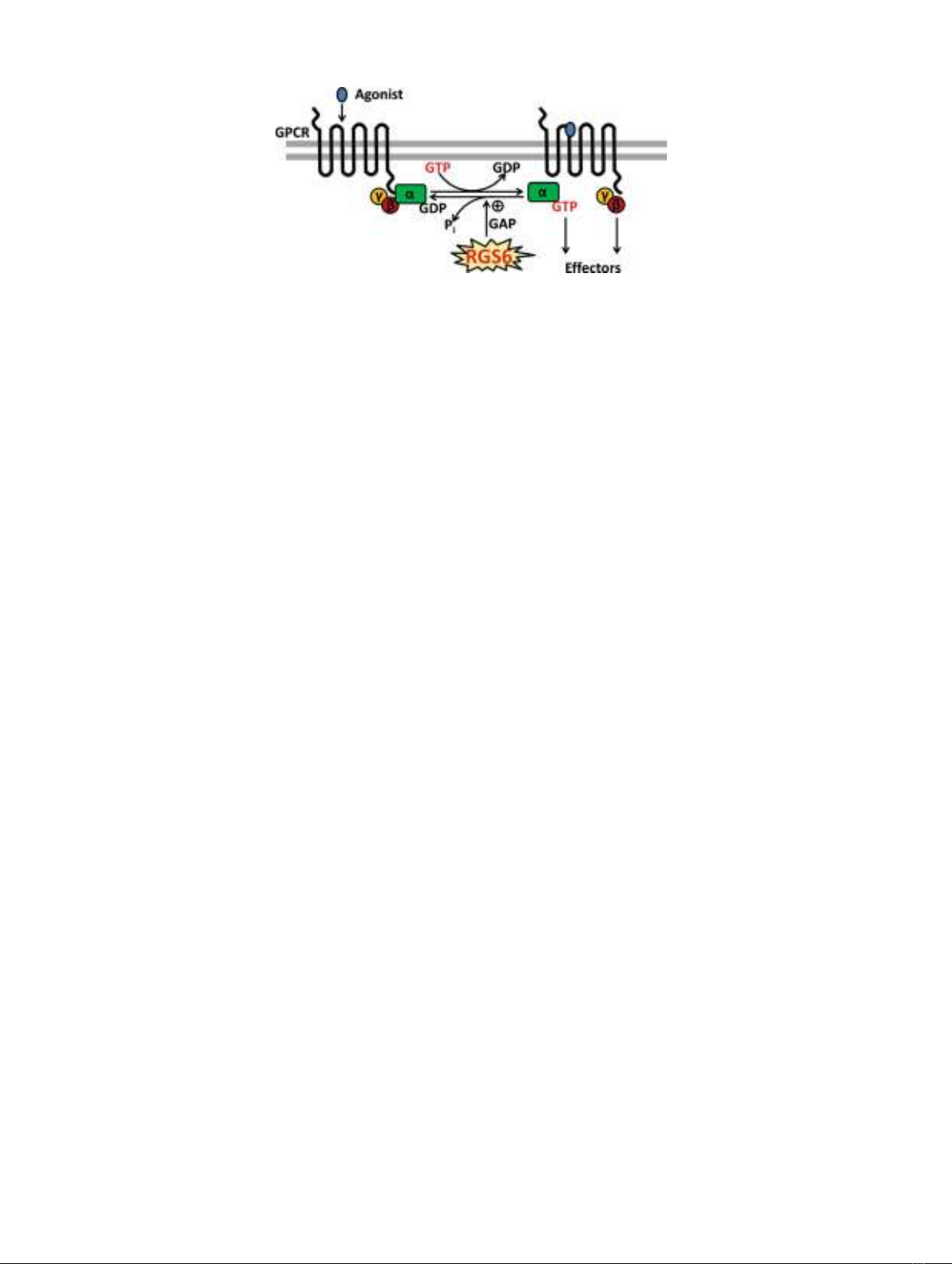
Gαand Gβγ signaling. Regulator of G protein signaling
(RGS) proteins act as GTPase-activating proteins (GAPs) for
Gαsubunits by stabilizing the transition state of the GTP
hydrolysis reaction by Gαsubunits. Therefore, RGS proteins
play a critical role in regulating the duration and magnitude
of signaling initiated by GPCRs by serving as gatekeepers of
signaling mediated by G protein Gαand Gβγ subunits (3–6)
(Fig. 1).
There are 20 canonical mammalian RGS proteins that
have been divided into four subfamilies based upon sequence
homology and protein domain structure. RGS6 is a member
of the R7 subfamily (RGS6, RGS7, RGS9, RGS11) of RGS
proteins that shares two unique domains outside of the RGS
domain (common to all RGS proteins): the disheveled EGL-
10, pleckstrin homology (DEP)/DEP helical extension
(DHEX) domain and the G gamma subunit-like (GGL)
domain (Fig. 2). Together, these three domains modulate
1
Department of Pharmacology, The Roy J. and Lucille A. Carver
College of Medicine, University of Iowa, 2-505 Bowen Science
Building, Iowa City, Iowa 52242, USA.
2
Department of Internal Medicine, The Roy J. and Lucille A. Carver
College of Medicine, University of Iowa, Iowa City, Iowa 52242,
USA.
3
To whom correspondence should be addressed. (e-mail: rory-
fisher@uiowa.edu)
The AAPS Journal, Vol. 18, No. 3, May 2016 ( #2016)
DOI: 10.1208/s12248-016-9899-9
5601550-7416/16/0300-0560/0 #2016 American Association of Pharmaceutical Scientists
RGS6 protein stability, localization, and function. In consid-
ering RGS6 protein stability, interaction of the GGL domain
and the atypical Gβsubunit, Gβ
5
, is a general requirement
for stabilization of the whole R7 protein subfamily (8–10). As
such, genetic ablation of the Gβ
5
gene (GNB5) is correlated
with the loss of the R7 protein subfamily in the retina and
striatum (11). However, the ability of Gβ
5
to stabilize RGS6
may not be solely dependent on its interaction with the GGL
domain, but may require a direct interaction with its DEP/
DHEX domain as well. In evidence of this, Gβ
5
has also been
shown (via crystal structure and pull-down experiments) to
interact with the DEP/DHEX domain of the R7 family
members RGS7 and RGS9, and mutation of Gβ
5
residues
mediating this interaction leads to the instability of both RGS
proteins (12–14). In addition to promoting protein stability,
both the GGL and DEP/DHEX domains are also important
for modulating RGS6 cellular localization. Experiments in
which COS-7 cells were transfected with GFP-tagged RGS6
splice variants demonstrated that the GGL domain promotes
cytoplasmic retention of RGS6. However, when the GGL
domain is lost due to alternative splicing (−GGL variants,
Figs. 2and 3), or when Gβ
5
is overexpressed to generate
RGS6:Gβ
5
complexes, GFP-tagged RGS6 moves into the
nucleus (7). Similarly, the DEP/DHEX domain also regulates
cytoplasmic-nuclear shuttling of RGS6. Indeed, further
experiments looking at the subcellular localization GFP-
tagged RGS6 protein variants in COS-7 cells demonstrated
that the RGS6 splice variants containing the DEP/DHEX
domain (RGS6 long (RGS6L) variants, Figs. 2and 3) were
largely cytoplasmic, whereas those lacking the domain (RGS6
short (RGS6S) variants, Figs. 2and 3) were primarily nuclear
(7). It is believed that this shuttling may in part be due to a
DEP/DHEX-mediated interaction of RGS6 with R7 family-
binding protein (R7BP) as it has been shown that R7BP is
reversibly palmitoylated promoting either a membrane
(palmitoylated) or nuclear (depalmitoylated) distribution of
another R7 family member, RGS7 (15). This differential
subcellular localization of RGS6 appears to be functionally
relevant as it can also be seen in native tissues. For example,
immunohistochemical analysis of RGS6 protein localization
in the mouse cerebellum, using an antibody that the Fisher
laboratory generated against the N-terminal protein domain,
common among all RGS6L isoforms, demonstrated that
RGS6L has distinct cytoplasmic and nuclear localization
patterns (7). In further support of the functional relevance
of this differential subcellular localization, other R7 family
members, in particular RGS7 and RGS9, as well as Gβ
5
have
also been shown to have both distinct cytoplasmic and
nuclear localization patterns (16–19). Finally, in terms of
RGS6 function in negatively regulating heterotrimeric G
protein signaling, the RGS domain is responsible for the
GAP activity of RGS6, and other RGS proteins, and allows it
to negatively regulate Gα
i/o
proteins (20). RGS6 specific
modulation of Gα
i/o
protein activity has been implicated in
the regulation of several disease states, particularly in the
central nervous system (CNS), including the following:
alcoholism (21), anxiety/depression (22), Parkinson’s disease
(23), and potentially Alzheimer’s disease (24), schizophrenia
(25), and vision (26). However, RGS6 is also unique in that it
remains the only member of the R7 protein family that has
been demonstrated to regulate G protein-independent path-
ways, as evidenced by its compelling pro-apoptotic and tumor
suppressor actions in cancer (27–30).
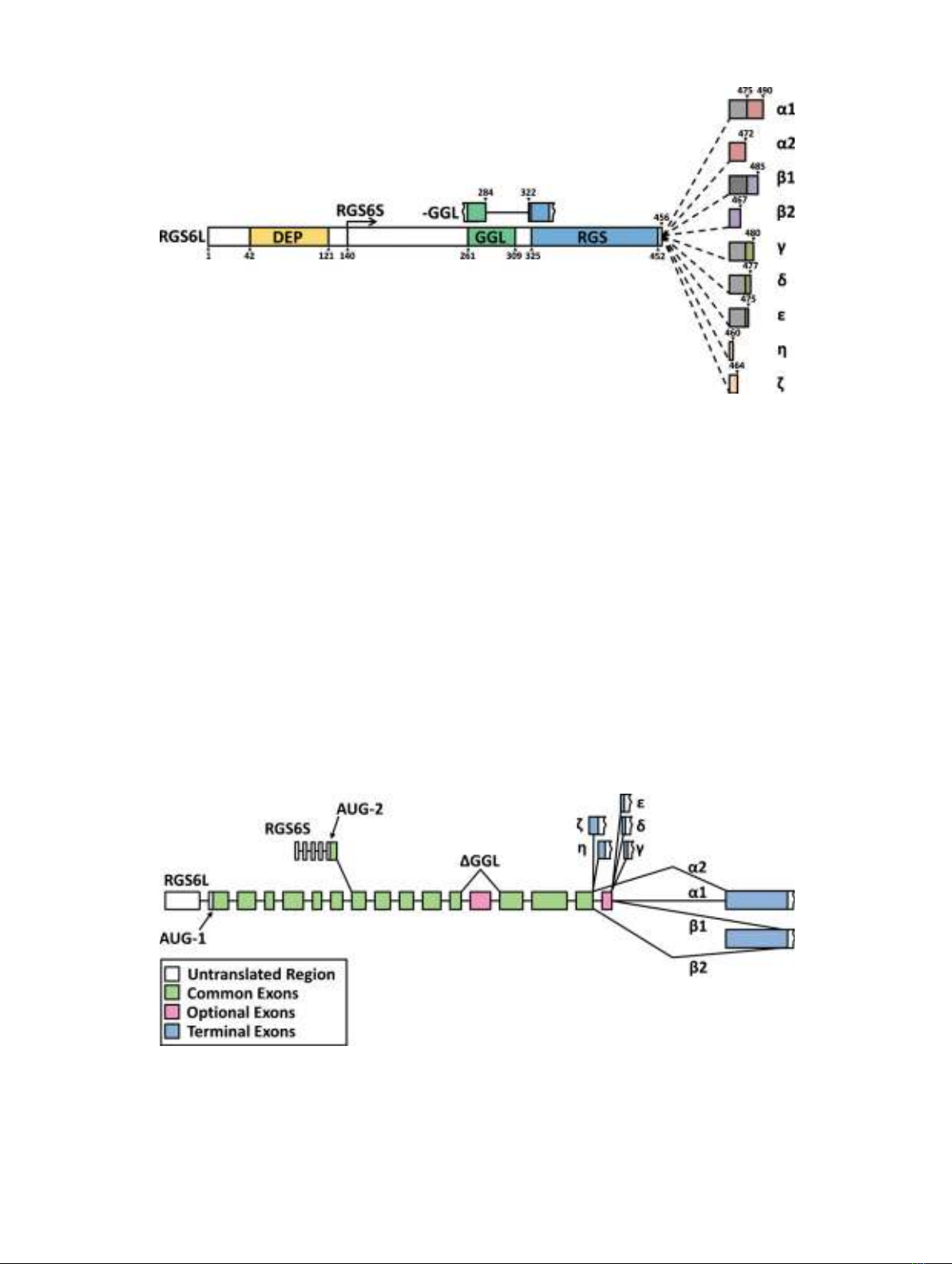
Potentially key to RGS6’s G protein-independent
signaling, as well as its modulation of G protein signaling,
are previously unidentified domains present in a subset of
RGS6 proteins. These domains may arise via alternative
splicing of RGS6 messenger RNA (mRNA) transcripts. In
support of this idea, when the Fisher laboratory first
cloned RGS6 from a Marathon-ready human brain cDNA
library (brain tissue is where RGS6 is most highly
expressed at the mRNA (31) and protein level (Fisher
Laboratory, unpublished)), they described 36 distinct
isoforms that could arise through complex splicing of two
primaryRGS6transcripts(
7)(Fig.3).These36distinct
splice forms are predicted to produce 18 long isoforms
(RGS6L) ranging from ∼49 to 56 kDa in size and 18 short
isoforms (RGS6S) ranging from 32 to 40 kDa. While the
various RGS6L and RGS6S splice forms are largely
similar in sequence, making it difficult to develop anti-
bodies to confirm their individual existence and determine
their individual function, the Fisher laboratory has had
some success in characterizing the proteins resulting from
Fig. 1. Regulation of G protein-coupled receptor (GPCR) signaling by
regulator of G protein signaling (RGS) proteins. RGS proteins act as
GTPase-activating proteins (GAPs) for specificGαsubunits and
thereby function to terminate GPCR signaling
561RGS6’s Role in CNS Diseases and Cancer
several of these splice variants. As mentioned earlier,
characterization of the differential subcellular localization
for multiple GFP-tagged RGS6 protein isoforms in COS-7
cellsdemonstratedthatanalterationinRGS6protein
structure can dictate whether the protein is primarily
localized in the cytoplasm (RGS6L and +GGL protein
isoforms) or nucleus (RGS6S and −GGL protein iso-
forms), suggesting that alternatively spliced RGS6 tran-
scripts may result in proteins with unique functions, and
indeed such differential localization of RGS6L was also
seen in native tissues (7,32). The Fisher lab has also
demonstrated using western blot that certain tissues
express multiple distinct RGS6 protein isoforms natively. For
example, the brain expresses at least two distinct RGS6 isoforms
that are larger (∼61 and 69 kDa) than ubiquitously expressed
smaller forms of the protein (21,33). Interestingly, western
blot analysis of brain tissue lysates using the antibody against the
N-terminal protein domain, common to all RGS6L proteins,
reveals a broad band of RGS6 immunoreactivity which
could be explained by the presence of multiple RGS6L
Fig. 2. Predicted protein structure of human RGS6 proteins. There are predicted to be numerous RGS6 protein
isoforms that differ in length due to the following: inclusion or exclusion of the disheveled EGL-10, pleckstrin
homology (DEP) domain at their N-terminus, inclusion or exclusion of a complete G gamma subunit-like (GGL)
domain, and in the inclusion of one of seven distinct C-termini. RGS6 proteins with either the long or the short N-
terminus are labeled as RGS6L or RGS6S, respectively. The C-terminal domains are labeled as α,β,γ,δ,ε,η, and
ζ. The αand βC-termini exist in two forms, either with (α1 and β1) or without (α2 and β2); an 18 amino acid
sequence encoded by exon 18 (grey square) of the RGS6 gene. Finally, proteins that lack the GGL domain are
designated as −GGL proteins. Amino acid numbers are included to specify where key regions of the protein begin
and end. Image adapted from reference (7)
Fig. 3. Diagram of the complex splicing of human RGS6 pre-mRNA to generate 36 splice variants. Two primary
transcripts encode the 5′-splice forms of RGS6; the AUG-1 start site produces a transcript that encodes the RGS6L
forms of the protein while the AUG-2 start site produces a transcript that encodes the RGS6S forms of the protein.
Retention or removal of exon 13 (first pink square) generates transcripts that encode for proteins containing or
lacking a complete GGL domain, respectively. 3′-splicing generates transcripts containing seven distinct 3′exons.
RGS6 αand βtranscripts exist in two forms that arise from either the retention (α1 and β1) or removal (α2 and β2)
of exon 18 (second pink square). Image adapted from reference (7)
562 Ahlers et al.

isoforms with different C-terminal domains and with or
without complete GGL domains (22,34). The functions for
these RGS6 variants and how they all arise (either through
protein modification or additional RNA splicing) are unknown.
RGS6 IN CNS DISEASES
Alcohol Use Disorders
Approximately 12% of the US population suffers from
alcoholism causing a substantial annual economic burden
(∼$223.5 billion). In light of these statistics, researchers
have sought to identify and understand the underlying
mechanisms of alcohol dependence, but have been met
with only limited success. As a result, there are few
therapeutic options available to reduce alcohol cravings
and withdrawal symptoms and there are no drugs that have
been approved to prevent/treat alcohol-related organ dam-
age. Part of the problem is that alcohol does not have a
specific molecular target in the brain, but instead induces
neuronal alterations in the mesolimbic pathway (implicated
in drug addiction (35–38)) by both inhibiting N-methyl-D-
aspartate (NMDA) receptor activity and enhancing gamma-
aminobutyric acid B (GABA
B
)receptoractivity(39).
Although alcohol disrupts mesolimbic neuronal signaling
via multiple mechanisms, the end result is an alteration in
neurotransmitter release. As the majority of neurotransmit-
ters in the mesolimbic pathway (e.g., dopamine (DA),
GABA, opioids, and serotonin (5-HT)) interact with GPCRs,
G protein-dependent signaling may offer a therapeutic target
in the treatment of alcohol abuse. With this in mind, multiple
drugs targeting these neurotransmitter receptors have been
recommended for the treatment of alcoholism (40–42). One
such drug, baclofen, a GABA
B
R agonist, has been approved
in Europe as a treatment for alcohol withdrawal symptoms
and cravings (43–45). However, despite the positive effects of
baclofen in the treatment of alcohol abuse, its use remains
limited as it compounds both the muscle relaxant and
sedative properties of alcohol.
In light of the fact that baclofen-mediated modulation
of the GABA
B
R is a viable treatment for alcoholism, RGS6
also became a protein of interest, as previous research had
demonstrated its ability to negatively regulate GABA
B
R
signaling in the cerebellum (33). In addition, there was also
evidence to suggest that RGS6 was capable of regulating
the signaling of other GPCRs, such as 5-HT
1A
Rs and μ-
opioid receptors (22,46), which had already been identified
as potential therapeutic targets in the treatment of alcohol-
ism (40,42). Both immunohistochemical and western blot
studies in wild type (RGS6+/+) mice subsequently demon-
strated that RGS6 protein expression was upregulated in
the ventral tegmental area (VTA) of the mesolimbic system
following prolonged alcohol exposure. Conversely, studies
performed in RGS6 knockout (RGS6−/−) mice established
that loss of RGS6 ameliorated not only alcohol seeking
behavior but also those behaviors associated with alcohol-
conditioned reward and withdrawal. Further inspection of
the RGS6−/−mice under control conditions revealed a
reduction in the striatal DA suggesting that RGS6 might
regulate DA production presynaptically, potentially through
its ability to inhibit GPCR signaling. In support of this
hypothesis, daily intraperitoneal (i.p.) administration of a
GABA
B
R antagonist, SCH-50911, or a dopamine 2 receptor
(D
2
R) antagonist, raclopride, was associated with an
increase in voluntary alcohol consumption in RGS6−/−
mice. Although it is not exactly clear how the GABA
B
Rs
and D
2
Rs regulate DA levels and thus alcohol seeking
behavior, it has been hypothesized that they may do so by
modulating the levels of the DA-synthesizing enzyme
tyrosine hydroxylase (TH), the vesicular monoamine trans-
porter 2 (VMAT2), and the dopamine transporter (DAT).
Indeed, levels of TH and VMAT2 mRNA were lower in the
VTA of RGS6−/−animals compared to RGS6+/+ mice
under basal conditions, and DAT mRNA levels were
upregulated in RGS6−/−mice following chronic alcohol
exposure. Furthermore, i.p. injection of RGS6−/−mice with
the DAT inhibitor GBR-12909 promoted voluntary alcohol
consumption in these mice to an even greater degree than
either of the GABA
B
RandD
2
R inhibitors (21). These
findings suggest that RGS6 inhibition of GPCR-mediated
signaling may prevent upregulation of DAT and assure the
normal synthesis and release of DA that is responsible for
alcohol reward behaviors (Fig. 4a).
The evidence presented thus far indicates that RGS6 is
critical for normal DA-mediated alcohol seeking behavior,
thus identifying it as a viable therapeutic target. However, the
advantages of RGS6 as a therapeutic target may not only
reside in its ability to mediate alcohol seeking behavior but
also in its ability to mediate signaling pathways that prevent
alcohol-induced organ damage. In evidence of this fact, RGS6
deficiency was not only associated with blunted alcohol
seeking behavior but also protection from the pathological
effects of chronic alcohol consumption on peripheral tissues.
In particular, RGS6−/−mice chronically exposed to alcohol
lacked alcohol-induced cardiac hypertrophy and fibrosis,
hepatic steatosis, and gastrointestinal barrier dysfunction
and endotoxemia. This reduction in alcohol-induced periph-
eral tissue damage is believed to involve RGS6’s direct or
indirect regulation of reactive oxygen species (ROS) produc-
tion and the apoptotic cascade (21) similar to its functions in
cancer suppression (27–30).
The results of this study, which describe RGS6 as a
critical mediator of alcohol-associated reward behaviors, have
established a foothold for RGS6 in the growing body of
evidence which speaks to the importance of the R7 subfamily
in modulating drug-induced reward behaviors and addiction.
Indeed, both RGS7 and RGS9 have been strongly linked to
these processes in models of morphine exposure and addic-
tion (46–51). In the context of morphine addiction, which also
involves modulation of neuronal signaling in the mesolimbic
reward pathway, RGS7 and RGS9 appear to act primarily
postsynaptically in neurons of the nucleus accumbens (NAc)
to modulate the μ-opioid receptor (MOR), although they
appear to have distinct functions (47,48). Interestingly, there
is also some preliminary evidence suggesting a potential role
for the two remaining R7 family members, RGS6 and RGS11,
in morphine responses (46,51).
Anxiety and Depression
Deficits in serotonergic neurotransmission within the
cortico-limbic-striatal neuronal circuit have been associated
563RGS6’s Role in CNS Diseases and Cancer
with both anxiety and depression. Many of the current
therapies for the treatment of these disorders (e.g., selective
serotonin reuptake inhibitors; SSRIs) seek to prolong
serotonin (5-HT) synaptic presence and postsynaptic sero-
tonergic signaling by inhibiting presynaptic 5-HT reuptake.
However, the limited efficacy of these drugs, their off target
effects, and the delay in their therapeutic onset (weeks to
months) have promoted investigation into new treatment
options.
Of particular interest, in the search for new antidepres-
sants and anxiolytics were the 5-HT
1A
receptors, which are
GPCRs located in the cortical and hippocampal neurons that
are believed to mediate the antidepressant and anxiolytic
effects of 5-HT (52–60). As such, it was hypothesized that
regulation of these receptors might represent a new thera-
peutic strategy. This hypothesis was supported by the finding
that mice expressing a knock-in mutation within Gα
i2
(G148S), which disrupts RGS-mediated regulation of the 5-
HT
1A
receptor, not only have increased 5-HT
1A
receptor signaling
but also display spontaneous anxiolytic and antidepressant behav-
iors (61). However, while this study demonstrated that RGS
modulation of 5-HT
1A
receptor signaling is important for its
antidepressant effects, it did not address which RGS protein was
responsible for this regulation. RGS6 was later discovered
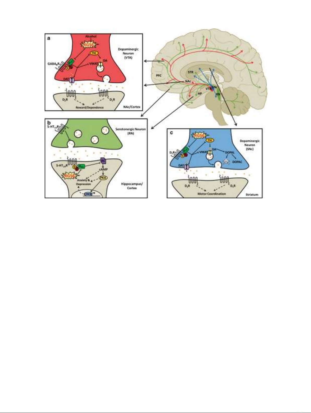
Fig. 4. RGS6 in central nervous system diseases. Schematic outlining the role of RGS6 in alcoholism (a), anxiety and
depression (b), and Parkinson’s disease (c). Red indicates neurons projecting from the ventral tegmental area (VTA) in the
meso-limbo-cortical pathway. Green indicates neurons projecting from the raphe nucleus (RN). Blue represents neurons
projecting from the substantia nigra pars compacta (SNc) in the nigro-striatal pathway. aIt is believed that RGS6 acts as a
critical mediator of alcohol-seeking behaviors by inhibiting GABA
B
R signaling which normally promotes upregulation of
the dopamine transporter (DAT) and inhibits vesicular monoamine transporter 2 (VMAT2) and tyrosine hydroxylase (TH).
Conversely, removal of RGS6, which is normally upregulated in the VTA following alcohol consumption, may ameliorate
alcohol reward and withdrawal by promoting GABA
B
R signaling decreasing dopamine (DA) bioavailability. bRGS6
promotes anxiety and depression by inhibiting the 5-HT
1A
heteroreceptors in cortical and hippocampal neurons that
synapse with serotonergic neurons of the RN. By blocking 5-HT
1A
heteroreceptor-mediated inhibition of adenylyl cyclase
(AC), RGS6 promotes the accumulation of cyclic AMP (cAMP) and the subsequent activation of protein kinase A (PKA)
and cAMP responsive element-binding protein (CREB), all of which contribute to anxiety and depression and counteract
the actions of antidepressant/anxiolytic medications. cRGS6 may also mediate the survival of dopaminergic SNc neurons by
inhibiting dopamine receptor (D
2
R) signaling in these neurons. By acting as a gatekeeper of D
2
R signaling, RGS6 is
believed to assure not only normal synaptic release of DA but may also prevent the accumulation of cytotoxic DA
byproducts that could contribute to neuronal degeneration. PFC prefrontal cortex, STR striatum, NAc nucleus accumbens,
HP hippocampus, DOPAL 3,4-dihydroxyphenylacetaldehyde, DOPAC 3,4-dihydroxyphenylacetic acid. Image adapted from
references (21–23)
564 Ahlers et al.


![Kỹ năng phân tích cận lâm sàng cơ bản: Tài liệu [mới nhất]](https://cdn.tailieu.vn/images/document/thumbnail/2025/20251231/vitobirama/135x160/2501767669821.jpg)


![Tài liệu triệu chứng học chấn thương [chuẩn nhất]](https://cdn.tailieu.vn/images/document/thumbnail/2025/20250806/tuhaolg/135x160/99781754535086.jpg)


![Cẩm nang Huyết học [Chuẩn nhất]](https://cdn.tailieu.vn/images/document/thumbnail/2026/20260513/baobinh_011/135x160/6151778671287.jpg)


%20--%3e%3cdefs%3e%3cstyle%3e%20.st0%20{%20fill:%20%23fff;%20}%20.st1%20{%20fill:%20%237800fa;%20}%20%3c/style%3e%3c/defs%3e%3cpath%20class='st1'%20d='M117.78,12.18H43.11c2.9,3.47,4.65,7.94,4.65,12.82,0,5.6-2.3,10.66-6.01,14.29h76.02l7.22-13.56-7.22-13.56Z'/%3e%3cg%3e%3cpath%20class='st0'%20d='M53.58,26.17h-.59v-1.46h.59v-4.96h2.83c1.78,0,2.67.94,2.67,2.82v5.76c0,1.87-.89,2.81-2.67,2.81h-2.83v-4.96ZM55.36,21.37v3.34h1.1v1.46h-1.1v3.34h1.01c.61,0,.91-.37.91-1.1v-5.93c0-.74-.3-1.1-.91-1.1h-1.01Z'/%3e%3cpath%20class='st0'%20d='M65.99,31.14h-1.8l-.31-2.07h-2.19l-.31,2.07h-1.64l1.82-11.39h2.62l1.82,11.39ZM65.28,18.04c-.25.46-.51.77-.75.94-.21.15-.47.22-.79.22-.26,0-.57-.07-.92-.22l-.38-.15c-.14-.05-.26-.07-.37-.07-.3,0-.53.18-.71.54l-.91-.68c.25-.46.51-.77.75-.94.21-.14.48-.21.79-.21.26,0,.57.07.92.21l.38.15c.14.05.26.07.37.07.3,0,.53-.18.71-.54l.91.68ZM61.91,27.52h1.73l-.87-5.76-.87,5.76Z'/%3e%3cpath%20class='st0'%20d='M74.53,26.89v1.52c0,1.91-.89,2.86-2.67,2.86s-2.67-.95-2.67-2.86v-5.93c0-1.91.89-2.86,2.67-2.86s2.67.95,2.67,2.86v1.11h-1.69v-1.22c0-.75-.31-1.12-.93-1.12s-.93.37-.93,1.12v6.15c0,.74.31,1.11.93,1.11s.93-.37.93-1.11v-1.63h1.69Z'/%3e%3cpath%20class='st0'%20d='M81.4,31.14h-1.8l-.31-2.07h-2.19l-.31,2.07h-1.64l1.82-11.39h2.62l1.82,11.39ZM75.9,19.2l1.52-1.91h1.71l1.51,1.91h-1.61l-.76-.95-.75.95h-1.61ZM77.32,27.52h1.73l-.87-5.76-.87,5.76ZM83.1,15.99l-1.76,1.91h-1.26l1.17-1.91h1.86Z'/%3e%3cpath%20class='st0'%20d='M84.86,19.75c1.78,0,2.67.94,2.67,2.82v1.48c0,1.87-.89,2.81-2.67,2.81h-.85v4.28h-1.79v-11.39h2.64ZM84.01,21.37v3.86h.85c.58,0,.87-.36.87-1.08v-1.71c0-.71-.29-1.07-.87-1.07h-.85Z'/%3e%3cpath%20class='st0'%20d='M93.51,19.75c1.78,0,2.67.94,2.67,2.82v1.48c0,1.87-.89,2.81-2.67,2.81h-.85v4.28h-1.79v-11.39h2.64ZM92.66,21.37v3.86h.85c.58,0,.87-.36.87-1.08v-1.71c0-.71-.29-1.07-.87-1.07h-.85Z'/%3e%3cpath%20class='st0'%20d='M98.8,31.14h-1.79v-11.39h1.79v4.88h2.03v-4.88h1.83v11.39h-1.83v-4.88h-2.03v4.88Z'/%3e%3cpath%20class='st0'%20d='M105.36,24.55h2.46v1.62h-2.46v3.34h3.09v1.63h-4.88v-11.39h4.88v1.63h-3.09v3.18ZM108.17,17.29l-1.76,1.91h-1.26l1.17-1.91h1.86Z'/%3e%3cpath%20class='st0'%20d='M112.2,19.75c1.78,0,2.67.94,2.67,2.82v1.48c0,1.87-.89,2.81-2.67,2.81h-.85v4.28h-1.79v-11.39h2.64ZM111.35,21.37v3.86h.85c.58,0,.87-.36.87-1.08v-1.71c0-.71-.29-1.07-.87-1.07h-.85Z'/%3e%3c/g%3e%3ccircle%20class='st1'%20cx='25'%20cy='25'%20r='20'/%3e%3cpath%20class='st0'%20d='M32.78,19.27c2.92,0,4.43,2.55,5.28,5.33l.71,2.17c.14.38-.33.75-.71.75h-5.61c.19-.33.24-.71.09-1.08l-.75-2.45c-.43-1.32-.99-2.64-1.79-3.77.75-.57,1.65-.94,2.78-.94h0ZM25,18.38c3.25,0,4.9,2.78,5.89,5.89l.76,2.45c.14.42-.33.8-.8.8h-11.69c-.42,0-.94-.38-.8-.8l.75-2.45c.99-3.11,2.64-5.89,5.89-5.89h0ZM25,11.35c1.74,0,3.11,1.37,3.11,3.11s-1.37,3.11-3.11,3.11-3.11-1.41-3.11-3.11,1.41-3.11,3.11-3.11h0ZM17.27,19.27c1.08,0,1.98.38,2.73.94-.8,1.13-1.37,2.45-1.74,3.77l-.8,2.45c-.14.38-.05.75.09,1.08h-5.56c-.42,0-.9-.38-.75-.75l.71-2.17c.9-2.78,2.41-5.33,5.33-5.33h0ZM17.27,12.91c1.51,0,2.78,1.27,2.78,2.83s-1.27,2.83-2.78,2.83-2.83-1.27-2.83-2.83,1.27-2.83,2.83-2.83h0ZM32.78,12.91c1.56,0,2.78,1.27,2.78,2.83s-1.23,2.83-2.78,2.83-2.83-1.27-2.83-2.83,1.27-2.83,2.83-2.83h0ZM27.07,28.56v.09c0,.57-.24,1.08-.61,1.46h0v.05c-.38.33-.9.57-1.46.57s-1.08-.24-1.46-.61h0c-.38-.38-.61-.9-.61-1.46v-.09h1.41v.09c0,.19.05.38.19.47v.05c.09.09.28.19.47.19s.38-.09.47-.19v-.05c.14-.09.24-.28.24-.47t-.05-.09h1.41ZM30.99,28.56v.09c0,1.65-.66,3.16-1.74,4.24-1.08,1.08-2.59,1.79-4.24,1.79s-3.16-.71-4.24-1.79l-.05-.05c-1.04-1.08-1.7-2.55-1.7-4.2v-.09h1.41v.09c0,1.27.47,2.4,1.27,3.25h.05c.85.85,1.98,1.37,3.25,1.37s2.4-.52,3.25-1.37c.85-.8,1.37-1.98,1.37-3.25v-.09h1.37ZM34.99,28.56v.09c0,2.78-1.13,5.28-2.92,7.07-1.79,1.79-4.29,2.92-7.07,2.92s-5.23-1.13-7.07-2.92c-1.79-1.79-2.92-4.29-2.92-7.07v-.09h1.41v.09c0,2.4.94,4.53,2.5,6.08,1.56,1.56,3.72,2.5,6.08,2.5s4.52-.94,6.08-2.5c1.56-1.56,2.5-3.68,2.5-6.08v-.09h1.41Z'/%3e%3c/svg%3e)