
CÔNG NGHỆ https://jst-haui.vn Tạp chí Khoa học và Công nghệ Trường Đại học Công nghiệp Hà Nội Tập 60 - Số 8 (8/2024)
128
KHOA H
ỌC
P
-
ISSN 1859
-
3585
E
-
ISSN 2615
-
961
9
PREPARATION OF ASTAXANTHIN/PYCNOGENOL NANOPARTICLES: A DELIVERY SYSTEM WITH IMPROVED ASTAXANTHIN STABILITY AND BIOAVAILABILITY
CHẾ TẠO HẠT NANO ASTAXANTHIN/PYCNOGENOL: MỘT HỆ THỐNG PHÂN PHỐI CẢI THIỆN ĐỘ ỔN ĐỊNH VÀ KHẢ DỤNG SINH HỌC CHO ASTAXANTHIN Ho Thi Oanh1, Hac Thi Nhung1,2, Nguyen Hong Tham1, Doan Tien Dat1,2, Nguyen Duc Tuyen1, Nguyen Thi Sang3, Nguyen Yen Thanh3, Truong Cong Doanh4, Hoang Mai Ha1,2,* DOI: http://doi.org/10.57001/huih5804.2024.276 ABSTRACT Astaxanthin (AST) and pycnogenol (PYC) are natural bioactive compounds commonly used in pharmaceuticals and functional foods
due to their highly
antioxidant activity and potential health benefits. However, the widespread application of astaxanthin is hinde
red by its poor water solubility and highly
unsaturated structure. In this study, nanoparticles co-
delivering astaxanthin and pycnogenol (NAP) were fabricated to enhance the dispersibility, stability, and
bioavailability of astaxanthin. The particle size,
dispersion index, zeta potential (ζ), and morphology of the nanoparticles were characterized, revealing that the
NAP were spherical, had a negative surface charge, and an average particle size in the range of 70 - 91nm with a narrow size distribution. X-ra
y diffraction (XRD)
demonstrated that AST existed in an amorphous form within the NAP. Notably, the excellent water dispersibility and high stabi
lity of the astaxanthin/pycnogenol
nanoparticles significantly enhanced cellular uptake, with absorption effici
ency being 12 times higher than that of pure astaxanthin. Furthermore, the
antioxidant activity was notably improved. These findings suggest that the astaxanthin/pycnogenol nanoparticles is an ideal f
ormulation of astaxanthin, offering
potential for the development of new functional foods. Keywords: Astaxanthin, pycnogenol, nanoparticles, cell uptake, antioxidant activity. TÓM TẮT Astaxanthin (AST) và pycnogenol (PYC) là các hợp chất có hoạt tính sinh học tự nhiên thường được sử dụng trong dược phẩm và th
ực phẩm chức năng bởi
hoạt tính chống oxy hóa cao và lợi ích tiềm năng của chúng đối với sức khoẻ. Tuy nhiên, việc ứng dụng rộng rãi astaxanthin bị hạn chế do khả năng h
òa tan trong
nước kém và cấu trúc không bão hòa cao. Trong nghiên cứu này, các hạt nano đồng phân phối astaxanthin và pycnogenol (NAP) đã đư
ợc chế tạo để nâng cao khả
năng phân tán, tính ổn định và sinh khả dụng cho astaxanthin. Kích thước hạt, chỉ số phân tán, thế zeta (ζ) và hình thái của các hạt nano đã đư
ợc xác định, cho
thấy rằng các hạt nano NAP có dạng hình cầu, có điện tích bề mặt âm và kích thước hạt trung bình trong khoảng từ 70 - 91nm với dải phân bố kích thư
ớc hẹp.
Giản đồ nhiễu xạ tia X đã chứng minh rằng AST tồn tại ở dạng vô định hình trong các hạt nano NAP. Đặc biệt, khả năng phân tán tốt và tính
ổn định cao trong
nước của các hạt nano astaxanthin/pycnogenol đã tăng cường khả năng hấp thu vào trong tế bào một cách đáng kể, với hiệu quả h
ấp thu gấp 12 lần so với
astaxanthin tự do. Ngoài ra, hoạt tính chống oxy hóa cũng được cải thiện đáng kể. Những kết quả này cho thấy rằng hệ nano astaxanthin/pycnogenol là m
ột
công thức bào chế lý tưởng của astaxanthin, mở ra tiềm năng phát triển các loại thực phẩm chức năng mới. Từ khóa: Astaxanthin, pycnogenol, hạt nano, hấp thu tế bào, khả năng chống oxy hoá. 1Institute of Chemistry, Vietnam Academy of Science and Technology, Vietnam 2Graduate University of Science and Technology, Vietnam Academy of Science and Technology, Vietnam 3Faculty of Engineering Physics and Nanotechnology, VNU University of Engineering and Technology, Vietnam 4Hanoi University of Industry, Vietnam *Email: hoangmaiha@ich.vast.vn Received: 10/6/2024 Revised: 23/8/2024 Accepted: 27/8/2024
P-ISSN 1859-3585 E-ISSN 2615-9619 https://jst-haui.vn SCIENCE - TECHNOLOGY Vol. 60 - No. 8 (Aug 2024) HaUI Journal of Science and Technology 129
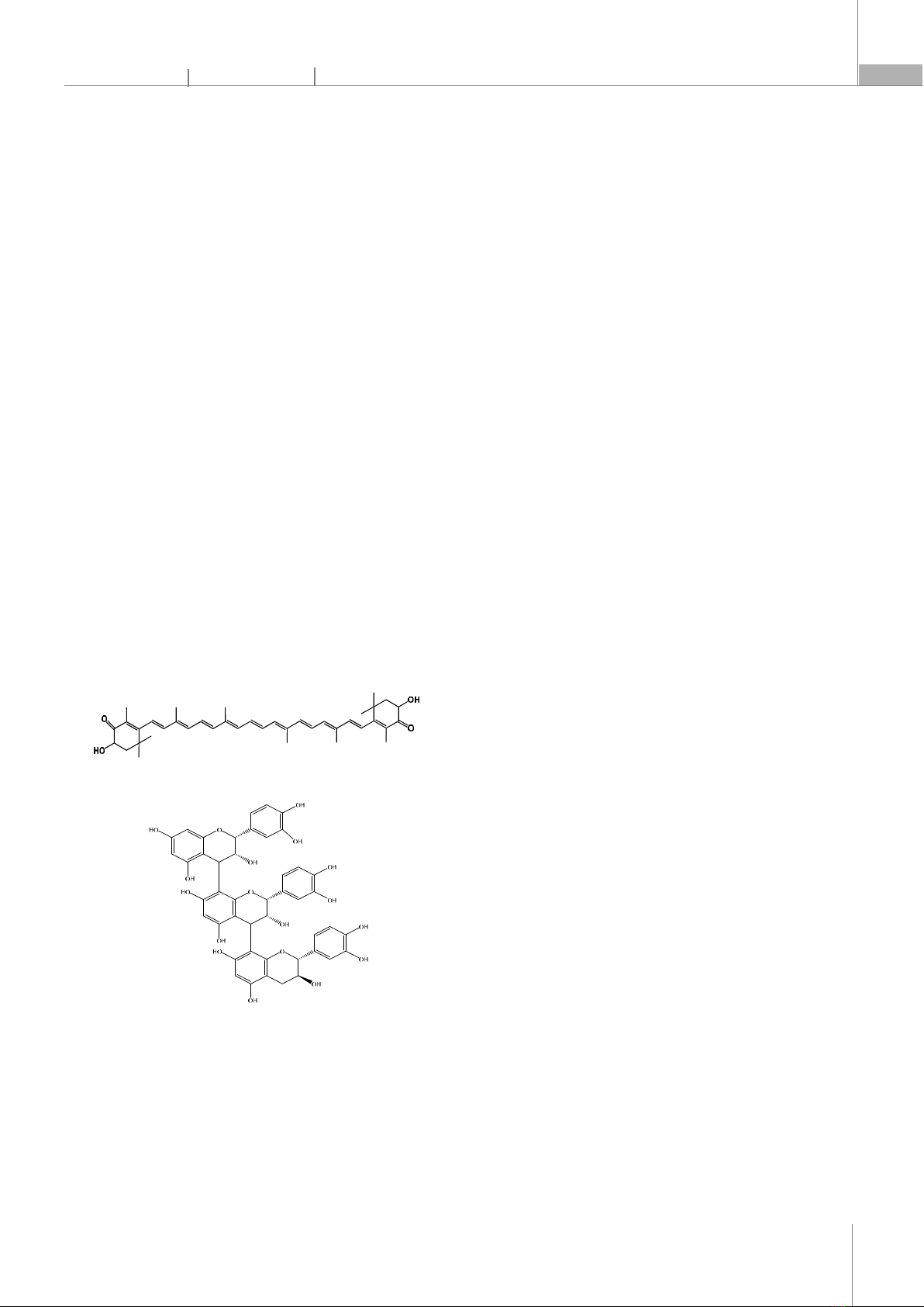
1. INTRODUCTION Nowadays, natural compounds like astaxanthin and pycnogenol are receiving significant attention for their roles in functional foods and nutraceuticals aimed at improving human health, well-being, and performance [1, 2]. Astaxanthin, a red pigment belonging to the carotenoid group, is found in red yeast, the microalga Haematococcus pluvialis, and aquatic organisms such as salmon, shrimp, crab, and lobster [3]. Astaxanthin has various advantageous biological activities, including strong antioxidant capacity, anti-inflammatory effects, inhibition of cancer cell proliferation, prevention of atherosclerosis, and reduction of symptoms related to cardiovascular, respiratory, and nervous system diseases [4-6]. Astaxanthin has a molecular formula of C40H52O4, with a structure comprising 11 conjugated double bonds, 2 β-ionone rings, and hydroxyl groups (Fig. 1) [7]. The conjugated double bond system is responsible for astaxanthin's red color and its strong antioxidant activity. However, this molecular structure with many conjugated double bonds makes astaxanthin unstable and easily degraded by oxidative agents, light, and temperature [8]. Moreover, astaxanthin is a hydrophobic molecule with low oral bioavailability, which also limits its application potential [9]. Astaxanthin Procyanidins (primary active ingredient in Pycnogenol) Fig. 1. Molecular structures of astaxanthin and pycnogenol Pycnogenol is a flavonoid extracted from the bark of the French maritime pine, carrying many beneficial pharmacological properties [10]. It is well-known for its excellent antioxidant and anti-inflammatory capabilities, which help enhance cardiovascular health, improve blood circulation, protect neural cells, manage pain, and eliminate free radicals [11]. The main component of pycnogenol is procyanidin (Fig. 1), accounting for 70 - 85%, which are biological polymers of catechin and epicatechin subunits, recognized as important components in human nutrition [12]. Several reports have demonstrated that the combination of natural bioactive compounds in composite products yields synergistic effects, enhancing stability and bioactivity compared to using individual compounds alone [13-15]. Flavonoid compounds such as pycnogenol, quercetin, and kaempferol can create synergistic effects, enhancing the stability of compounds with many conjugated double bonds, such as astaxanthin, lycopene, and beta-carotene. However, research on the fabrication of nanoparticle systems containing both carotenoid and flavonoid compounds remains limited, particularly in successfully preparing nanoparticles co-delivering astaxanthin and pycnogenol. Therefore, the application of nanotechnology to prepare astaxanthin/pycnogenol nanoparticles to improve the dispersibility, stability, absorption efficiency, and antioxidant activity of astaxanthin is a novel and necessary scientific direction. In this study, astaxanthin/pycnogenol nanoparticles were prepared using two surfactants, tween 80 and lecithin, with beta-cyclodextrin as the encapsulating agent, followed by freeze-drying to obtain the nanopowder. The total content of the natural compounds astaxanthin and pycnogenol in the nanoparticle system reached up to 10%. Subsequently, the nanoparticles were analyzed for their structure, morphology, and stability, while also evaluating biological activities such as cellular uptake capability and antioxidant potential. 2. MATERIALS AND METHODS 2.1. Material Astaxanthin is a compound extracted from algal biomass Haematococcus pluvialis, with a purity of over 96% [16]. Pycnogenol (containing 70 - 85% procyanidins) is a commercial product extracted from the bark of the French maritime pine. Surfactants: tween 80 and lecithin from Sigma-Aldrich, USA. Encapsulating agent: beta-cyclodextrin purchased from AKscientific, USA. All solvents used in this study were of analytical or HPLC grade.
CÔNG NGHỆ https://jst-haui.vn Tạp chí Khoa học và Công nghệ Trường Đại học Công nghiệp Hà Nội Tập 60 - Số 8 (8/2024)
130
KHOA H
ỌC
P
-
ISSN 1859
-
3585
E
-
ISSN 2615
-
961
9
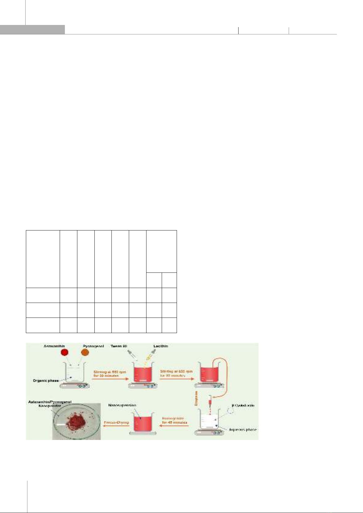
2.2. Preparation of nanoparticles co-delivering astaxanthin and pycnogenol (NAP) Astaxanthin and pycnogenol were mixed at different ratios (Table 1). These active ingredients were then dissolved in tetrahydrofuran solvent using a magnetic stirrer (RCT basic IKAMAG® safety control, Germany) at 550rpm for 30 minutes. Subsequently, tween 80 and lecithin, the two surfactants, were simultaneously added, and the stirring speed was increased to 650rpm. After stirring for 30 minutes, the organic phase was slowly dripped into the aqueous phase containing the carrier beta-cyclodextrin. The resulting solution is further homogenized under an Ultra-Turrax (T18 IKA, Germany) for 45 minutes to obtain a uniform nanoparticle system. The LABCONCO freeze-dryer was then used to obtain astaxanthin/pycnogenol nanopowder. Freeze drying was performed with a collector temperature of -50°C and a vacuum of 0.050mBar. The schematic fabrication procedure for the astaxanthin/pycnogenol nanopowder is presented in Fig. 2. Table 1. Composition for each of the prepared nanoparticle formulations Formulation AST (mg) PYC (mg) T80 (mg) Lec (mg) β- CD (mg) Loading Bioactive Compound (%) AST PYC NAP1 10 30 80 40 240 2.5 7.5 NAP2 20 20 80 40 240 5.0 5.0 NAP3 30 10 80 40 240 7.5 2.5 Abbreviations: T80 (Tween 80), Lec (Lecithin), and β-CD (β-cyclodextrin). Fig. 2. Schematic illustration of astaxanthin/pycnogenol nanopowder formation 2.3. Characterization of nanoparticles co-delivering astaxanthin and pycnogenol 2.3.1. Fourier transform infrared spectroscopy (FTIR) The FT-IR spectra of astaxanthin, pycnogenol and astaxanthin/pycnogenol nanoparticle systems were measured using a PerkinElmer GX instrument (USA) over the range of 400 - 4000cm⁻1. 2.3.2. X-ray Diffraction The crystalline states of astaxanthin, pycnogenol, and the astaxanthin/pycnogenol nanoparticle systems were assessed by a D8-ADVANCE diffractometer (Bruker, Germany) with a 2θ angle range from 10 to 70°, utilizing a CuKα radiation source operated at 30mA and 40kV. 2.3.3. Particle size, polydispersity index (PDI), and zeta potential The mean particle diameter, polydispersity index and surface potential (ζ-potential) of the astaxanthin/pycnogenol nanoparticles in the dispersions were measured by dynamic light scattering (DLS) using a LitesizerTM 500 instrument (Anton Paar, Austria). All measurements were conducted at 25°C and each sample was analyzed in triplicate. 2.3.4. Morphology Transmission electron microscopy (TEM) was used to determine the shape and surface morphology of the produced nanoparticles. The morphological investigation of nanoparticles co-delivering astaxanthin and pycnogenol was carried out according to the method described by Fuguo Liu et al. (2018). The samples were observed directly without any staining. 2.4. Cell viability assay The toxicity of the astaxanthin/pycnogenol nanosystems at different concentrations on HepG2 cells was evaluated using the MTT ((3-[4,5-dimethylthiazol-2-yl]-2,5 diphenyl tetrazolium bromide) assay. HepG2 cells were seeded into 96-well plates at a density of 0.5 × 10⁵ cells per well and incubated overnight at 37°C with 5% CO2. After incubation, the old medium was replaced with fresh medium containing different concentrations (20 and
P-ISSN 1859-3585 E-ISSN 2615-9619 https://jst-haui.vn SCIENCE - TECHNOLOGY Vol. 60 - No. 8 (Aug 2024) HaUI Journal of Science and Technology 131
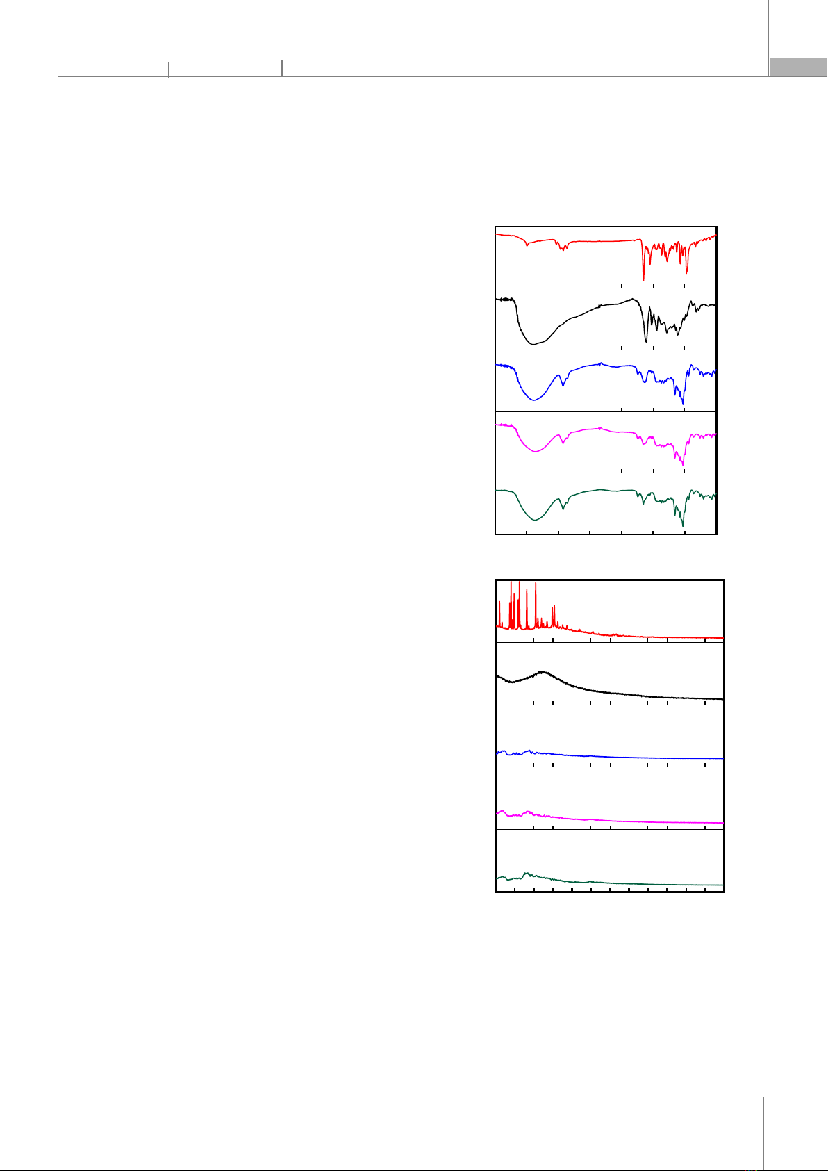
100µg/mL) of astaxanthin (AST), pycnogenol (PYC), and the nanosystem co-delivering astaxanthin and pycnogenol (NAP2). Untreated cells in wells were used as controls. HepG2 cells were incubated with AST, PYC, and NAP2 for 2 hours. Subsequently, 5mg/mL MTT reagent was added to the samples, and the cells were incubated for an additional 3 hours until purple formazan products developed. Formazan crystals formed in the cells were dissolved by adding 100µL of DMSO, followed by further incubation for 2 hours. The absorbance was measured using a Multiskan SkyHigh microplate spectrophotometer (Thermo Scientific) at a wavelength of 570nm. Cell viability was calculated using the following expression: Cell Viability = (OD570nm sample/OD570nm control) * 100% 2.5. Antioxidant activity studies 1,1-Diphenyl-2-picrylhydrazyl (DPPH) is a free radical generator used to screen the antioxidant effects of substances under study. Antioxidant activity is demonstrated by the reduction of DPPH color, determined spectrophotometrically at a wavelength of 517nm. In summary, 100μL of a 0.2mM DPPH solution was prepared in methanol (MeOH), and then 100μL of nanoparticles co-delivering astaxanthin and pycnogenol (at concentrations of 4, 20, and 100µg/mL) was added. The reaction was allowed to proceed for 30 minutes at 37°C, after which the absorbance of DPPH was measured using a microplate reader (Thermo Fisher Scientific, USA). Ascorbic acid was used as a positive control. The % DPPH radical scavenging activity of the test sample was calculated using the following formula: % SA = (ODcontrol - ODsample)*100/ODcontrol 2.6. Cellular uptake The absorption capability of astaxanthin/pycnogenol nanoparticles was indirectly determined by comparing the absorption capacity of astaxanthin within nanoparticles to pure astaxanthin in HepG2 cells. The method for evaluating the absorption capacity of astaxanthin in cells was carried out according to the procedure described by Hien et al. [17]. Specifically, HepG2 cells were cultured for 24 hours at a density of 1×10⁶ cells per dish and then treated with 5µg/mL of free astaxanthin or 100µg/mL of nano astaxanthin/pycnogenol (containing an equivalent amount of 5µg/mL astaxanthin) for 4 hours. After the incubation period, the cells were washed three times with phosphate-buffered saline (PBS), lysed, and centrifuged at 2000rpm for 3 minutes. The concentration of astaxanthin in the cells was determined by high-performance liquid chromatography (HPLC), as previously reported by Hien et al. [17]. 3. RESULTS AND DISCUSSION 3.1. FTIR and XRD analysis of astaxanthin/pycnogenol nanoparticles Fig. 3. The FT-IR spectrum (A) and XRD patterns (B) of astaxanthin (AST), pycnogenol (PYC), and astaxanthin/pycnogenol nanoparticles (NAP1, NAP2, and NAP3) The nano systems were prepared with astaxanthin/pycnogenol ratios of 1/3, 1/1, and 3/1, respectively. Specifically, we examined samples NAP1 (2.5
4000 3500 3000 2500 2000 1500 1000 500
Wavenumber (cm
-1
)
Transmittance (%)
NAP2
NAP3
NAP1
AST
PYC
(A)
10 15 20 25 30 35 40 45 50 55 60 65 70
Intensity (a.u.)
2
q
(degree)
NAP3
NAP2
NAP1
AST
PYC
(B)
CÔNG NGHỆ https://jst-haui.vn Tạp chí Khoa học và Công nghệ Trường Đại học Công nghiệp Hà Nội Tập 60 - Số 8 (8/2024)
132
KHOA H
ỌC
P
-
ISSN 1859
-
3585
E
-
ISSN 2615
-
961
9
wt% AST and 7.5 wt% PYC), NAP2 (5 wt% AST and 5 wt% PYC), and NAP3 (7.5 wt% AST and 2.5 wt% PYC). The FTIR spectra of pure astaxanthin, pure pycnogenol, and the nanosystems encapsulating astaxanthin and pycnogenol are shown in Fig. 3A. The strong absorption band of pure astaxanthin at 1651cm⁻1 is assigned to the C=O stretching vibration peak. Additionally, bands at 1549cm⁻1 and 975cm⁻1 correspond to the C=C stretching vibration in aromatic rings and the C–H in the C and C conjugate system, respectively. The characteristic absorption peak at 2920cm⁻1 indicates the stretching vibration of –CH3. Similar results were also reported in previous studies [18]. The characteristic peak of pycnogenol observed in the broad range of wavenumbers from 3600 to 3015cm⁻1 is assigned to the ‒OH stretching. Additionally, the peak at 1612cm⁻1 is characteristic of the C=C stretching vibration in aromatic rings. The FTIR spectrum of pycnogenol obtained is completely consistent with previously published studies [19]. All characteristic functional groups of astaxanthin and pycnogenol are present in the FTIR spectra of nanoparticles co-delivering astaxanthin and pycnogenol (NAP1, NAP2, and NAP3). The results indicated that the two active compounds, astaxanthin and pycnogenol, have been successfully physically encapsulated in the nanoparticles. The XRD pattern was performed on astaxanthin, pycnogenol, and nanoastaxanthin/pycnogenol to obtain information about their crystalline states (Figure 3B). The XRD pattern of astaxanthin shows multiple characteristic peaks with high intensity at 11.03o; 13.6o; 14.9o; 16.3o; 18.1o; 20.6o; 23.4o; 24.8o; 25.6o and 27.5o, confirming that pure astaxanthin is in crystalline form, consistent with previous reports [20]. In contrast, the X-ray diffraction pattern of pycnogenol does not exhibit clear diffraction peaks. This can be explained by the chemical composition of pycnogenol, which includes a mixture of phenolic compounds: flavonoids (procyanidins account for 70 - 85%), monomers (catechin, epicatechin, taxifolin), phenolic or cinnamic acids, and their glycosides. For the NAP1, NAP2, and NAP3 nanoparticles, the sharp peaks of astaxanthin also disappeared in the XRD pattern of these systems. This phenomenon indicated that astaxanthin has been loaded into the nanoparticles and has transitioned from a crystalline state to an amorphous form. Changing the crystalline nature through nano formulation is often an ideal way to enhance the solubility of astaxanthin, thereby improving the practical applications of the active ingredient. 3.2. Morphology and particle size analysis of nanoparticles co-delivering astaxanthin and pycnogenol In this study, the morphology, particle size distribution, polydispersity index (PDI), and zeta potential of the nanoparticles co-delivering astaxanthin and pycnogenol were also investigated. The particle size distribution and polydispersity index of the astaxanthin/pycnogenol nanoparticles (Fig. 4A) showed that the average particle diameters for NAP1, NAP2, and NAP3 samples were 70, 78, and 91nm, respectively, with PDI values of 0.164, 0.168, and 0.195. The co-encapsulated astaxanthin and pycnogenol nano systems exhibited a narrow distribution with average particle sizes below 100nm. The zeta potential of the astaxanthin/pycnogenol nanoparticles is presented in Figures 4B-D. The NAP1, NAP2, and NAP3 nanoparticles had a negative charge with relatively high absolute zeta potential values of 48.5, 44.9, and 42.9mV, respectively. According to previous studies, nanoparticles with absolute zeta potential values above 20mV were considered to have high physical stability [21].
0 50 100 150 200 250 300
0
2
4
6
8
10
12
NAP1
NAP2
NAP3
Particle Diameter (nm)
Distribution (%)
NAP1 NAP2 NAP3
0.00
0.05
0.10
0.15
0.20
0.25
Polydispersity index (PDI)
0.168
0.160
0.195
(A)
-200 -100 0 100 200
0.0
0.5
1.0
1.5
2.0
2.5
3.0
Rel. frequency [%]
Zeta potential distribution [mV]
NAP1
-48.5 mV
(B)


![Câu hỏi ôn thi Hóa lý dược [năm] chuẩn nhất](https://cdn.tailieu.vn/images/document/thumbnail/2025/20250714/kimphuong1001/135x160/4391752479246.jpg)


![Tài liệu Vi sinh vật môi trường [Mới nhất]](https://cdn.tailieu.vn/images/document/thumbnail/2025/20251123/ngkimxuyen/135x160/21891763953413.jpg)
![Sổ tay truyền thông Phân loại chất thải rắn sinh hoạt trên địa bàn tỉnh Quảng Nam [Chuẩn nhất]](https://cdn.tailieu.vn/images/document/thumbnail/2025/20251114/kimphuong1001/135x160/1701763094001.jpg)

![Quản lý chất thải nguy hại: Sổ tay Môi trường [Chuẩn nhất]](https://cdn.tailieu.vn/images/document/thumbnail/2025/20251029/kimphuong1001/135x160/9011761720170.jpg)