
BioMed Central
Page 1 of 8
(page number not for citation purposes)
Journal of Translational Medicine
Open Access
Research
Correlation between expression of p53, p21/WAF1, and MDM2
proteins and their prognostic significance in primary hepatocellular
carcinoma
Mei-Fang Zhang1,2, Zhi-Yi Zhang1,2, Jia Fu1,2, Yu-Feng Yang1,2 and
Jing-Ping Yun*1,2
Address: 1State Key Laboratory of Oncology in Southern China, Cancer Center of Sun Yat-Sen University, Guangzhou, China and 2Department of
Pathology, Cancer Center, Sun Yat-Sen University, Guangzhou 510060, China
Email: Mei-Fang Zhang - mf.zhang@live.cn; Zhi-Yi Zhang - ls01zzy@yahoo.com.cn; Jia Fu - fujia81@126.com; Yu-
Feng Yang - y313yang@sina.com; Jing-Ping Yun* - yunjp@mail.sysu.edu.cn
* Corresponding author
Abstract
Background: Tumor Protein p53 (p53), cyclin-dependent kinase inhibitor 1A (p21/WAF1), and
murine double minute 2 (MDM2) participate in the regulation of cell growth. Altered expression
of these gene products has been found in malignant tumors and has been associated with poor
prognosis. Our aim was to investigate the expression of the 3 proteins in hepatocellular carcinoma
(HCC) and their prognostic significance.
Methods: We examined p53, p21/WAF1, and MDM2 expression in 181 pairs of HCC tissues and
the adjacent hepatic tissues by performing immunohistochemistry and examined the expression of
the 3 proteins in 7 pairs of HCC tissues and the adjacent hepatic tissues by using western blot
analysis.
Results: The expression of p53, p21/WAF1, and MDM2 in the HCC tissues was significantly higher
than those in the adjacent hepatic tissues (P < 0.05). A statistical correlation was observed between
p53 and p21/WAF1 expression in HCC tissues (R = 0.195, P = 0.008). A statistical correlation was
observed between expression of p53 and p21/WAF1 (R = 0.380, P = 0.000), p53 and MDM2 (R =
0.299, P = 0.000), p21/WAF1 and MDM2 (R = 0.285, P = 0.000) in 181 liver tissues adjacent to the
tumor. Patients with a low pathologic grade HCC (I+II) had a higher tendency to express p53 on
tumor cells than the patients with high pathologic grade HCC (III+IV) (P = 0.007). Survival analysis
showed that positive p21/WAF1 expression or/and negative MDM2 expression in HCC was a
predictor of better survival of patients after tumor resection (P < 0.05).
Conclusions: The proteins p53, p21/WAF1, and MDM2 were overexpressed in all the HCC cases
in this study, and p53 and p21/WAF1 overexpression were positively correlated. The expression
of p21/WAF1 and MDM2 can be considered as 2 useful indicators for predicting the prognosis of
HCC.
Published: 22 December 2009
Journal of Translational Medicine 2009, 7:110 doi:10.1186/1479-5876-7-110
Received: 9 October 2009
Accepted: 22 December 2009
This article is available from: http://www.translational-medicine.com/content/7/1/110
© 2009 Zhang et al; licensee BioMed Central Ltd.
This is an Open Access article distributed under the terms of the Creative Commons Attribution License (http://creativecommons.org/licenses/by/2.0),
which permits unrestricted use, distribution, and reproduction in any medium, provided the original work is properly cited.

Journal of Translational Medicine 2009, 7:110 http://www.translational-medicine.com/content/7/1/110
Page 2 of 8
(page number not for citation purposes)
Background
Hepatocellular carcinoma (HCC) is the fifth most com-
mon malignancy worldwide and is the third most com-
mon cause of cancer-related deaths [1]. HCC develops in
patients with chronic liver diseases, and its etiopathogen-
esis includes viral infection (hepatitis B and C), alcohol,
and aflatoxin B1 consumption. The majority of HCC
patients have associated cirrhosis and impaired liver func-
tion, making the treatment of HCC more difficult than
that of many other cancers. Surgery, including transplan-
tation, remains the only potential curative modality for
HCC.
Prognosis of HCC remains unsatisfactory even after surgi-
cal resection and liver transplantation. Considerable
interest has been generated in identifying factors that
influence the prognosis of HCC. Several staging systems
have been developed to predict survival period after the
diagnosis of HCC [2]. The most widely studied prognostic
factors are related to the pathological characteristics of the
neoplasm, including tumor size, grade, stage, and vascular
invasion. However, several biological molecules that can
predict the survival period of HCC patients have been
reported in recent years; however, the results are contro-
versial.
Previous studies have explored the molecular alterations
in HCC, including changes in the expression of p53, cyc-
lin-dependent kinase inhibitor 1A (p21/WAF1), and
murine double minute 2 (MDM2). The tumor suppressor
gene p53 plays a key role in regulating the cell cycle and
serves as a principal mediator of growth arrest, senes-
cence, and apoptosis in response to a broad array of cellu-
lar damage [3]. The p21/WAF1 protein is encoded by the
human WAF1/CIP1 gene and its expression is directly
induced by the wild-type p53 protein [4]. This protein
binds to a variety of cyclin-dependent kinases and inhibits
their activity, regulates DNA repair, and directly blocks
DNA replication by inhibiting the proliferating cell
nuclear antigen [5], thus inhibiting cell-cycle progression
and decreasing cell growth. MDM2 is the product of a
p53-inducible gene and inhibits the p53 activity by ubiq-
uitinating p53 and creating a negative-feedback loop [5-
8]. Altered expression of these gene products has been
found in malignant tumors including HCC and correlated
with poor prognosis. In HCC, the prognostic value of p53
is controversial, since several studies show an association
of p53 with patient survival [9-12], while other investiga-
tions report no association [13,14]. The predictive value
of the p21/WAF1 expression level in HCC is also ambigu-
ous [10,11,15]. However, few studies pertaining to the
expression of the 3 proteins p53, p21/WAF1, and MDM2
in HCC cases have reported different results [11,16].
We determined the expression of p53, p21/WAF1, and
MDM2 in a relatively large sample size of 181 pairs of
human HCC tissues and the corresponding adjacent
hepatic tissues obtained after resection by performing
immunohistochemistry (IHC). In addition, we performed
western blot analysis in 7 such pairs. Further, we
attempted to address the correlation among their expres-
sion and the relationship between their expression and
the clinical parameters, including overall survival.
Methods
Clinical samples
Samples from 181 Chinese patients with HCC and their
clinical records from 1997 to 2007 were collected from
the Cancer Center of Sun Yat-Sen University, Guangzhou,
China. Tissue blocks prepared from HCC tissues and the
adjacent liver tissues were sectioned for performing IHC
of p53, p21/WAF1, and MDM2; in addition, for 7 cases,
we collected the tissue samples inclusive of the HCC and
its adjacent tissues from the tissue bank department of
this cancer center and subjected these samples to western
blot analyses. The collection of the human specimens in
the study was approved by the Independent Ethics Com-
mittee of the Cancer Center of Sun Yat-Sen University.
Western blot analysis
For immunolabeling, lysates were prepared from the tis-
sues as described previously [17,18]. We separated 100 μg
of each lysate by sodium dodecyl sulfate-polyacrylamide
gel electrophoresis (SDS-PAGE). The proteins were trans-
ferred onto blotting membranes. After blocking, the
membranes were incubated overnight with rabbit poly-
clonal antibody against p53 (Clone: FL-393; Cat No. sc-
6243; Santa Cruz, CA); mouse monoclonal antibody
against p21/WAF1 (Clone: SX118; Cat No. 556430; BD
Pharmigen, CA) and MDM2 (Clone: N-20; Cat No. sc-
813; Santa Cruz, CA); and mouse monoclonal antibody
against glyceraldehydes 3-phosphate dehydrogenase
(GAPDH) (Kangchen Biotech; Shanghai, China) (p53,
1:500; p21/WAF1, 1:250; MDM2, 1:2000; and GAPDH,
1:1000), followed by incubation with horseradish perox-
idase-conjugated immunoglobulin G (IgG). The blots
were then visualized by using an ECL kit (Amersham Life
Science; Piscataway, NH, USA) and exposed for 1 min to
an X-ray film.
Immunohistochemistry
For immunohistochemistry studies, a labeled-streptavi-
din-biotin (LAB-SA) method was performed with Histo-
stain®-Plus Bulk Kit Zymed® 2nd generation LAB-SA
detection system (CAT. NO. 85-9043, Zymed Laborato-
ries, CA) as previously described [18,19]. All the primary
antibodies (p53, p21/WAF1, and MDM2; mouse mono-
clonal antibody, Cat No. ZM-0408, ZM-0206, and ZM-
0425, respectively, Zymed, CA) were ready to use without
dilution. Each paraffin-embedded tissue section (4 μm in
thickness) was deparaffinized, hydrated, and incubated in
3% H2O2 and microwaved for 3 minutes to block endog-
Journal of Translational Medicine 2009, 7:110 http://www.translational-medicine.com/content/7/1/110
Page 3 of 8
(page number not for citation purposes)
enous peroxidase activity. The tissue sections were sub-
jected to antigen retrieval by microwaving in 10 mM
citrate buffer for 30 min. The sections were incubated with
serum blocking solution (Reagent A) for 10 minutes to
block nonspecific binding and then with the primary anti-
bodies in moist chamber for 60 minutes. After rinsed with
PBS for 2 minutes, the sections were incubated with the
biotinylated secondary antibody (Reagent B) for 10 min-
utes and rinsed with PBS. The sections followed by incu-
bation with enzyme conjugate (Reagent C) for 10
minutes. Subsequently, the sections were stained with
DAB and counterstained with hematoxylin. Serum block-
ing solution (Reagent A) in place of the primary antibody
was used as a negative control. A brown particle in nuclei
was considered as positive labeling. Immunostaining
labeling intensities were defined as: +, less than 10% of
the tumor cells were positive; ++, 10%-50% of the tumor
cells were positive; +++, more than 50% the tumor cells
were positive; -, negative staining. These sections were
observed under light microscopy and the staining intensi-
ties were assessed by 2 pathologists--Dr JP Yun and Dr MF
Zhang.
Statistical analysis
Statistical analysis was performed to determine the rela-
tionship between the clinical parameters of gender, age,
tumor size, number of tumors, hepatitis B surface antigen
(HBsAg), pathologic grade, serum level of alpha-fetal pro-
tein (AFP), and the 3 immunohistochemical markers by
Peason's chi-square test. The Spearman correlation was
employed to examine the relationship between the
expression of p53, p21/WAF1, and MDM2. Survival was
assessed by the Kaplan-Meier method, and log-rank test
was used to analyze survival curves. Statistical significance
was initially set at P < 0.05. All statistical analysis was per-
formed using the SPSS 13.0 software for Windows.
Results
Increase in the expression of p53, p21/WAF1, and MDM2
in HCC
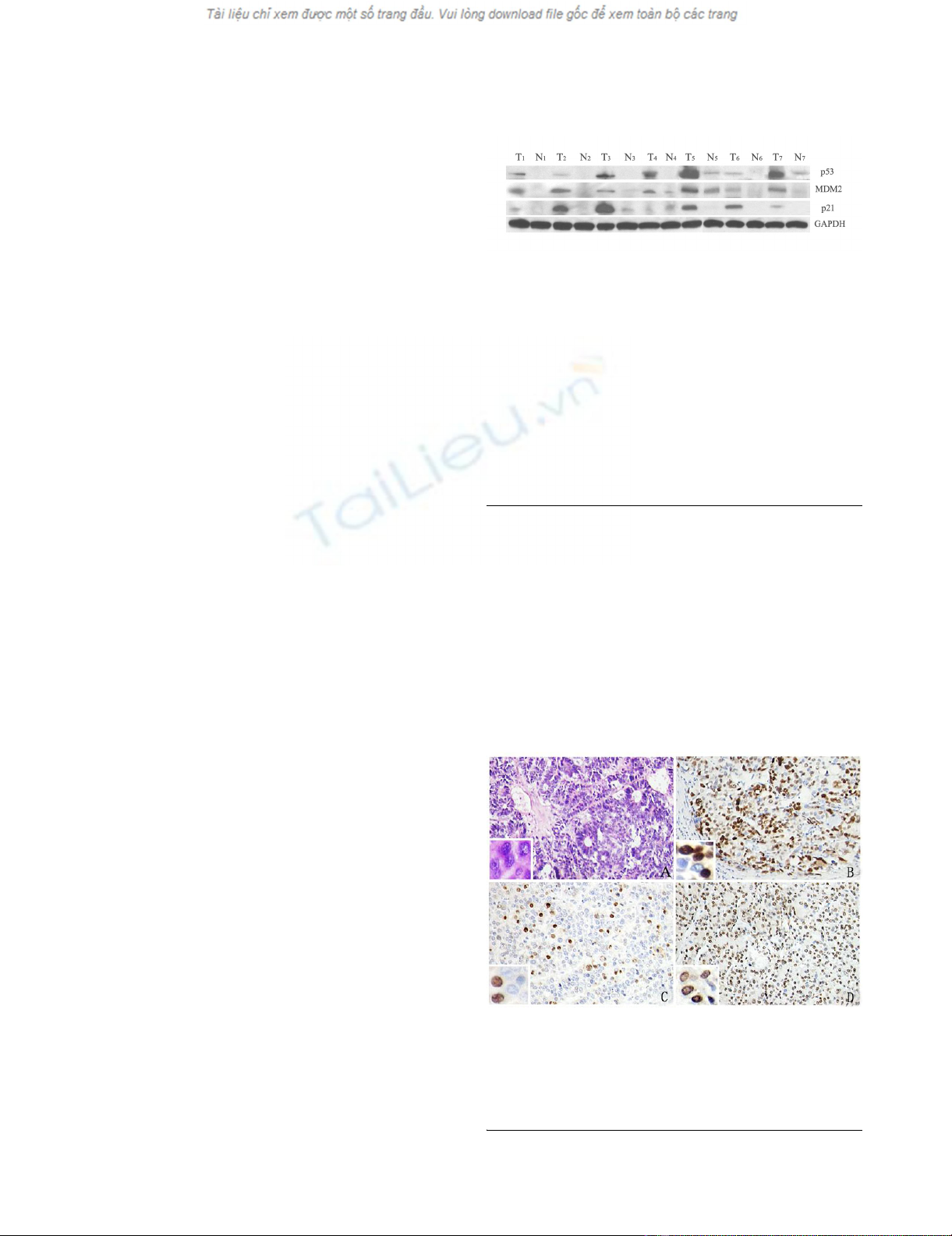
The expression of p53 and MDM2 in the 7 pairs was
higher in the HCC tissues than in the adjacent hepatic tis-
sues (tissues 1-7), as determined by western blot (Figure
1). In 6 out of 7 pairs, p21/WAF1 expression was higher in
the HCC tissues than in the adjacent hepatic tissues (tis-
sues 1-3 and 5-7). In 1 case, the expression of p21/WAF1
in the HCC tissue was lower than that in adjacent hepatic
tissue (tissue 4). These results indicated that the expres-
sion levels of p53, p21/WAF1, and MDM2 were higher in
the HCC tissues than those in the adjacent hepatic tissues.
The expression of p53, p21/WAF1, and MDM2 in the 181
pairs of tissues was analyzed by IHC. As shown in Figure
2, p53, p21/WAF1, and MDM2 were mainly located in the
nuclei of the cancer cells and highly expressed in the HCC
tissue. Statistical analysis showed that positive propor-
tions of p53, p21/WAF1, and MDM2 expression in HCC
tissues were 70.7% (128/181), 33.1% (60/181), and
52.5% (95/181), respectively. Positive proportions of
p53, p21/WAF1, and MDM2 expression in the corre-
sponding adjacent hepatic tissues were 16.6% (30/175),
15.5% (28/178), and 32.6% (59/179), respectively. The
expression of p53, p21/WAF1, and MDM2 in HCC was
significantly higher than that in adjacent hepatic tissues (P
< 0.05 for each protein).
Expression of p53, p21/WAF1 and MDM2 in HCC by West-ern blotFigure 1
Expression of p53, p21/WAF1 and MDM2 in HCC by
Western blot. The expression of p53, p21/WAF1, and
MDM2 was detected in hepatocellular carcinoma (HCC) tis-
sues by western blot analysis. We used 7 pairs of HCC tis-
sues and the adjacent hepatic tissues. Tissues T1-7 were
HCC tissues and N1-7 were the adjacent hepatic tissues. The
expression of the housekeeping gene, glyceraldehydes 3-
phosphate dehydrogenase (GAPDH), served as a control.
The expression of p53 was higher in the HCC tissues (T1-7)
than in the adjacent hepatic tissues (N1-7). The MDM2
expression followed a similar trend in both the tissues. The
expression of p21/WAF1 was higher in HCC tissues (T1-3,
T5-7) than the adjacent hepatic tissues (N1-3, N5-7).
Expression of p53, p21/WAF1 and MDM2 in HCC by IHCFigure 2
Expression of p53, p21/WAF1 and MDM2 in HCC by
IHC. The hematoxylin and eosin (H&E) stained sections
show a solid area of hepatocellular carcinoma (HCC) (A).
Immunohistochemical staining for p53 (B), p21/WAF1 (C),
and MDM2 (D) in HCC. (Mag. ×400).

Journal of Translational Medicine 2009, 7:110 http://www.translational-medicine.com/content/7/1/110
Page 4 of 8
(page number not for citation purposes)
Statistically significant correlation between p53, p21/
WAF1, and MDM2 expression in HCC tissues
We calculated the correlation between p53, p21/WAF1,
and MDM2 expression in 181 HCC tissues by Spearman
correlation analysis (Table 1). Statistical correlation was
observed between p53 and p21/WAF1 expression in HCC
(R = 0.195, P = 0.008). No statistical correlations were
observed between p53 and MDM2 expression in HCC (P
= 0.058) and between p21/WAF1 and MDM2 expression
in HCC (P = 0.431). Interestingly, statistical correlations
were observed between the expressions of p53 and p21/
WAF1 (R = 0.380, P = 0.000), p53 and MDM2 (R = 0.299,
P = 0.000), p21/WAF1 and MDM2 (R = 0.285, P = 0.000)
in 181 liver tissues adjacent to the tumor (Table 2).
We further investigated the differences between the
expression of p53, p21/WAF1, and MDM2 in 181 pairs of
HCC on the basis of different clinical parameters, includ-
ing the gender, age, tumor size, number of tumors,
HBsAg, pathologic grade, and serum level of AFP of the
patient. We observed a statistical correlation between p53
and the pathologic grade in HCC tissues (P = 0.007).
Patients with a low pathologic grade (I+II) had a higher
tendency to express p53 on tumor cells than patients with
high pathologic grade (III+IV). No statistical significance
was found between p53, p21/WAF1, and MDM2 expres-
sion and the other clinical parameters (Table 3).
Positive p21/WAF1 expression or/and negative MDM2
expression in HCC tissues associated with better survival in
patients
The associations between survival time and the 3 immu-
nohistochemical markers (p53, p21/WAF1, and MDM2)
in HCC were analyzed with Kaplan-Meier survival analy-
sis (Figure 3). The survival curve for p21/WAF1-positive
patients tended to be better than that for p21/WAF1-neg-
ative patients (P = 0.026). The survival curve for MDM2-
negative patients tended to be better than that for MDM2-
positive patients (P = 0.043). There was no significant cor-
relation between p53 expression and the survival time of
the patients (P = 0.275). Further analysis of the prognostic
value of combining p21 and MDM2 expression in HCC
was undertaken. It can be divided into 4 groups: p21+/
MDM2-, p21+/MDM2+, p21-/MDM2- and p21-/MDM2+.
The survival curve for p21+/MDM2- patients tended to be
better than that for p21-/MDM2+ patients (P = 0.012),
and there was no significant difference between the other
groups. These results indicated that the expression of p21/
WAF1 and MDM2 were associated with survival in
patients with HCC.
Discussion
The results from our study revealed a significant increase
in the expression of p53, p21/WAF1, and MDM2 in HCC
tissues than the corresponding adjacent hepatic tissues;
the expression levels of the 3 proteins in the former was
70.7%, 33.1%, and 52.5%, respectively and those in the
later were 16.6%, 15.5%, and 32.6%, respectively. These
results indicated that these proteins play important roles
in hepatocarcinogenesis.
Several IHC-based studies have reported the proportion of
p53-positive HCC cases to vary in the range of 22% to
81% [20]. The cause for the variation in p53 expression
can be partly attributed to the lack of a consistent cutoff
value among different studies for determining positive
p53 expression. In some studies, the HCC was regarded as
p53-positive when ≥10% of the tumor cells expressed
p53, while in others, this cutoff value was defined as ≥5%
of the tumor cells being positive for p53; further, the
majority of studies have not defined the lower limit for
p53-positive tumor cells. Another cause of the discrepancy
in the reported percentage of p53-positive tumors is the
differences in the p53 expression with the prevalent carci-
nogenic factors and certain unknown molecular mecha-
nisms. The tumor suppressor gene, p53, has been reported
to be mutated in 24-69% of HCCs. Mutations of p53
result in unregulated replication of defective DNA,
Table 1: Correlation among p53, p21/WAF1, and MDM2 expression in HCC tissues
n p53 positive p21/WAF1 positive MDM2 positive
nP value n P value n P value
p53 181
positive 128 50 0.008* 73 0.058
negative 53 10 22
p21/WAF1 181
positive 60 50 0.008* 34 0.431
negative 121 78 61
MDM2 181
positive 95 73 0.058 24 0.431
negative 86 55 18
*Statistically significant (P < 0.05, 2-sided probability)

Journal of Translational Medicine 2009, 7:110 http://www.translational-medicine.com/content/7/1/110
Page 5 of 8
(page number not for citation purposes)
genomic instability, and cancer progression because of the
loss of the tumor-suppressive activity of the wild-type p53
gene. Wild-type p53 has a short half-life and is therefore
undetectable by IHC. Mutations in the p53 gene result in
stabilization of the protein, permitting nuclear accumula-
tion, and immunohistochemical detection. A number of
previous studies have focused on the incidence of p53
gene mutations or p53 protein expression in HCC and
have reported that there is a large variation among geo-
graphical areas because of the differences in the prevalent
carcinogenic factors and some unknown molecular mech-
anisms. However, few of studies have investigated the p53
protein expression in the liver tissues adjacent to the
tumor in the same group of HCC patients. On the basis of
our results, the comparison between p53 expression in
HCC tissues and the corresponding adjacent liver tissues
indicate that IHC can be used to assess the status of p53
expression in HCC and that p53 plays important roles in
hepatocarcinogenesis.
The protein p21/WAF1 plays a key role in the p53-medi-
ated cell cycle arrest in response to DNA damage [5,21-
23]. Its expression varies in different malignancies; it is
overexpressed in non-small cell lung carcinoma [24] and
cutaneous squamous cell carcinoma [25], but is decreased
in colorectal carcinoma [26] and ovarian carcinoma [27].
Table 2: Correlation among p53, p21/WAF1, and MDM2 expression in the adjacent hepatic tissues
n p53 positive p21/WAF1 positive MDM2 positive
nP value n P value n P value
P53 175
positive 30 14 0.000* 19 0.000
negative 145 14 40
p21/WAF1 178
positive 28 13 0.000* 18 0.000
negative 150 17 41
MDM2 179
positive 59 18 0.000 18 0.000
negative 120 12 10
*Statistically significant (P < 0.05, 2-sided probability)
Table 3: The expression of p53, p21/WAF1, and MDM2 in HCC tissues and clinical parameters
Cases(n) p53 positive p21/WAF1 positive MDM2 positive
n (%) P value n (%) P value n (%) P value
Sex 181
Male 165 117(70.9) 0.857 53(32.1) 0.348 84(50.9) 0.174
Female 16 11(68.8) 7(43.8) 11(68.8)
Age 181
<45 y 75 55(73.3) 0.518 23(30.7) 0.553 38(50.7) 0.682
≥45 y 106 73(68.9) 37(34.9) 57(53.8)
Tumor size 181
<5 cm 61 45(73.8) 0.523 21(34.4) 0.796 33(54.1) 0.758
≥5 cm 120 83(69.2) 39(32.5) 62(51.7)
Tumor amount 181
1 145 100(69.0) 0.301 46(31.7) 0.417 74(51.0) 0.435
≥2 36 28(77.8) 14(38.9) 21(58.3)
HbsAg 181
Positive 161 116(72.0) 0.267 53(32.9) 0.853 85(52.8) 0.815
Negative 20 12(60.0) 7(35.0) 10(50.0)
Histological gradeΔ181
I+II 143 108(75.5) 0.007* 49(34.3) 0.627 75(52.4) 0.940
III+IV 38 20(52.6) 11(28.9) 206(52.6)
Serum AFP 181
<20 ng/ml 52 37(71.2) 0.935 22(42.3) 0.098 26(50.0) 0.673
≥20 ng/ml 129 91(70.5) 38(29.5) 69(53.5)
ΔHistological grade was with reference to the World Health Organization classification published in 2002.
*Statistically significant (P < 0.05, 2-sided probability)


%20--%3e%3cdefs%3e%3cstyle%3e%20.st0%20{%20fill:%20%23fff;%20}%20.st1%20{%20fill:%20%237800fa;%20}%20%3c/style%3e%3c/defs%3e%3cpath%20class='st1'%20d='M117.78,12.18H43.11c2.9,3.47,4.65,7.94,4.65,12.82,0,5.6-2.3,10.66-6.01,14.29h76.02l7.22-13.56-7.22-13.56Z'/%3e%3cg%3e%3cpath%20class='st0'%20d='M53.58,26.17h-.59v-1.46h.59v-4.96h2.83c1.78,0,2.67.94,2.67,2.82v5.76c0,1.87-.89,2.81-2.67,2.81h-2.83v-4.96ZM55.36,21.37v3.34h1.1v1.46h-1.1v3.34h1.01c.61,0,.91-.37.91-1.1v-5.93c0-.74-.3-1.1-.91-1.1h-1.01Z'/%3e%3cpath%20class='st0'%20d='M65.99,31.14h-1.8l-.31-2.07h-2.19l-.31,2.07h-1.64l1.82-11.39h2.62l1.82,11.39ZM65.28,18.04c-.25.46-.51.77-.75.94-.21.15-.47.22-.79.22-.26,0-.57-.07-.92-.22l-.38-.15c-.14-.05-.26-.07-.37-.07-.3,0-.53.18-.71.54l-.91-.68c.25-.46.51-.77.75-.94.21-.14.48-.21.79-.21.26,0,.57.07.92.21l.38.15c.14.05.26.07.37.07.3,0,.53-.18.71-.54l.91.68ZM61.91,27.52h1.73l-.87-5.76-.87,5.76Z'/%3e%3cpath%20class='st0'%20d='M74.53,26.89v1.52c0,1.91-.89,2.86-2.67,2.86s-2.67-.95-2.67-2.86v-5.93c0-1.91.89-2.86,2.67-2.86s2.67.95,2.67,2.86v1.11h-1.69v-1.22c0-.75-.31-1.12-.93-1.12s-.93.37-.93,1.12v6.15c0,.74.31,1.11.93,1.11s.93-.37.93-1.11v-1.63h1.69Z'/%3e%3cpath%20class='st0'%20d='M81.4,31.14h-1.8l-.31-2.07h-2.19l-.31,2.07h-1.64l1.82-11.39h2.62l1.82,11.39ZM75.9,19.2l1.52-1.91h1.71l1.51,1.91h-1.61l-.76-.95-.75.95h-1.61ZM77.32,27.52h1.73l-.87-5.76-.87,5.76ZM83.1,15.99l-1.76,1.91h-1.26l1.17-1.91h1.86Z'/%3e%3cpath%20class='st0'%20d='M84.86,19.75c1.78,0,2.67.94,2.67,2.82v1.48c0,1.87-.89,2.81-2.67,2.81h-.85v4.28h-1.79v-11.39h2.64ZM84.01,21.37v3.86h.85c.58,0,.87-.36.87-1.08v-1.71c0-.71-.29-1.07-.87-1.07h-.85Z'/%3e%3cpath%20class='st0'%20d='M93.51,19.75c1.78,0,2.67.94,2.67,2.82v1.48c0,1.87-.89,2.81-2.67,2.81h-.85v4.28h-1.79v-11.39h2.64ZM92.66,21.37v3.86h.85c.58,0,.87-.36.87-1.08v-1.71c0-.71-.29-1.07-.87-1.07h-.85Z'/%3e%3cpath%20class='st0'%20d='M98.8,31.14h-1.79v-11.39h1.79v4.88h2.03v-4.88h1.83v11.39h-1.83v-4.88h-2.03v4.88Z'/%3e%3cpath%20class='st0'%20d='M105.36,24.55h2.46v1.62h-2.46v3.34h3.09v1.63h-4.88v-11.39h4.88v1.63h-3.09v3.18ZM108.17,17.29l-1.76,1.91h-1.26l1.17-1.91h1.86Z'/%3e%3cpath%20class='st0'%20d='M112.2,19.75c1.78,0,2.67.94,2.67,2.82v1.48c0,1.87-.89,2.81-2.67,2.81h-.85v4.28h-1.79v-11.39h2.64ZM111.35,21.37v3.86h.85c.58,0,.87-.36.87-1.08v-1.71c0-.71-.29-1.07-.87-1.07h-.85Z'/%3e%3c/g%3e%3ccircle%20class='st1'%20cx='25'%20cy='25'%20r='20'/%3e%3cpath%20class='st0'%20d='M32.78,19.27c2.92,0,4.43,2.55,5.28,5.33l.71,2.17c.14.38-.33.75-.71.75h-5.61c.19-.33.24-.71.09-1.08l-.75-2.45c-.43-1.32-.99-2.64-1.79-3.77.75-.57,1.65-.94,2.78-.94h0ZM25,18.38c3.25,0,4.9,2.78,5.89,5.89l.76,2.45c.14.42-.33.8-.8.8h-11.69c-.42,0-.94-.38-.8-.8l.75-2.45c.99-3.11,2.64-5.89,5.89-5.89h0ZM25,11.35c1.74,0,3.11,1.37,3.11,3.11s-1.37,3.11-3.11,3.11-3.11-1.41-3.11-3.11,1.41-3.11,3.11-3.11h0ZM17.27,19.27c1.08,0,1.98.38,2.73.94-.8,1.13-1.37,2.45-1.74,3.77l-.8,2.45c-.14.38-.05.75.09,1.08h-5.56c-.42,0-.9-.38-.75-.75l.71-2.17c.9-2.78,2.41-5.33,5.33-5.33h0ZM17.27,12.91c1.51,0,2.78,1.27,2.78,2.83s-1.27,2.83-2.78,2.83-2.83-1.27-2.83-2.83,1.27-2.83,2.83-2.83h0ZM32.78,12.91c1.56,0,2.78,1.27,2.78,2.83s-1.23,2.83-2.78,2.83-2.83-1.27-2.83-2.83,1.27-2.83,2.83-2.83h0ZM27.07,28.56v.09c0,.57-.24,1.08-.61,1.46h0v.05c-.38.33-.9.57-1.46.57s-1.08-.24-1.46-.61h0c-.38-.38-.61-.9-.61-1.46v-.09h1.41v.09c0,.19.05.38.19.47v.05c.09.09.28.19.47.19s.38-.09.47-.19v-.05c.14-.09.24-.28.24-.47t-.05-.09h1.41ZM30.99,28.56v.09c0,1.65-.66,3.16-1.74,4.24-1.08,1.08-2.59,1.79-4.24,1.79s-3.16-.71-4.24-1.79l-.05-.05c-1.04-1.08-1.7-2.55-1.7-4.2v-.09h1.41v.09c0,1.27.47,2.4,1.27,3.25h.05c.85.85,1.98,1.37,3.25,1.37s2.4-.52,3.25-1.37c.85-.8,1.37-1.98,1.37-3.25v-.09h1.37ZM34.99,28.56v.09c0,2.78-1.13,5.28-2.92,7.07-1.79,1.79-4.29,2.92-7.07,2.92s-5.23-1.13-7.07-2.92c-1.79-1.79-2.92-4.29-2.92-7.07v-.09h1.41v.09c0,2.4.94,4.53,2.5,6.08,1.56,1.56,3.72,2.5,6.08,2.5s4.52-.94,6.08-2.5c1.56-1.56,2.5-3.68,2.5-6.08v-.09h1.41Z'/%3e%3c/svg%3e)