Effect of ecdysone receptor gene switch ligands on
endogenous gene expression in 293 cells
Siva K. Panguluri
1
, Bing Li
2
*, Robert E. Hormann
2
and Subba R. Palli
1
1 Department of Entomology, College of Agriculture, University of Kentucky, Lexington, KY, USA
2 Intrexon Corporation, Norristown, PA, USA
Gene therapy is used to correct a defect in the expres-
sion of a gene by transferring a gene expression cas-
sette containing a promoter, a terminator, and the
coding region of a gene whose absence or defect causes
a disease. Current technology uses constitutive promot-
ers, such as the cytomegalovirus promoter, for expres-
sion of transgenes. Such an ‘always on’ arrangement is
not desirable because it can exacerbate pleiotropic
effects and also leaves no option for remediation in the
event of medical complications due to transgene
expression. To address this issue, regulated expression
of the additive or corrective gene becomes attractive
for various gene therapy applications. Despite extra
complexity, the regulated expression system can
Keywords
diacylhydrazine; ecdysone; gene therapy;
microarray; RSL-1
Correspondence
S. R. Palli, Department of Entomology,
College of Agriculture, University of
Kentucky, Lexington, KY 40546 USA
Fax: +1 859 323 1120
Tel: +1 859 257 4962
E-mail: rpalli@uky.edu
*Present address
MicroBiotiX, Inc., Worcester, MA, USA
(Received 24 June 2007, revised 13 August
2007, accepted 4 September 2007)
doi:10.1111/j.1742-4658.2007.06089.x
Regulated gene expression may substantially enhance gene therapy. Corre-
lated with structural differences between insect ecdysteroids and mamma-
lian steroids, the ecdysteroids appear to have a benign pharmacology
without adversely interfering with mammalian signaling systems. Conse-
quently, the ecdysone receptor-based gene switches are attractive for appli-
cation in medicine. In the present study, the effect of inducers of ecdysone
receptor switches on the expression of endogenous genes in HEK 293 cells
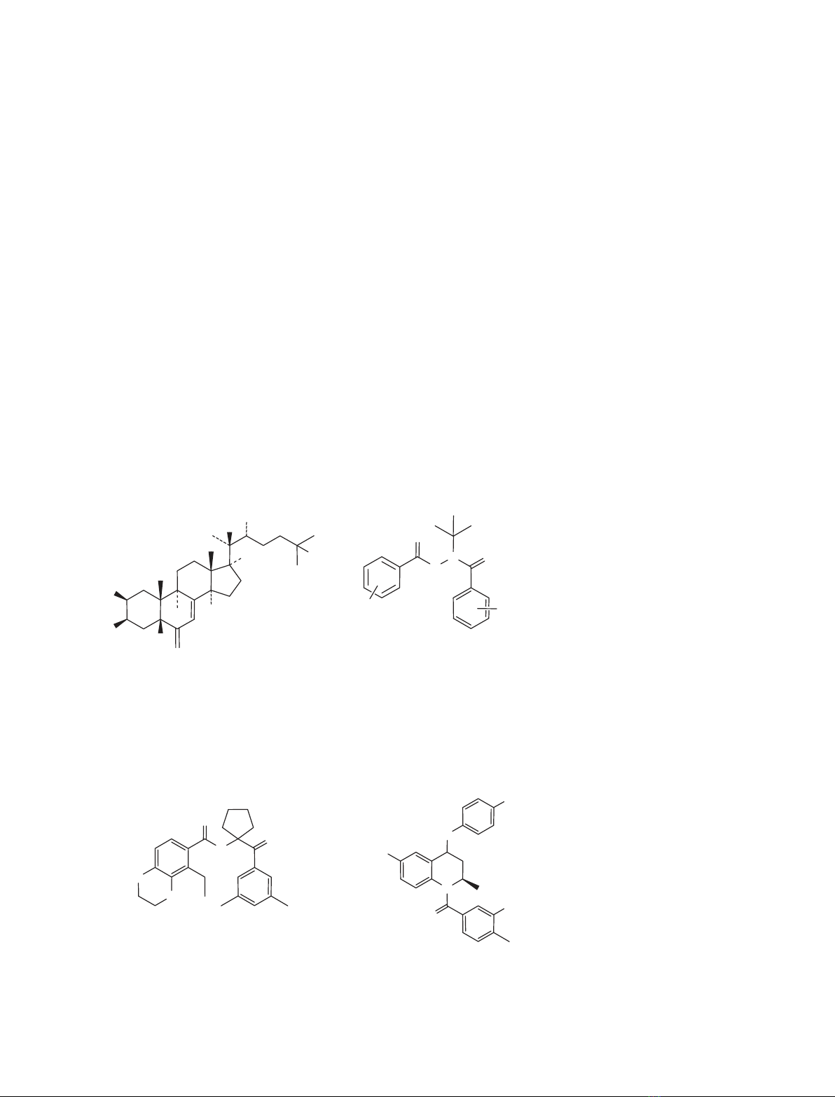
was determined. Four ligand chemotypes, represented by a tetrahydroquin-
oline (RG-120499), one amidoketone (RG-121150), two ecdysteroids
[20-hydroxyecdysone (20E) and ponasterone A (Pon A)], and four diacyl-
hydrazines (RG-102240, RG-102277, RG-102398 and RG-100864), were
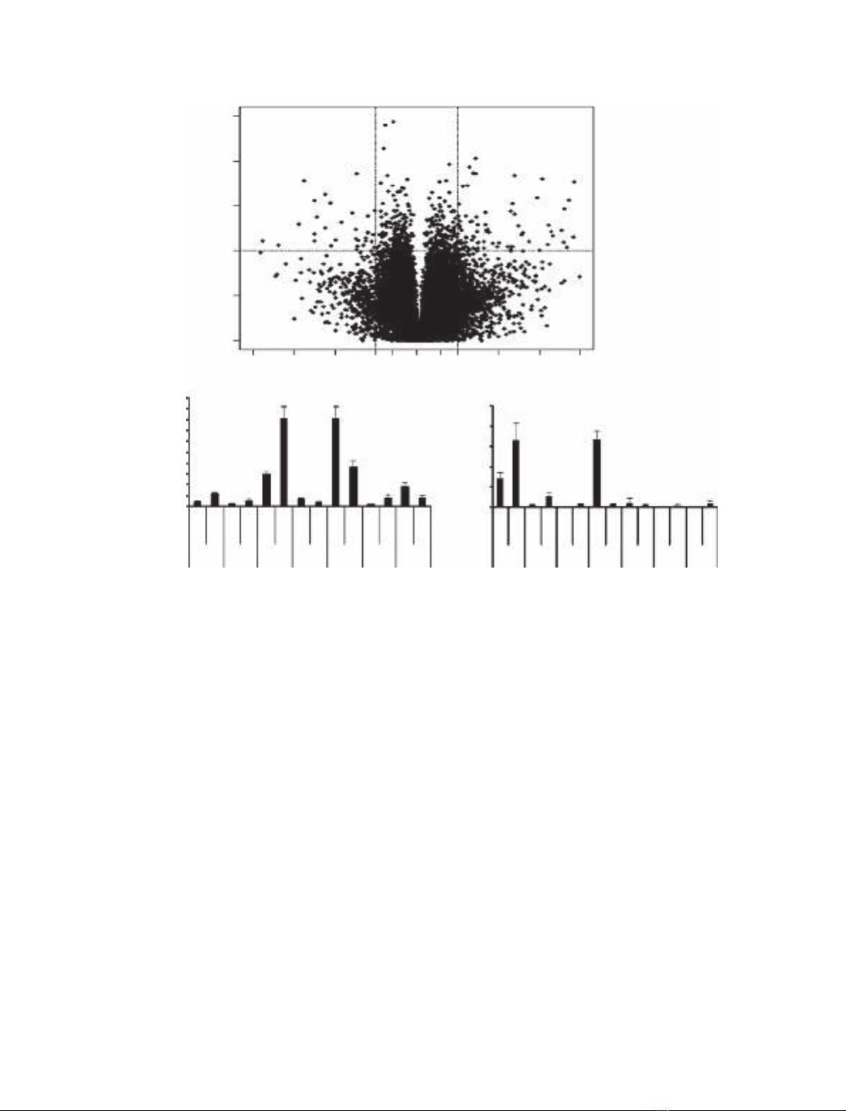
tested in HEK 293 cells. The cells were exposed to ligands at concentra-
tions of 1 lm(RG-120499) or 10 lm(all others) for 72 h and the total
RNA was isolated and analyzed using microarrays. Microarray data
showed that the tetrahydroquinoline ligand, RG-120499 caused cell death
at concentrations 10 lm.At1lm, this ligand caused changes in the
expression of genes such as TNF,MAF,Rab and Reprimo.At10lm, the
amidoketone, RG-121150, induced changes in the expression of genes such
as v-jun,FBJ and EGR, but was otherwise noninterfering. Of the two ste-
roids tested, 20E did not affect gene expression, but Pon A caused some
changes in the expression of endogenous genes. At lower concentrations
pharmacologically relevant for gene therapy, intrinsic gene expression
effects of ecdysteroids and amidoketones may actually be insignificant.
A fortiori, even at 10 lm, the four diacylhydrazine ligands did not cause
significant changes in expression of endogenous genes in 293 cells and
therefore should have minimum pleiotropic effects when used as ligands
for the ecdysone receptor gene switch.
Abbreviations
AMK, amidoketone; qRT-PCR, quantitative real-time reverse transcription PCR; STAT 6, signal transducer and activator of transcription 6;
THQ, tetrahydroquinoline.
FEBS Journal 274 (2007) 5669–5689 ª2007 The Authors Journal compilation ª2007 FEBS 5669
substantially raise the level of safety and might even
be essential to control the expression levels of proteins
that have narrow therapeutic indices, such as cyto-
kinins and hormones.
The main purpose of a regulated gene expression
system is to control the timing and levels of transgene
expression in vivo. Whether the expressed protein
remains within the cell or, more commonly, is secreted
and or distributed in extracellular compartments, it
will undergo elimination according to pharmacokinetc
principles [1]. Thus, too little protein will be subthera-
peutic, too much will be potentially toxic. Therefore, a
successful therapy would be characterized by regulated
expression of the transgene finely tuned to the chang-
ing clinical state of a patient.
Several gene switches have been developed for regu-
lating the expression of transgenes in humans [2].
More specifically, ecdysone receptor (EcR) gene
switches for medicinal purposes have been reported by
several laboratories [3–9]. Among these switches, EcR-
based gene switches display particularly low basal
activity in the absence of an inducer and strong induc-
ible activity in the presence of an inducer [2,8,10].
The relative structural dissimilarity of ecdysteroids
and mammalian steroids might suggest that binding of
the former to vertebrate steroid receptors would be too
weak for pharmacological effects, particularly adverse
ones. In support of this proposition, humans consume
significant amounts of phytoecdysteroids contained in
dietary vegetables seemingly without any apparent
detrimental effects [11]. However, Oehme et al. [12]
reported recently that ecdysteroids and ecdysone
mimics can induce and or suppress endogenous genes
in RKO and other mammalian cells and promote
apoptosis.
A more comprehensive understanding of possible
pleiotropic effects of ligand inducers and or the switch
components is essential for successful use of the EcR
gene switch for in vivo applications such as gene ther-
apy. The diacylhydrazine [13,14] nonsteroidal ecdysone
agonists, such as Rheoswitch ligand 1(RSL-1; Fig. 1),
are reported to be an excellent inducer for EcR gene
switches, supporting up to 9000-fold induction of
reporter activity [15]. Other steroidal ligands, such as
ponasterone A (Pon A; Fig. 1), have also been
reported as potential inducers of EcR-based gene
HO
HO H
HOH
OH
RH
OH
Ecdysteroids (ECD)
20-hydroxyecdysone
ponasterone
N
H
N
O
O
O
Diacylhydrazines (DAH)
RG-100864 R
1
=Cl R
2
=H
R = OH
R = H
RG-102398 R
1
=2-CH
3
, 3-OCH
3
R
2
=3,5-di-CH
3
RG-102240 R
1
=2-CH
2
CH
3
, 3-OCH
3
R
2
=3,5-di-CH
3
RG-102277 R
1
=2-CH
2
CH
3
, 3-OCH
3
R
2
=3-CH
3
, 5-C(O)NH
2
N
O
HN
F
F
F
N
H
O
O
O
O
Amidoketone (AMK)
RG-121150
Tetrahydroquinoline (THQ)
RG-120499
R1R2
Fig. 1. EcR ligands analyzed by microarray
and qRT-PCR analysis.
Ecdysone receptor gene switch ligands S. K. Panguluri et al.
5670 FEBS Journal 274 (2007) 5669–5689 ª2007 The Authors Journal compilation ª2007 FEBS
switches [8,14]. In addition, other chemotypes, such
as amidoketones (AMK) and tetrahydroquinolines
(THQ), are also being developed as inducers of EcR
gene switches [16–20] (Fig. 1).
The main goal of the present study was to determine
the intrinsic gene expression effects of EcR switch in-
ducers in mammalian cells. We studied the effect of
eight EcR ligands: four diacylhydrazines, two ecdyster-
oids, one THQ and one AMK ligand (Fig. 1) on the
expression of endogenous genes in HEK 293 cells
using microarray and quantitative real-time reverse
transcription PCR (qRT-PCR). THQ ligand caused
changes in the expression of genes such as TNF,MAF,
Rab and Reprimo. The AMK ligand induced changes
in the expression of genes such as v-jun,FBJ and
EGR. 20-Hydroxyecdysone (20E) did not affect gene
expression, but Pon A caused some changes in the
expression of endogenous genes. At lower ligand con-
centrations applicable for therapeutic use, potential
pleiotropic effects may or may not be observed. The
four diacylhydrazine ligands did not cause significant
changes in the expression of endogenous genes.
Results and Discussion
THQ ligand, RG-120499, affects 293 cells via many
pathways
Incubation of 293 cells with the THQ compound RG-
120499 at 10 lmfor 3 days resulted in the death of
70% of the cells as indicated by observation of cell
morphology. To determine the possible genes and path-
ways that are affected by this ligand, we performed a
microarray experiment using RNA isolated from
293 cells treated with 1 lmRG-120499. A total of 1171
genes were up-regulated and 443 genes were down-
regulated in 293 cells treated with this compound
compared to the cells treated with dimethylsulfoxide
(Fig. 2A). Among these, 115 genes showed P£0.01
with a two-fold or more greater in expression levels in
ligand-treated cells compared to the levels in dimethyl-
sulfoxide-treated cells (Table 1). Among these 115
genes, 55 genes showed signal detection values of more
than 100. We selected v-maf,TNF,PNAS13,Rab,Rep-
rimo,DNAH and KIF9 genes for qRT-PCR. The qRT-
PCR data (Fig. 2B,C) showed that v-maf,TNF,
PNAS13 and DNAH mRNA levels increased, and Rab
and Reprimo mRNA levels decreased in RG-120499
treated cells, compared to the levels in dimethylsulfox-
ide-treated cells. The microarray and qRT-PCR data
showed perfect correlation for these six genes. The two-
fold down-regulation of the KIF9 gene observed in
microarray analyses was not confirmed by qRT-PCR
because this method showed a ten-fold up-regulation of
this gene in the presence of RG-102499.
MAF [v-maf musculoaponeurotic fibrosarcoma
oncogene homolog (avian)] is a basic-leucine zipper
transcription factor that plays crucial roles in gene reg-
ulation, differentiation, oncogenesis and development
in many organisms [21]. v-maf is a viral oncogene
encoding a leucine zipper motif that forms heterodi-
mers with the protein products of maf-related genes or
other proteins such as fos,jun and myc oncogenes that
have leucine zipper motifs [22]. Our microarray and
qRT-PCR data showed that, in 293 cells, RG-120499
up-regulates v-maf gene expression by 2.4- and
2.3-fold, respectively. Nishizawa et al. [22] have dem-
onstrated that the human cellular counterpart of the
v-maf (c-maf) gene is conserved across species. Addi-
tionally, Massrieh et al. [21] reported that the MAF
transcription factor transcript levels are induced by
proinflammatory cytokines in PHM1-31 myometrial
cells and that MAF transcription factor mRNA is
rapidly induced by IL-1B and TNF in primary
myometrial and PHM1-13 cells. Our data also indicate
up-regulation of the TNF gene in microarray and
qRT-PCR data (by 2.2- and 5.5-fold, respectively), an
observation consistent with TNF-induced up-regulation
of MAF transcript levels. Zheng et al. [23] reported
that the tumor necrosis factor (TNF) can mediate
mature T-cell receptor-induced apoptosis through the
p75 TNF receptor. This may be the possible reason
for the death of 293 cells when treated with 10 lm
RG-120499 ligand.
In addition to the aforementioned up-regulated
genes, two genes, namely, Rab and Reprimo, were
down-regulated by this THQ ligand in 293 cells, as
observed by both microarray and qRT-PCR tech-
niques. The Rab proteins constitute a subfamily of
Ras-related GTP-binding proteins that are localized in
distinct intracellular compartments [24]. Mutations in
the Rab gene can alter the morphology of entire organ-
elles by blocking protein transport along the exocytic
and endocytic pathways because Rab proteins plays a
key role in membrane trafficking [25]. Barbosa et al.
[26] reported that mutations in the Rab gene(s) can
cause irregularities in the protein transport machinery
leading to the formation of giant lysosomes in mouse
beige (bg) mutant and other mutant mice. It has been
suggested that Chediak–Higashi syndrome, a rare
autosomal recessive disorder in humans, is the conse-
quence of a mutation to a homolog of bg. Our micro-
array and qRT-PCR data indicate that the rab gene was
down-regulated in 293 cells by two- and 23-fold, respec-
tively, by the THQ, RG-120499. Mutation studies of the
Rab gene suggest that the Rab gene down-regulation by
S. K. Panguluri et al.Ecdysone receptor gene switch ligands
FEBS Journal 274 (2007) 5669–5689 ª2007 The Authors Journal compilation ª2007 FEBS 5671
this ligand may be a reason for cell death in addition to
modulation of the v-maf and TNF genes.
Tumor suppressor genes that encode transcriptional
factors can affect a variety of cellular mechanisms
underlying growth, differentiation, and apoptosis
[27,28]. Also, when cells were exposed to DNA dam-
age-inducing agents or other noxious stress, the p53
protein, which is the most commonly mutated gene in
human cancer, is induced and or activated, resulting in
cell cycle arrest or apoptosis [29–31]. Reprimo is a
highly glycosylated protein, which will localize in the
cytoplasm and induce G2 arrest of the cell cycle when
expressed ectopically [32]. In the present study, it was
observed that RG-120499 down-regulates the Reprimo
gene by two-fold as measured by both microarray and
qRT-PCR techniques. From these observations and
previous reports, we suggest that the down-regulation
of the Reprimo gene may cause loss of DNA repair,
which, in turn, is passed on to the next generations,
thereby accumulating DNA defects. RG-120499 modu-
lates the expression of other genes as well. The
PNAS123 gene (transformation related protein 11) is
up-regulated by 2.7- and 4.3-fold as measured by
microarray and qRT-PCR, respectively. The pathways
in which these genes are involved are not known. RG-
120499 also triggers five- and 23-fold up-regulation of
the DNAH (human axonemal dynein heavy chain)
gene as measured by microarray and qRT-PCR tech-
niques, respectively. DNAH is a microtubule associ-
ated motor protein that moves cilia and flagella
[33,34]. Afzelius [35,36] showed that patients suffering
from Kartagener syndrome have cilia lacking dynein
A
BC
1E-051E-041E-03
Significance (t test p-value)
1E-021E-011E-00
/16
5000 0.025
0.02
0.015
0.01
0.005
0
2.7
2.3
5.5
4.3 -23 -2 23 10
2.4
v-Maf
DMSO
THQ
DMSO
THQ
DMSO
THQ
DMSO
THQ
DMSO
THQ
DMSO
THQ
DMSO
THQ
TNF PNAS RAB Reprimo DNAH KIF v-Maf
DMSO
THQ
DMSO
THQ
DMSO
THQ
DMSO
THQ
DMSO
THQ
DMSO
THQ
DMSO
THQ
TNF PNAS RAB Reprimo DNAH KIF
2.2 -2
-2
5.1 -2
4500
4000
3500
3000
2500
2000
Signal Values
Relative Expression
1500
1000
500
0
/8 /4 /2 /1.5
Fold Suppression/Induction
1x1.5 x4 x8 x16
Fig. 2. The THQ ligand RG-120499 affects 293 cells via many pathways. (A) The V-plot of differentially expressed genes from microarray
data. The P-values of t-test are plotted against fold suppression or induction. The horizontal bar in the plot represents the nominal significant
level 0.001 for the t-test under the assumption that each gene has a unique variance. The vertical bars represent the genes that are mini-
mum of two-fold up- or down-regulated compared to the control dimethylsulfoxide (DMSO). (B) The signal values of v-maf,TNF,PNAS,Rab,
Reprimo,DNAH and KIF9 genes from the microarray. The signal values from the microarray analysis were plotted for each gene are
indicated as mean ± SD (n¼3). The numbers above the bar represents the fold changes with this ligand against dimethylsulfoxide. (C) The
relative expressions of v-maf,TNF,PNAS,Rab,Reprimo,DNAH and KIF9 gene transcripts in qRT-PCR. The relative expression values from
the qRT-PCR analysis were plotted for each gene are indicated as mean ± SD (n¼3). The numbers above the bar represents the fold
changes with this ligand against dimethylsulfoxide.
Ecdysone receptor gene switch ligands S. K. Panguluri et al.
5672 FEBS Journal 274 (2007) 5669–5689 ª2007 The Authors Journal compilation ª2007 FEBS
arms. This disease is characterized by chronic respira-
tory tract infections, altered position of internal
organs, and infertility arising from immotile sperm.
Milisav and Affara [37] reported that the human
dynein-related gene DNEL2 may play an important
role specifically in sperm motility and is not involved
in the movement of cilia. In summary, the use of the
THQ gene switch ligand RG-120499 may cause signifi-
cant changes in the expression of host genes.
AMK, RG-121150, affects gene expression in
293 cells
The affect of the AMK RG-121150 on gene expression
in 293 cells was analyzed using microarray and qRT-
PCR. In microarray analysis, a total of 636 genes were
up-regulated and 604 genes were down-regulated by
this ligand (Fig. 3A). Among these genes, 71 genes
showed P£0.01 and a two-fold or greater change in
expression (Table 1). Among 71 genes, 24 genes
showed signal detection values greater than 100.
Among these are hypothetical proteins, nuclear pro-
teins, transcriptional factors, glycogen phosphorylase,
hormone degrading enzymes, kinases and some solute
carrier proteins. To validate microarray data with
qRT-PCR, the primers for early growth response gene
(EGR), FBJ,v-jun, and a hypothetical protein gene
were designed. For three of the four genes, the micro-
array data was confirmed by qRT-PCR. The data
showed that the hypothetical protein gene was up-regu-
lated by two- and 1.5-fold in microarray and qRT-PCR
experiments, respectively. EGR and v-jun showed sup-
pression of their expression by the ligand in analyses by
both methods (Fig. 3B,C). The gene FBJ mRNA levels
were down-regulated in microarray experiments and up-
regulated in qRT-PCR experiments.
The EGR gene product is a transcription factor that
plays a role in differentiation and growth. EGR genes
are transiently and coordinately induced upon activa-
tion of peripheral blood T lymphocytes [38,39]. These
EGR genes are also expressed in a wide range of cell
types, including lymphoid cells, myeloid cells such as
thymocytes, B cells, monocytes, and nonlymphoid cells
such as fibroblasts, kidney cells and neurons [40,41].
Huang et al. [42,43] showed that the expression of the
EGR gene exogenously in various tumor cells unex-
pectedly and markedly reduces growth and tumorige-
nicity, whereas the suppression of endogenous EGR
by antisense RNA enhances growth and promotes
phenotypic transformation. From our microarray and
qRT-PCR data, the AMK RG-121150 caused down-
regulation of this EGR gene by five- and six-fold,
Table 1. Differentially expressed genes in 293 cells treated with different ligands.
Ligand P-value
Total genes Genes with signal values 100 or more
Up-regulated Down-regulated Total Up-regulated Down-regulated Total
RG-120499 (THQ) < 0.0001 1 0 1 1 0 1
< 0.001 12 5 17 8 0 8
< 0.01 71 26 97 39 7 46
RG-121150 (AMK) < 0.0001 0 1 1 0 1 1
< 0.001 4 9 13 0 6 6
< 0.01 26 31 57 6 11 17
20E (steroid) < 0.0001 0 0 0 0 0 0
< 0.001 1 5 6 0 1 1
< 0.01 15 30 45 0 4 4
Pon A (steroid) < 0.0001 0 1 1 0 0 0
< 0.001 2 4 6 0 0 0
< 0.01 20 15 35 1 2 3
RG-100864
(halofenozide, diacylhydrazine)
< 0.0001 0 0 0 0 0 0
< 0.001 1 3 4 0 0 0
< 0.01 19 15 34 2 1 3
RG-102398
(methoxyfenozide, diacylhydrazine)
< 0.0001 0 0 0 0 0 0
< 0.001 3 4 7 0 0 0
< 0.01 20 25 45 1 2 3
RG-102240
(RSL-1, diacylhydrazine)
< 0.0001 0 1 1 0 1 1
< 0.001 2 2 4 0 0 0
< 0.01 27 14 41 3 2 5
RG-102277 (diacylhydrazine) < 0.0001 0 0 0 0 0 0
< 0.001 3 3 6 1 1 2
< 0.01 21 18 39 0 3 3
S. K. Panguluri et al.Ecdysone receptor gene switch ligands
FEBS Journal 274 (2007) 5669–5689 ª2007 The Authors Journal compilation ª2007 FEBS 5673