
BioMed Central
Page 1 of 12
(page number not for citation purposes)
Virology Journal
Open Access
Research
The directionality of the nuclear transport of the influenza A
genome is driven by selective exposure of nuclear localization
sequences on nucleoprotein
Winco WH Wu and Nelly Panté*
Address: Department of Zoology, University of British Columbia, 6270 University Boulevard, Vancouver, British Columbia, V6T 1Z4, Canada
Email: Winco WH Wu - winco@zoology.ubc.ca; Nelly Panté* - pante@zoology.ubc.ca
* Corresponding author
Abstract
Background: Early in infection, the genome of the influenza A virus, consisting of eight complexes
of RNA and proteins (termed viral ribonucleoproteins; vRNPs), enters the nucleus of infected cells
for replication. Incoming vRNPs are imported into the nucleus of infected cells using at least two
nuclear localization sequences on nucleoprotein (NP; NLS1 at the N terminus, and NLS2 in the
middle of the protein). Progeny vRNP assembly occurs in the nucleus, and later in infection, these
are exported from the nucleus to the cytoplasm. Nuclear-exported vRNPs are different from
incoming vRNPs in that they are prevented from re-entering the nucleus. Why nuclear-exported
vRNPs do not re-enter the nucleus is unknown.
Results: To test our hypothesis that the exposure of NLSs on the vRNP regulates the
directionality of the nuclear transport of the influenza vRNPs, we immunolabeled the two NLSs of
NP (NLS1 and NLS2) and analyzed their surface accessibility in cells infected with the influenza A
virus. We found that the NLS1 epitope on NP was exposed throughout the infected cells, but the
NLS2 epitope on NP was only exposed in the nucleus of the infected cells. Addition of the nuclear
export inhibitor leptomycin B further revealed that NLS1 is no longer exposed in cytoplasmic NP
and vRNPs that have already undergone nuclear export. Similar immunolabeling studies in the
presence of leptomycin B and with cells transfected with the cDNA of NP revealed that the NLS1
on NP is hidden in nuclear exported-NP.
Conclusion: NLS1 mediates the nuclear import of newly-synthesized NP and incoming vRNPs.
This NLS becomes hidden on nuclear-exported NP and nuclear-exported vRNPs. Thus the
selective exposure of the NLS1 constitutes a critical mechanism to regulate the directionality of
the nuclear transport of vRNPs during the influenza A viral life cycle.
Background
The influenza A virus exploits the cellular nuclear trans-
port machinery several times during infection (reviewed
in [1]). Early in infection, the influenza A viral genome –
consisting of eight complexes of RNA and proteins (ribo-
nucleoproteins; vRNPs) – is released into the cytoplasm
and imported into the nucleus for replication. Subse-
quently, newly-synthesized viral proteins from the cyto-
plasm enter the nucleus to form newly-synthesized
vRNPs. Later in infection, newly-assembled vRNPs are
Published: 2 June 2009
Virology Journal 2009, 6:68 doi:10.1186/1743-422X-6-68
Received: 9 April 2009
Accepted: 2 June 2009
This article is available from: http://www.virologyj.com/content/6/1/68
© 2009 Wu and Panté; licensee BioMed Central Ltd.
This is an Open Access article distributed under the terms of the Creative Commons Attribution License (http://creativecommons.org/licenses/by/2.0),
which permits unrestricted use, distribution, and reproduction in any medium, provided the original work is properly cited.

Virology Journal 2009, 6:68 http://www.virologyj.com/content/6/1/68
Page 2 of 12
(page number not for citation purposes)
exported from the nucleus to the cytoplasm to allow for
their packaging into progeny virions. The vRNPs contain
multiple copies (up to 97) of viral nucleoprotein (NP; 56
kDa) forming a core around which the RNA is helically
wrapped (reviewed in [2]). Each NP monomer has at least
two nuclear localization sequences (NLS1, spanning resi-
dues 1–13 at the N terminus, and NLS2, spanning resi-
dues 198–216 in the middle of the protein) that mediate
the nuclear import of NP and vRNPs [3-7]. We have pre-
viously found that both NLS1 and NLS2 on NP are
responsible for mediating the nuclear import of vRNPs
purified from influenza A virions in permeabilized cells
[7]. We also found that NLS1 of NP is the principal medi-
ator of the nuclear import of incoming vRNPs because
NLS1 has higher surface accessibility than NLS2, both
within each vRNP molecule and on a greater number of
vRNP molecules [8].
Within the nucleus, the original incoming and newly-syn-
thesized negative-sense vRNAs act as templates to tran-
scribe the positive mRNA strand, which is selectively
exported into the cytoplasm and used to translate new
viral proteins (reviewed in [9]). Some of the newly-syn-
thesized viral proteins (NP; the RNA polymerases PA,
PB1, and PB2; the nonstructural protein NS1; the matrix
protein M1) are then imported into the nucleus through
their respective NLSs. In the nucleus, the newly-synthe-
sized NP, PB1, PB2, PA, and the vRNA assemble into new
vRNPs (reviewed in [10]). Subsequently, the newly-
assembled vRNPs use the cellular export receptor CRM1
to exit the nucleus through the nuclear pore complexes
[11-13].
Nuclear-exported vRNPs are different from incoming
vRNPs in that they are somehow prevented from being
imported back into the nucleus. It has been demonstrated
that association of the vRNPs with the viral protein M1
regulates nuclear trafficking of influenza vRNPs [14,15].
However details of how M1 prevents newly-assembled
vRNPs from re-entering the nucleus is unknown. Our
hypothesis is that the NLSs on NP are the key determi-
nants for the nuclear transport directionality of the vRNPs
by possessing differential exposure. To test this hypothe-
sis, we analyzed the exposure of the NLSs on NP in tissue
culture cells infected with influenza A virus. We found
that an exposed NLS1 on NP allows newly-synthesized NP
to enter the nucleus, but NLS1 becomes masked or hidden
once the progeny vRNPs undergo nuclear export. Hidden
NLSs on the nuclear-exported vRNPs prevents the nuclear
re-entry of the progeny vRNPs. This selective exposure and
masking of NLS1 on vRNPs thus constitutes a critical
mechanism to regulate the directionality of the nuclear
transport of the influenza vRNPs.
Results
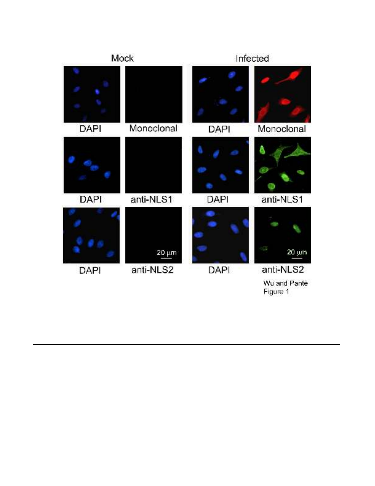
Specificity of NP antibodies
We have previously generated and characterized two pol-
yclonal anti-peptide antibodies that specifically recognize
NLS1 and NLS2 on NP [7,8]. In this study, we used these
anti-NLS antibodies to analyze the exposure of these NLSs
within cells infected with influenza A virus or transfected
with the cDNA of NP. Total NP was detected by using a
monoclonal antibody specific for NP. To ensure that all
three of the NP monoclonal, anti-NLS1, and anti-NLS2
antibodies were specific for NP and not for components
of the cell, we first compared the antibody labeling in
infected cells with that in mock-infected cells. We found
that each of the respective antibodies gave a strong signal
in infected cells compared with mock-infected cells in
which no virus was added (Fig. 1). A similar specificity of
the anti-NP monoclonal, anti-NLS1, and anti-NLS2 anti-
bodies was observed in cells transfected with the cDNA of
NP compared with mock-transfected cells (results not
shown).
Besides testing for the specificity of the anti-NP antibod-
ies, the results from Fig. 1 also indicated that NLS1 was
generally more exposed than NLS2, and exposed in a
greater number of influenza A virus-infected cells. This is
in agreement with our previous studies examining the
immunogold labeling of purified vRNPs with the anti-
NLS1 or anti-NLS2 antibodies [8], and with our conclu-
sion that NLS1 is stronger that NLS2 in mediating the
nuclear import of the influenza vRNPs [7].
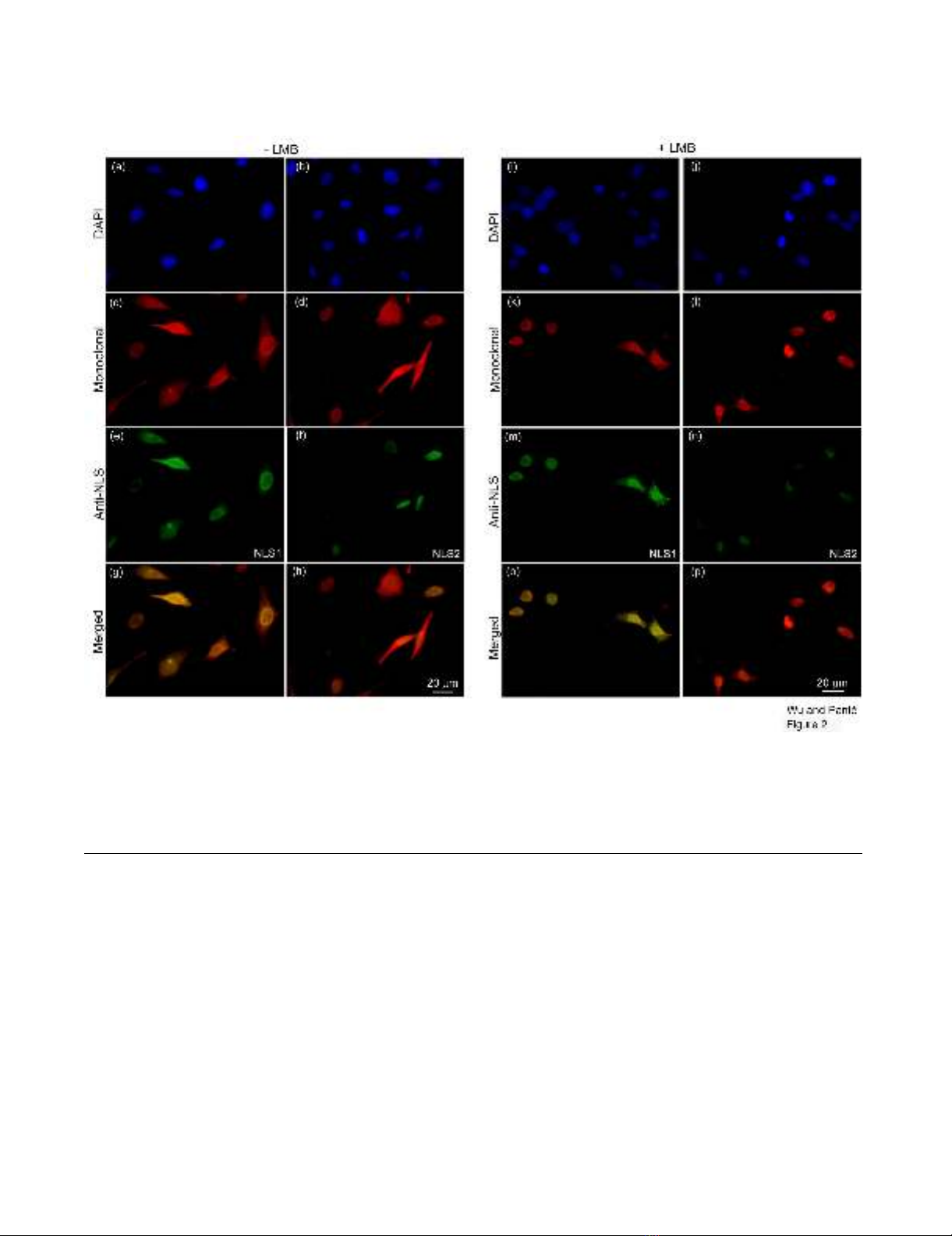
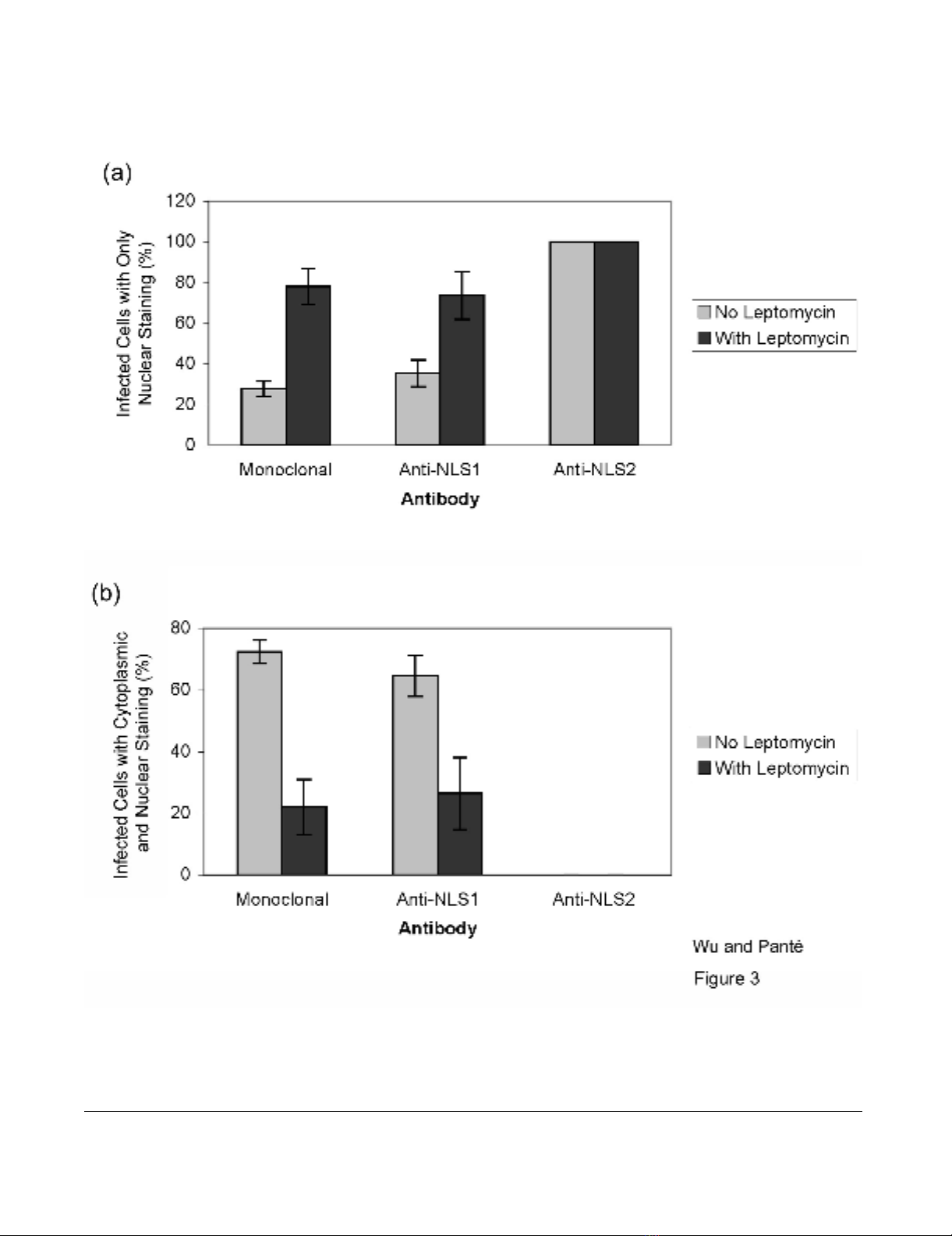
Exposure of NLS1 and NLS2 in influenza-infected cells
We performed double-immunolabeling studies with the
monoclonal NP antibody in conjunction with either the
polyclonal NP anti-NLS1 or with the polyclonal NP anti-
NLS2 antibody to analyze the exposure of the NLSs in
cells infected with the influenza A virus. As illustrated in
Fig. 2, the NP monoclonal antibody detected NP in both
the nucleus and cytoplasm of infected cells (Fig. 2c–d),
with 28% of the infected cells showing only nuclear stain-
ing (Fig. 3a). Similarly, the NLS1 epitope on NP was
exposed in both the nucleus and cytoplasm (Fig. 2e). In
contrast, the NLS2 epitope was only exposed in the
nucleus of the infected cells (Fig. 2f). Quantitative analy-
sis showed that 100% of the infected cells labeled with the
anti-NLS2 antibody had only nuclear staining of anti-
NLS2, while 35% of the infected cells labeled with the
anti-NLS1 antibody had only nuclear staining of anti-
NLS1 (Fig. 3a).
To distinguish between incoming vRNPs and newly syn-
thesized NP and progeny vRNPs, we next performed a
similar double-immunolabeling experiment with cells
infected with influenza A virus in the presence of
cycloheximide (a protein synthesis inhibitor). As illus-
Virology Journal 2009, 6:68 http://www.virologyj.com/content/6/1/68
Page 3 of 12
(page number not for citation purposes)
trated in Fig. 4, there was no NP fluorescence signal in
cells treated with cycloheximide. This indicates that the
NP being labeled in the infected cells (Fig. 2) represents
indeed newly-synthesized NP. Therefore, this limits the
type of cytoplasmic NP detected in infected cells to be
either newly-synthesized NP or newly-assembled vRNPs
that have undergone nuclear export.
From the above results, it was unclear why these infected
cells did not contain an exposed NLS2 in the cytoplasm
even though the cells contained NP in the cytoplasm. The
experiment with cycloheximide helped us to conclude
that the cytoplasmic NP does not represent incoming
vRNPs. To distinguish whether the cytoplasmic NP is
newly-synthesized NP or nuclear-exported vRNPs, we
used leptomycin B (LMB) to inhibit the nuclear export of
vRNPs. These experiments with LMB detect newly synthe-
sized vRNPs that is trapped in the nucleus. LMB has been
successfully used in the past to inhibit the nuclear export
of vRNPs in infected cells [11,13]. We repeated these
experiments in the presence of LMB, to block vRNP
nuclear export and to determine whether the cytoplasmic
NP in the infected cells represented newly-synthesized NP
or nuclear-exported vRNPs. As documented in Fig. 2k–l,
and Fig. 3a, we found that in the presence of LMB 78% of
the infected cells showed only nuclear, and no cytoplas-
Specificity of NP antibodiesFigure 1
Specificity of NP antibodies. Immunofluorescence microscopy of HeLa cells infected with the influenza A virus and immu-
nolabeled with the monoclonal NP antibody, or the polyclonal anti-peptide antibodies that recognize the NLS1 and the NLS2
epitopes of NP. DAPI, a DNA marker, was used to determine the total number of cells present. As a control, a mock infection
without influenza A virus was also performed. Cells were fixed and prepared for immunofluorescence microscopy 17 hours
after infection.
Virology Journal 2009, 6:68 http://www.virologyj.com/content/6/1/68
Page 4 of 12
(page number not for citation purposes)
mic, NP. Quantitative analysis showed that 22% of the
infected cells, however, also still showed cytoplasmic NP
in addition to nuclear NP accumulation (Fig. 3b). Because
we were inhibiting nuclear export, this cytoplasmic NP
represents newly-synthesized NP that had not yet under-
gone nuclear import.
Consistent with the notion that there were two pools of
cytoplasmic NP in infected cells untreated with LMB
(newly-synthesized NP and newly-assembled vRNPs that
have undergone nuclear export), the experiment in the
presence of LMB yielded cells in which the fluorescence
intensity of the cytoplasmic NP was less intense than from
cells without LMB. Of particular note, this cytoplasmic NP
contained an exposed NLS1 (Fig. 2m). In fact, quantita-
tive analysis showed that 26% of infected cells in the pres-
ence of LMB still contained both cytoplasmic and nuclear
immunostaining with the anti-NLS1 antibody (Fig. 3b).
This indicates that newly-synthesized cytoplasmic NP that
had not yet undergone nuclear import contains an
exposed NLS1 epitope.
A longer time point in infected cells (30 hours instead of
17 hours) was also performed, and there was even less,
but still a small amount of cytoplasmic NP staining from
both the monoclonal and the anti-NLS1 antibodies
(results not shown), indicating that more NP had under-
gone nuclear import. Taken together, these results indi-
Exposure of NLS1 and NLS2 in influenza-infected cellsFigure 2
Exposure of NLS1 and NLS2 in influenza-infected cells. HeLa cells infected with influenza A virus for 17 hours, in the
absence (a-h) or presence (i-p) of the nuclear export inhibitor LMB, were immunolabeled with DAPI (a-b and i-j; blue), a
monoclonal anti-NP antibody (c-d and k-l; red), and either the polyclonal anti-NLS1 antibody (e and m; green) or the polyclo-
nal anti-NLS2 antibody (f and n; green). Merged images depict merge of the red and green channels for each respective set of
cells.
Virology Journal 2009, 6:68 http://www.virologyj.com/content/6/1/68
Page 5 of 12
(page number not for citation purposes)
Quantification of the exposure of NLS1 and NLS2 in influenza-infected cellsFigure 3
Quantification of the exposure of NLS1 and NLS2 in influenza-infected cells. Bar graphs of the percentage of
infected cells showing fluorescent staining only in the nucleus (a) or both in the cytoplasm and the nucleus (b) for the experi-
mental conditions described in Fig. 2. Data shows the mean values and standard error scored from 152 and 82 infected cells in
the absence and presence of LMB, respectively.


