
BioMed Central
Page 1 of 9
(page number not for citation purposes)
World Journal of Surgical Oncology
Open Access
Research
The importance of rectal cancer MRI protocols on iInterpretation
accuracy
Chikako Suzuki1, Michael R Torkzad*2,3, Soichi Tanaka4, Gabriella Palmer4,
Johan Lindholm5, Torbjörn Holm4 and Lennart Blomqvist6
Address: 1Department of Diagnostic Radiology, Institution for Molecular Medicine and Surgery, Karolinska University Hospital Solna and
Karolinska Institute, Stockholm, Sweden, 2Department of Radiology, Uppsala University Hospital, Uppsala, Sweden, 3Dept. of Oncology,
Radiology and Clinical Immunology Section of Radiology Uppsala University Hospital and Karolinska Institute, Uppsala, Sweden, 4Department
of Surgery, Institution for Molecular Medicine and Surgery, Karolinska University Hospital Solna and Karolinska Institute, Stockholm, Sweden,
5Department of Pathology, Karolinska University Hospital Solna and Karolinska Institute, Stockholm, Sweden and 6Department of radiology,
Danderyd Hospital, Stockholm, and Karolinska Institute, Stockholm, Sweden
Email: Chikako Suzuki - chikasakit@yahoo.co.jp; Michael R Torkzad* - mictor@ki.se; Soichi Tanaka - soh368@hotmail.com;
Gabriella Palmer - gabriella.jansson-palmer@karolinska.se; Johan Lindholm - johan.lindholm@karolinska.se;
Torbjörn Holm - torbjorn.holm@karolinska.se; Lennart Blomqvist - lennart.k.blomqvist@ki.se
* Corresponding author
Abstract
Background: Magnetic resonance imaging (MRI) is used for preoperative local staging in patients
with rectal cancer. Our aim was to retrospectively study the effects of the imaging protocol on the
staging accuracy.
Patients and methods: MR-examinations of 37 patients with locally advanced disease were
divided into two groups; compliant and noncompliant, based on the imaging protocol, without
knowledge of the histopathological results. A compliant rectal cancer imaging protocol was defined
as including T2-weighted imaging in the sagittal and axial planes with supplementary coronal in low
rectal tumors, alongside a high-resolution plane perpendicular to the rectum at the level of the
primary tumor. Protocols not complying with these criteria were defined as noncompliant.
Histopathological results were used as gold standard.
Results: Compliant rectal imaging protocols showed significantly better correlation with
histopathological results regarding assessment of anterior organ involvement (sensitivity and
specificity rates in compliant group were 86% and 94%, respectively vs. 50% and 33% in the
noncompliant group). Compliant imaging protocols also used statistically significantly smaller voxel
sizes and fewer number of MR sequences than the noncompliant protocols
Conclusion: Appropriate MR imaging protocols enable more accurate local staging of locally
advanced rectal tumors with less number of sequences and without intravenous gadolinium
contrast agents.
Published: 20 August 2008
World Journal of Surgical Oncology 2008, 6:89 doi:10.1186/1477-7819-6-89
Received: 27 May 2008
Accepted: 20 August 2008
This article is available from: http://www.wjso.com/content/6/1/89
© 2008 Suzuki et al; licensee BioMed Central Ltd.
This is an Open Access article distributed under the terms of the Creative Commons Attribution License (http://creativecommons.org/licenses/by/2.0),
which permits unrestricted use, distribution, and reproduction in any medium, provided the original work is properly cited.

World Journal of Surgical Oncology 2008, 6:89 http://www.wjso.com/content/6/1/89
Page 2 of 9
(page number not for citation purposes)
Background
Total mesorectal excision (TME) is the standard surgical
treatment used for patients with primary rectal cancer.
TME involves removal of a distinct anatomic compart-
ment, the mesorectum, containing the rectal tumor, all
local draining nodes and the mesorectal fat, by means of
sharp dissection along the mesorectal fascia [1-3]. There is
substantial evidence for efficacy of neoadjuvant therapy in
combination with TME as being important to reduce local
tumor recurrence rates [4-7]. When performing TME,
knowledge of the relationship of the tumor to the circum-
ferential resection margin (CRM) is of importance. When
CRM is involved by the tumor, the risk of local recurrence
is high [8-16]. The local prognostic factors assessed at pre-
operative magnetic resonance imaging (MRI) of rectal
cancer include the extent of extramural tumor spread,
involvement of the lateral resection margin, involvement
of neighboring organs in the pelvis, presence of local
lymph node metastases, extramural lymphovascular infil-
tration and peritoneal involvement [15,17]. This informa-
tion helps select patients who should receive neoadjuvant
treatment. This applies especially to cases with locally
advanced rectal cancer, in order to maximize the chances
of a complete resection and survival [18,19], and at the
same time, to minimize morbidity and loss of quality of
life. It is therefore of paramount interest to provide
detailed anatomic knowledge of tumor and tumor inva-
sion toward neighboring organs before treatment.
Although evaluated in several studies during the past two
decades, it is only during recent years that MRI gained
wide acceptance as a valuable method for assessment in
patients with rectal cancer [20-33].
As a tertiary referral center responsible for patients with
advanced rectal cancer, we assess magnetic resonance
(MR) examinations from other institutions and hospitals
at multidisciplinary team (MDT) meetings. When demon-
strating these examinations at MDT meetings, variations
in imaging sequences among different centers are noted.
These differences may be related to both different equip-
ments and level of dedicated experience in pelvic MRI.
To our knowledge, no study has reported the importance
of the imaging protocol for assessment of tumor involve-
ment of neighboring organs in locally advanced rectal
cancer. The aim of the present study was to compare the
equivalence between MRI and histopathology in patients
with locally advanced rectal cancer based on the effects of
using different MRI protocols.
Patients and methods
Forty-one patients assessed as clinically suspicious for
locally advanced primary rectal cancer by surgeons from
2000 to 2005, were included. 37 patients, 27 male and 10
female, with a mean age of 60.1 ± 9.8 (mean ± SD, range
28–79) who had available MRI of the pelvis were studied
further. The surgeon's decision that a cancer might be
advanced was based on findings at diagnostic laparotomy
and/or by means of digital rectal examination.
Radiological assessment
All examinations were provided from ten different hospi-
tals or institutions (two of which were university hospi-
tals). Each MR examination (all done on 1.5 T) was
assessed by two or three radiologists (C.T., M.R.T. and
L.B.) in consensus without knowledge of the clinical and
histopathological results prior to this study according to a
standard evaluation looking specifically at which organs
and/or structures had been involved. However, the radiol-
ogists were aware of the high suspicion for locally
advanced tumors by the clinicians. Radiologists had eval-
uated the morphological characteristics of the primary
tumor, local prognostic factors including threatening or
involvement of the mesorectal fascia, and adjacent organs
in each patient.
For the part of this study, anterior organs were defined as
those positioned ventral to the rectum and included the
seminal vesicles, the prostate gland, the perineal body,
uterus, vagina, ovaries, the small and large intestines, and
the urinary bladder. Inferior and posterior organs had
been defined as those that were located inferior and dorsal
to the rectum, respectively, and included the levator ani
muscles, obturator muscles, piriformis muscles and the
sacral bone. Involvement of the abovementioned organs
was defined as T4-tumor stage.
The imaging protocol of each MR-examination was
recorded by one author (C.T.). Those examinations that
showed the following prerequisites were defined as com-
pliant rectal imaging protocol vs. those that did not dem-
onstrate the same sequences (called henceforth
noncompliant):
1. Sagittal and axial T2-weighted images of the pelvis per-
formed,
2. T2-weighted images with equal to or less than 3 mm
slice thickness perpendicular to the rectal length at the
level of the tumor with a 16–20 cm field of view and at
least a 256 × 256 matrix, otherwise called 'high resolution
imaging' [20,21,25,34].
3. For low rectal tumors, coronal imaging obtained.
If the patients underwent MR examinations twice but at
two different institutions, with different protocols, one
compliant and the other non compliant; these were noted
separately as combination protocol but categorized with

World Journal of Surgical Oncology 2008, 6:89 http://www.wjso.com/content/6/1/89
Page 3 of 9
(page number not for citation purposes)
the compliant group regarding some aspects. The number
of other sequences and different types of artifacts (if dis-
tinguishable) were also noted.
The common denominators of all MR examinations,
whether compliant or otherwise, were that they had to be
performed on the request of a surgeon or oncologist for
assessment of local extension of the rectal tumor preoper-
atively, and that the radiologist at the primary institution
had not called the examination incomplete.
Histopathological examination
All evaluations were performed according to the protocol
of Quirke, et al [16,35], by one pathologist (J.L.) with
more than 10 years of experience in gastrointestinal
pathology. The pathologist was blinded to the MRI study
protocol. The tumor site was sliced transversely at 0.5–
1.0-cm intervals. The extent of tumor spread into mes-
orectal fascia and other structures or organs was assessed
both macroscopically and with high magnification.
Tumor extension into the surrounding structures and
organs at microscopical examination were used as the
standard of reference against which MRI findings were
compared. The extension of tumor cells into mesorectal
fascia and other structures or organs was assessed from
inspection of the histological macrosection by light
microscopy at 20× – 200× magnification.
Statistical analysis
All MRI findings including the size of tumor, the name
and number of involved fascia(e) and organ(s), the pat-
tern of tumor involvement according to MRI and histopa-
thology as well as the MR imaging protocol were recorded
using Microsoft Excel 2003 and Microsoft Access 2000.
Sensitivity and specificity of MRI between different groups
were compared and 95% confidence interval (CI) was cal-
culated with P-value < 0.05 considered significant using
Stat View J-5.0 (SAS Institute. Inc., Cary, NC).
Ethical considerations
The study was approved by the local ethical committee.
No separate informed consent was obtained for this retro-
spective study.
Results
Tumor staging according to MRI
Nineteen patients were evaluated as T4 rectal tumors
based on MRI. The remaining 18 were evaluated as T3
tumors without obvious invasion of neighboring organ.
Assessment of imaging quality
Eleven patients were assessed as having compliant (D)
protocols and 13 patients as combination protocols (C)
and 13 patients a noncompliant imaging (N).
Regarding imaging parameters, compliant imaging proto-
cols were used with smaller field of view (FOV) (D, 201.7
± 77.0 mm; N, 263.5 ± 129.8 mm; mean ± SD, p = 0.03),
thinner slice thickness (D, 3.8 ± 1.4 mm; N, 5.3 ± 1.9 mm;
mean ± SD, p < 0.01), smaller slice gap (D, 0.2 ± 0.9 mm;
N 2.0 ± 2.4 mm; mean ± SD, p < 0.01) and smaller voxel
size (D, 1.3 ± 1.5 mm3; N, 6.7 ± 6.0 mm3; mean ± SD, p <
0.01). The total number of MR sequences performed in
each patient was also larger in the N group (N, 9.2 ± 3.2
sequences vs. D, 5.2 ± 0.7 sequences; mean ± SD, p < 0.01
(table 1). One patient from the noncompliant group had
some motion artifacts.
Involvement of the anterior organs
In the group with compliant protocols and the group with
combination protocol, preoperative MRI indicated tumor
involvement of anterior pelvic organs in seven out of the
24 patients. Compared to pathological examination, six
cases were true positives and one was false positive.
Among the remaining 17 patients without organ involve-
ment on MRI, pathological examination revealed one
false negative case and 16 true negatives (table 2). Figure
Table 1: Comparison of various MR imaging parameters, average number of sequences in each group and imaging protocols.
Compliant protocol (D) Noncompliant protocol (N) P-value
Parameters on T2-WI*
Field of view
Mean ± SD (mm) 201.7 ± 77.0 263.5 ± 129.8 0.03
Slice thickness
Mean ± SD (mm) 3.8 ± 1.4 5.3 ± 1.9 < 0.01
Gap
Mean ± SD (mm) 0.2 ± 0.9 2.0 ± 2.4 < 0.01
Matrix size
Mean (mm × mm) 0.5 × 0.5 0.9 × 1.1 0.02
Voxel size
Mean ± SD (mm3) 1.3 ± 1.5 6.7 ± 6.0 < 0.01
No. of sequence
Mean ± SD (mm) 5.2 ± 0.7 9.2 ± 3.2 < 0.01
*T2 weighted image;

World Journal of Surgical Oncology 2008, 6:89 http://www.wjso.com/content/6/1/89
Page 4 of 9
(page number not for citation purposes)
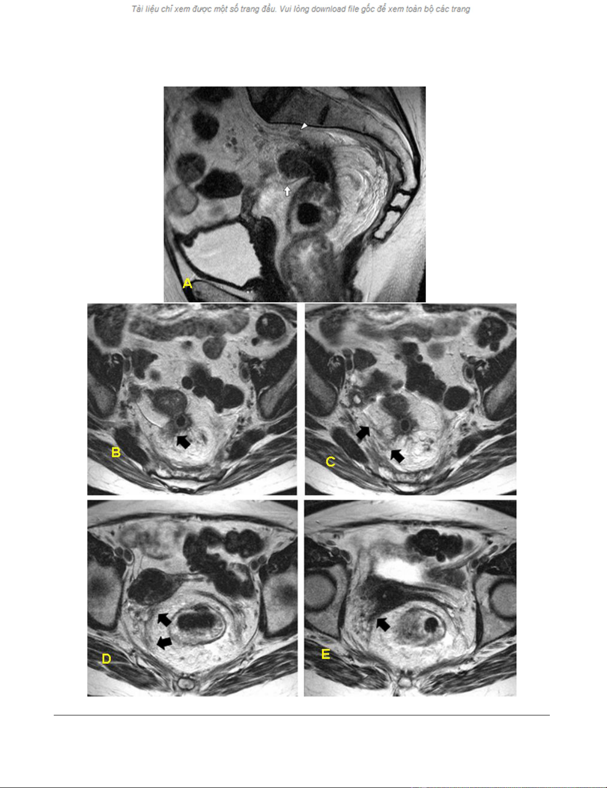
1 demonstrates the false-negative case. In this case, there
appears to be no continuity between the tumor and the
uterus. However, histopathological examination showed
tumor invasion along the fascia, reaching the posterior
wall of the uterus and the left adnexa. The radiologist
failed to ascertain the anterior extension of the tumor cor-
rectly.
In the noncompliant imaging group, preoperative MRI
was indicative of organ involvement in eight cases. Patho-
logical examination revealed two as true positives and six
as false positives (Figure 2). Among the remaining five
patients without organ involvement, pathological exami-
nation revealed two false negatives and three true nega-
tives.
Sensitivity, specificity, positive predictive value (PPV) and
negative predictive value (NPV) in the compliant and
combination protocol group were 85.7%, 94.1%, 85.7%,
and 94.1%, respectively. On the other hand, in the group
with non-compliant protocol, the sensitivity, specificity,
PPV and NPV were 50.0%, 33.3%, 25.0%, and 60%,
respectively. Statistically significant difference (p < 0.05)
was observed regarding measured specificity (95% CI; 7–
70 for group N vs. 95% CI; 71–99 for the other two
groups, D and C). The difference in sensitivity in the two
groups did not reach statistical significance levels (Table
2).
Posterior or inferior organ involvement
Only three out of the present 19 patients with locally
advanced tumor, showed involvement of an inferior
organ (levator ani muscle, piriformis muscle) or a poste-
rior organ (Os sacrum) by the tumor, without simultane-
ous involvement of any anterior organ. Two of these
patients used compliant imaging, and pathological exam-
ination revealed both to be true positives. In one patient
with noncompliant imaging an inferior organ involve-
ment was suspected but pathological examination proved
no obvious tumor infiltration or fibrosis in that organ
(false-positive). The number of cases was too few to make
any meaningful statistical analysis.
Discussion
The results of this study indicate considerable differences
in correlation between preoperative imaging and histopa-
thology depending on the imaging protocol. Using com-
pliant imaging, despite fewer imaging sequences, a
considerably better prediction of tumor invasion towards
anterior pelvic organs is seen. On the contrary, this study
also indicates that MRI performed with noncompliant
imaging protocol does not allow accurate prediction. One
other observation is that the radiologist tends to over-
stage when the imaging protocol is not optimal. This
could be due to the fear of positive resection margins
caused by a false negative assessment and partial volume
effect observed with thick slices not obtained in the
appropriate planes. This could of course be due to nature
of the study as well. The radiologists assessing the MR
exams were aware of the selection criteria and might have
felt compelled to over-stage.
The lack of compliant imaging, and as we suspect the lack
of high resolution T2-weighted imaging, probably forced
the radiologists to rely on images with considerable vol-
ume averaging. Compared to the compliant imaging,
both slice thickness including gap and voxel size were sig-
nificantly larger in the noncompliant imaging group (P <
0.05). Larger slice thickness and gap yield more partial
volume effect, thus leading the radiologists to make over-
estimation of tumor extent. In areas of the pelvis where
there are small interfaces between tissues, such as in the
anterior and low part of the rectum, this is probably of
particular importance. In the compliant and combination
groups, there was one false positive and one false negative
finding of anterior organ involvement out of 24 cases.
In the noncompliant imaging group, there were six false
positive and two false negative cases out of 13 cases. This
means that one patient out of 24 from D and C groups
and six patients out of 13 from the N group might receive
unnecessary extensive surgery and prolonged, preopera-
tive chemoradiotherapy. Anterior pelvic organs are closely
related to urinary and sexual function, and anterior organ
surgery has great impact on the patient's quality of life
after surgery. By contrast at least partially because of false
negative assessments by radiologists, one out of 24 cases
from D and C groups, and two out of 13 cases from the N
group had involved resection margins.
Although the low number of cases prohibits any meaning-
ful analysis to be done regarding accuracy of MRI for
assessment of organs inferior or dorsal to rectum, our
findings suggest that compliant imaging might be supe-
rior to noncompliant imaging also for these patients. This
low frequency could be due to less likelihood of involve-
ment of posterior organs compared to anterior organs due
to more distance between rectum and these neighboring
organs [36].
The number of MR sequences was different between vari-
ous groups with larger numbers observed in the noncom-
pliant imaging group. It seems that whenever the
compliant sequences were not employed, there was a ten-
dency to conduct several other sequences. One of the
most widely used sequences in the N group was the one
with usage of gadolinium intravenous contrast. Recently,
Vliegen and others have shown that gadolinium-
enhanced MRI does not improve the diagnostic accuracy
in local staging of rectal cancer [37]. Unnecessary use of
World Journal of Surgical Oncology 2008, 6:89 http://www.wjso.com/content/6/1/89
Page 5 of 9
(page number not for citation purposes)
Figure 1


%20--%3e%3cdefs%3e%3cstyle%3e%20.st0%20{%20fill:%20%23fff;%20}%20.st1%20{%20fill:%20%237800fa;%20}%20%3c/style%3e%3c/defs%3e%3cpath%20class='st1'%20d='M117.78,12.18H43.11c2.9,3.47,4.65,7.94,4.65,12.82,0,5.6-2.3,10.66-6.01,14.29h76.02l7.22-13.56-7.22-13.56Z'/%3e%3cg%3e%3cpath%20class='st0'%20d='M53.58,26.17h-.59v-1.46h.59v-4.96h2.83c1.78,0,2.67.94,2.67,2.82v5.76c0,1.87-.89,2.81-2.67,2.81h-2.83v-4.96ZM55.36,21.37v3.34h1.1v1.46h-1.1v3.34h1.01c.61,0,.91-.37.91-1.1v-5.93c0-.74-.3-1.1-.91-1.1h-1.01Z'/%3e%3cpath%20class='st0'%20d='M65.99,31.14h-1.8l-.31-2.07h-2.19l-.31,2.07h-1.64l1.82-11.39h2.62l1.82,11.39ZM65.28,18.04c-.25.46-.51.77-.75.94-.21.15-.47.22-.79.22-.26,0-.57-.07-.92-.22l-.38-.15c-.14-.05-.26-.07-.37-.07-.3,0-.53.18-.71.54l-.91-.68c.25-.46.51-.77.75-.94.21-.14.48-.21.79-.21.26,0,.57.07.92.21l.38.15c.14.05.26.07.37.07.3,0,.53-.18.71-.54l.91.68ZM61.91,27.52h1.73l-.87-5.76-.87,5.76Z'/%3e%3cpath%20class='st0'%20d='M74.53,26.89v1.52c0,1.91-.89,2.86-2.67,2.86s-2.67-.95-2.67-2.86v-5.93c0-1.91.89-2.86,2.67-2.86s2.67.95,2.67,2.86v1.11h-1.69v-1.22c0-.75-.31-1.12-.93-1.12s-.93.37-.93,1.12v6.15c0,.74.31,1.11.93,1.11s.93-.37.93-1.11v-1.63h1.69Z'/%3e%3cpath%20class='st0'%20d='M81.4,31.14h-1.8l-.31-2.07h-2.19l-.31,2.07h-1.64l1.82-11.39h2.62l1.82,11.39ZM75.9,19.2l1.52-1.91h1.71l1.51,1.91h-1.61l-.76-.95-.75.95h-1.61ZM77.32,27.52h1.73l-.87-5.76-.87,5.76ZM83.1,15.99l-1.76,1.91h-1.26l1.17-1.91h1.86Z'/%3e%3cpath%20class='st0'%20d='M84.86,19.75c1.78,0,2.67.94,2.67,2.82v1.48c0,1.87-.89,2.81-2.67,2.81h-.85v4.28h-1.79v-11.39h2.64ZM84.01,21.37v3.86h.85c.58,0,.87-.36.87-1.08v-1.71c0-.71-.29-1.07-.87-1.07h-.85Z'/%3e%3cpath%20class='st0'%20d='M93.51,19.75c1.78,0,2.67.94,2.67,2.82v1.48c0,1.87-.89,2.81-2.67,2.81h-.85v4.28h-1.79v-11.39h2.64ZM92.66,21.37v3.86h.85c.58,0,.87-.36.87-1.08v-1.71c0-.71-.29-1.07-.87-1.07h-.85Z'/%3e%3cpath%20class='st0'%20d='M98.8,31.14h-1.79v-11.39h1.79v4.88h2.03v-4.88h1.83v11.39h-1.83v-4.88h-2.03v4.88Z'/%3e%3cpath%20class='st0'%20d='M105.36,24.55h2.46v1.62h-2.46v3.34h3.09v1.63h-4.88v-11.39h4.88v1.63h-3.09v3.18ZM108.17,17.29l-1.76,1.91h-1.26l1.17-1.91h1.86Z'/%3e%3cpath%20class='st0'%20d='M112.2,19.75c1.78,0,2.67.94,2.67,2.82v1.48c0,1.87-.89,2.81-2.67,2.81h-.85v4.28h-1.79v-11.39h2.64ZM111.35,21.37v3.86h.85c.58,0,.87-.36.87-1.08v-1.71c0-.71-.29-1.07-.87-1.07h-.85Z'/%3e%3c/g%3e%3ccircle%20class='st1'%20cx='25'%20cy='25'%20r='20'/%3e%3cpath%20class='st0'%20d='M32.78,19.27c2.92,0,4.43,2.55,5.28,5.33l.71,2.17c.14.38-.33.75-.71.75h-5.61c.19-.33.24-.71.09-1.08l-.75-2.45c-.43-1.32-.99-2.64-1.79-3.77.75-.57,1.65-.94,2.78-.94h0ZM25,18.38c3.25,0,4.9,2.78,5.89,5.89l.76,2.45c.14.42-.33.8-.8.8h-11.69c-.42,0-.94-.38-.8-.8l.75-2.45c.99-3.11,2.64-5.89,5.89-5.89h0ZM25,11.35c1.74,0,3.11,1.37,3.11,3.11s-1.37,3.11-3.11,3.11-3.11-1.41-3.11-3.11,1.41-3.11,3.11-3.11h0ZM17.27,19.27c1.08,0,1.98.38,2.73.94-.8,1.13-1.37,2.45-1.74,3.77l-.8,2.45c-.14.38-.05.75.09,1.08h-5.56c-.42,0-.9-.38-.75-.75l.71-2.17c.9-2.78,2.41-5.33,5.33-5.33h0ZM17.27,12.91c1.51,0,2.78,1.27,2.78,2.83s-1.27,2.83-2.78,2.83-2.83-1.27-2.83-2.83,1.27-2.83,2.83-2.83h0ZM32.78,12.91c1.56,0,2.78,1.27,2.78,2.83s-1.23,2.83-2.78,2.83-2.83-1.27-2.83-2.83,1.27-2.83,2.83-2.83h0ZM27.07,28.56v.09c0,.57-.24,1.08-.61,1.46h0v.05c-.38.33-.9.57-1.46.57s-1.08-.24-1.46-.61h0c-.38-.38-.61-.9-.61-1.46v-.09h1.41v.09c0,.19.05.38.19.47v.05c.09.09.28.19.47.19s.38-.09.47-.19v-.05c.14-.09.24-.28.24-.47t-.05-.09h1.41ZM30.99,28.56v.09c0,1.65-.66,3.16-1.74,4.24-1.08,1.08-2.59,1.79-4.24,1.79s-3.16-.71-4.24-1.79l-.05-.05c-1.04-1.08-1.7-2.55-1.7-4.2v-.09h1.41v.09c0,1.27.47,2.4,1.27,3.25h.05c.85.85,1.98,1.37,3.25,1.37s2.4-.52,3.25-1.37c.85-.8,1.37-1.98,1.37-3.25v-.09h1.37ZM34.99,28.56v.09c0,2.78-1.13,5.28-2.92,7.07-1.79,1.79-4.29,2.92-7.07,2.92s-5.23-1.13-7.07-2.92c-1.79-1.79-2.92-4.29-2.92-7.07v-.09h1.41v.09c0,2.4.94,4.53,2.5,6.08,1.56,1.56,3.72,2.5,6.08,2.5s4.52-.94,6.08-2.5c1.56-1.56,2.5-3.68,2.5-6.08v-.09h1.41Z'/%3e%3c/svg%3e)