MET H O D O LO G Y Open Access
Engineered artificial antigen presenting cells
facilitate direct and efficient expansion of tumor
infiltrating lymphocytes
Qunrui Ye
1
, Maria Loisiou
1
, Bruce L Levine
2
, Megan M Suhoski
3
, James L Riley
2
, Carl H June
2
, George Coukos
1,2
and Daniel J Powell Jr
1,2*
Abstract
Background: Development of a standardized platform for the rapid expansion of tumor-infiltrating lymphocytes
(TILs) with anti-tumor function from patients with limited TIL numbers or tumor tissues challenges their clinical
application.
Methods: To facilitate adoptive immunotherapy, we applied genetically-engineered K562 cell-based artificial
antigen presenting cells (aAPCs) for the direct and rapid expansion of TILs isolated from primary cancer specimens.
Results: TILs outgrown in IL-2 undergo rapid, CD28-independent expansion in response to aAPC stimulation that
requires provision of exogenous IL-2 cytokine support. aAPCs induce numerical expansion of TILs that is statistically
similar to an established rapid expansion method at a 100-fold lower feeder cell to TIL ratio, and greater than
those achievable using anti-CD3/CD28 activation beads or extended IL-2 culture. aAPC-expanded TILs undergo
numerical expansion of tumor antigen-specific cells, remain amenable to secondary aAPC-based expansion, and
have low CD4/CD8 ratios and FOXP3+ CD4+ cell frequencies. TILs can also be expanded directly from fresh
enzyme-digested tumor specimens when pulsed with aAPCs. These “young”TILs are tumor-reactive, positively
skewed in CD8+ lymphocyte composition, CD28 and CD27 expression, and contain fewer FOXP3+ T cells
compared to parallel IL-2 cultures.
Conclusion: Genetically-enhanced aAPCs represent a standardized, “off-the-shelf”platform for the direct ex vivo
expansion of TILs of suitable number, phenotype and function for use in adoptive immunotherapy.
Introduction
Adoptive immunotherapy using tumor-reactive T lym-
phocytes has emerged as a powerful approach for the
treatment of bulky, refractory cancer [1], however the
ability to generate large numbers of TILs for therapy is a
challenge that has significant regulatory hurdles, and
requires technically sophisticated cell processing and
extended in vitro lymphocyte culturing periods. Long-
term culture of tumor-derived T cells in high-dose inter-
leukin-2 (IL-2) allows for the generation of high numbers
of TILs (>1 × 10
11
) but with preferential expansion of
CD4+ lymphocytes [2-4]. Initial IL-2-based TIL
expansion followed by a “rapid expansion method”
(REM) [5-9] is a more time and labor efficient method,
requiring an excess of irradiated allogeneic peripheral
blood mononuclear cells (PBMC) as feeder cells, anti-
CD3 antibody and high doses of IL-2, that can result in
a 1,000-fold expansion of TILs over a 14-day period [9].
While routinely used, the REM has introduced technical,
regulatory, and logistic challenges that have prevented
larger and randomized clinical trials as a prelude to
widespread application. First, large numbers of allogeneic
feeders (200-fold excess), often from multiple donors, are
required for clinical expansions. Second, allogeneic feeder
cells harvested by large-volume leukapheresis from
healthy donors exhibit donor to donor variability in their
viability after cryopreservation and capacity to support
TIL expansion, and thus test expansions are often
* Correspondence: poda@mail.med.upenn.edu
1
Ovarian Cancer Research Center, Department of Obstetrics and Gynecology,
Perelman School of Medicine, University of Pennsylvania, Philadelphia, PA,
USA
Full list of author information is available at the end of the article
Ye et al.Journal of Translational Medicine 2011, 9:131
http://www.translational-medicine.com/content/9/1/131
© 2011 Ye et al; licensee BioMed Central Ltd. This is an Open Access article distributed under the terms of the Creative Commons
Attribution License (http://creativecommons.org/licenses/by/2.0), which permits unrestricted use, distribution, and reproduction in
any medium, provided the original work is properly cited.

required. Finally, this process necessitates additional
extensive and costly laboratory testing of each individual
donor cell product to confirm sterility.
Artificial antigen presenting cells (aAPCs) expressing
ligands for the T cell receptor and costimulatory mole-
cules can activate and expand T cells for transfer, while
improving their potency and function. The first genera-
tion of aAPC consisted of anti-CD3 and anti-CD28
monoclonal antibodies (mAbs) covalently bound to
magnetic beads (CD3/CD28 beads) which crosslink CD3
and CD28 on T cells, enabling efficient polyclonal
expansion of circulating T cells (50 to 1000-fold) over
10-14 days of ex vivo culture with preferential expansion
of naïve and memory CD4+ T cells [10], however their
efficiency in TIL expansion has not been examined.
Second generation cell-based aAPCs can substitute for
natural APCs, mediate efficient expansion of antigen-
specific T cells from peripheral blood [11-16] and stably
express multiple gene inserts, including CD64 (the high-
affinity Fc receptor), CD32 (the low-affinity Fc receptor),
and CD137L (4-1BBL), among others [13,15]. Compared
to beads, cell-based aAPCs bearing the costimulatory
ligand CD137L can more efficiently induce the prolifera-
tion of antigen-experienced CD8+ CD28
-
Tcellsfrom
peripheral blood and improve their in vivo persistence
and antitumor activity upon adoptive transfer to tumor-
bearing mice [15,17]. In these studies, enhanced prolif-
eration of antigen-experienced CD8+ CD28
-
T cells
mediated by aAPCs is dependent on CD137 ligation
[15,17].
Unlike peripheral blood lymphocytes (PBL), most tumor
antigen-specific CD8+ TILs derivedfromsolidtumors
express low levels of CD28 [18,19]. Together, the above
studies suggest that approaches utilizing CD137 ligation
may support ex vivo TIL expansion. In a trial of adoptive
TIL transfer with REM generated cells, the persistence of
TILs in vivo after infusion represented a major limitation
to successful therapy [20]. In vivo persistence and clinical
response were both associated with expression of the cost-
imulatory molecules CD28 and CD27 by TILs, as well as
their telomere length [18,21-24]. The REM requires
extended duration TIL culture which results in telomere
length shortening and reduced expression of CD28 and
CD27 [18,25], thus there remains a need for the develop-
ment of improved, standardized methods and materials
for generating TILs rapidly for adoptive transfer with
greater potency and engraftment capability.
Here we investigate the use of engineered K562 cell-
based aAPCs as an “off-the-shelf”platform for ex vivo
TIL expansion. K562 aAPCs that express CD137L offer
the potential to expand antigen-experienced TILs and
represent a potential new cell-based platform for the
standardization of ex vivo TIL expansion. Ovarian can-
cer and melanoma biospecimens were used to test the
notion that aAPC can stimulate TIL expansion in differ-
ent tumor histotypes [26,27], based on the knowledge
that TILs from these cancers can recognize autologous
tumor as well as known tumor antigens in vitro [28-32],
and exhibit tumor-specific reactivity ex vivo [33,34] and
in vivo [5,7,35]. We found that aAPCs efficiently expand
IL-2 cultured TILs from solid tumor specimens of ovar-
ian cancer similar to the REM, resulting in a favorable
CD4/8 T cell ratio, and low FOXP3+ CD4 T cell com-
position. aAPC-based TIL expansion depends on the
provision of exogenous IL-2 cytokine support in culture
and is largely CD28-independent. Under these condi-
tions, tumor antigen-specific TILs with demonstrated
anti-tumor reactivity can be expanded. Further, aAPC
can induce the rapid and efficient expansion of TILs
directly from freshly digested tumor samples, reducing
overall culture time, and output TILs are highly skewed
in CD8+ lymphocyte composition, possess high levels of
CD28 and CD27 expression after activation and are
amenable to secondary aAPC-based expansion. The
aAPC platform as described here thus establishes a stan-
dardized methodology for the rapid, clinical-grade
expansion of TILs for therapy.
Materials and methods
Generation of TILs
Patients were entered into an Institutional Review
Board-approved clinical protocol and signed an
informed consent prior to initiation of lymphocyte cul-
tures. Generation of TILs was performed as described
elsewhere [9]. Briefly, 2 mm
3
tumor fragments were cul-
tured in complete media (CM) comprised of AIM-V
medium (Invitrogen Life Technologies, Carlsbad, CA)
supplemented with 2 mM glutamine (Mediatech, Inc.
Manassas, VA), 100 U/ml penicillin (Invitrogen Life
Technologies), 100 μg/ml streptomycin (Invitrogen Life
Technologies), 5% heat-inactivated human AB serum
(Valley Biomedical, Inc. Winchester, VA) and 600 IU/
mL rhIL-2 (Chiron, Emeryville, CA). TILs established
from fragments were grown for 3-4 weeks in CM and
expanded fresh or cryopreserved in heat-inactivated
HAB serum with 10% DMSO and stored at -180°C until
the time of study. Tumor associated lymphocytes (TAL)
obtained from ascites collections were seeded at 3e6
cells/well of a 24 well plate in CM. TIL growth was
inspected about every other day using a low-power
inverted microscope. Each initial well was considered to
be an independent TIL culture and was maintained
accordingly. For enzymatic digestion of solid tumors,
tumor specimen was diced into RPMI-1640, washed and
centrifuged at 800 rpm for 5 minutes at 15-22°C, and
resuspended in enzymatic digestion buffer (0.2 mg/ml
Collagenase and 30 units/ml of DNase in RPMI-1640)
followed by overnight rotation at room temperature.
Ye et al.Journal of Translational Medicine 2011, 9:131
http://www.translational-medicine.com/content/9/1/131
Page 2 of 13

aAPC preparation
KT64/BBL and KT32/BBL aAPCs were generated, cul-
tured and prepared for co-culture as previously
described [13,15]. Briefly, Fc-binding receptors on
KT64/BBL aAPCs were pre-cleared of serum immuno-
globulins by culture in serum free AIM-V medium
(SFM) overnight and then irradiated at 10,000 rad. Anti-
CD3 (OKT-3) with or without anti-CD28 (clone 9.3)
mAbs were loaded on aAPCs at 0.5 ug/10
6
cells at 4°C
for30minutes.Beforeuse,aAPCswerewashedtwice
with SFM. For KT32/BBL aAPCs, anti-CD3 and anti-
CD28 antibodies were not washed out of culture med-
ium, per established protocol [13,15]. For expansion of
IL-2 cultured TILs, an optimal 2:1 aAPC to TIL ratio
was established and used in all experiments.
Expansion of TILs and TALs in vitro using aAPCs
10
6
heterogonous TILs or TALs were co-cultured with
KT64/BBL or KT32/BBL aAPCs loaded with anti-CD3
with or without anti-CD28 antibody in one well of a 24
well plate. rhIL-2 (100 IU/ml) was added into co-cultures
at day 2. Every other day the cell number was counted by
on a Coulter Multisizer and adjusted to a concentration of
0.5-1 × 10
6
cells/ml until day 8. Expanding cocultures
were transferred into an appropriately sized flask and sus-
pended in CM containing rhIL-2 100 IU/ml depending on
total cell numbers. Confirmatory hemacytometer counts
including Trypan Blue exclusion were performed. After
day 9, phenotypes of expanded TILs or TALs were exam-
ined by flow cytometry. Final expanded products were uni-
formly comprised by CD3+ TILs, TALs or PBLs, without
aAPC contamination, as verified by cell sizing, morphology
and flow cytometry. The total duration of cell expansion
culture was between 9 and 14 days. At the end of culture,
all remaining cells were frozen in 90% HAB serum and
10% DMSO for continued analysis. For comparison to
other methods of T cell expansion, TILs or TALs were
cultured in three conditions: with rhIL-2 (600 IU/ml) in
CM; with anti-CD3/CD28 magnetic beads (3:1 beads to T
cells) in rhIL-2 (100 IU/ml) (Chiron); or in a “rapid expan-
sion method”condition (200:1 allogeneic PBMC:TILs,
30 ng/ml of OKT-3 anti-CD3 mAb and 6000 IU/ml rhIL-
2 in 20 mL of CM in a T75 flask). For stimulation of fresh
tumor digests, 10
6
total cells from tumor digested pro-
ducts were stimulated using an equivalent number of irra-
diated aAPC loaded with anti-CD3 mAb in media
supplemented with 100 IU/mL IL-2.
Antibodies and flow cytometric immunofluorescence
analysis
Antibodies against human CD3, CD4, CD8, CD16,
CD25, CD32, CD64 and CD137 were purchased from
BD Bioscience. 7-AAD antibody for viability staining
was purchased from BD Bioscience (San Jose, CA).
HER2:369-377 peptide (KIFGSLAFL) and MART-1:26-
35(27L) peptide (ELAGIGILTV) containing HLA-A2010
tetramers were purchased from Beckman Coulter, Inc.
(Brea, CA). Anti-FOXP3 antibody (clone 259D) was
obtained from BioLegend (San Diego, CA). Fresh TILs
or TALs were resuspended in FACS buffer consisting of
PBS with 2% FBS (Gemini Bioproducts) at 10
7
cells/ml
and blocked with 10% normal mouse Ig (Caltag Labora-
tories) for 10 min on ice. A total of 10
6
cells in 100 μl
were stained with fluoro-chrome-conjugated mAbs at
4°C for 40 min in the dark. In some cases, cells were
briefly stained with 7-AAD antibody for nonviable cell
exclusion after washing twice and subsequently analyzed
in a FACSCanto II (BD Biosciences). FOXP3 staining
was performed using the eBioscience fixation and per-
meablization kits according to the manufacturer’s
instructions and cells stained with the anti-FOXP3 anti-
body from BioLegend. K562 aAPCs antibody loading
was performed using anti-CD3 (OKT3) purchased from
eBioscience (San Diego, CA) and anti-CD28 mAbs
(clone 9.3). For cell division assays, TILs or PBLs were
labeled with 128 nM of carboxyfluorescein succinimidyl
ester (CFSE). CFSE labeled TILs or PBLs were expanded
with aAPCs, CD3/28 beads, rhIL-2 (600 IU/ml) or REM
as described above. At day 6, the cells were stained with
anti-CD3, anti-CD4 and anti-CD8 and examined for
CFSE division by FACS. Statistical significance of phe-
notypic differences was determined using paired two-
tailed T-test.
ELISA assay for T cell function
Stimulation of TILs by tumor cells was assessed by IFN-
gsecretion. 1 × 10
5
TILs were cultured with 1 × 10
5
tar-
get cells in triplicate overnight in a 96 well U bottom
plate in 200 uL of CM containing 5% heat-inactivated
human AB serum. Supernatants were harvested and
analyzed for IFN-gby ELISA, according to manufac-
turer’s instruction (Biolegend, San Diego, CA). Values
represent the mean cytokine concentration (pg/mL) ±
SD of triplicate wells.
Results
KT64/BBL aAPCs-based expansion TILs
K562 cells expressing CD64, CD137L and CD28 ligands
CD80 and CD86, pulsed with anti-CD3 antibody effi-
ciently activate and expand CD8+ CD28- T cells and
antigen-specific T cells from peripheral blood when co-
cultured at a 0.5:1 aAPC to T cell ratio in the absence
of exogenous IL-2 and in a CD137L dependent manner
[15]. We therefore hypothesized that tumor infiltrating
lymphocytes (TILs) derived from cancer lesions could
be efficiently expanded to therapeutic treatment num-
bers using a K562 cell-based aAPC platform. To gener-
ate cell-based aAPCs, the parental K562 cell line was
Ye et al.Journal of Translational Medicine 2011, 9:131
http://www.translational-medicine.com/content/9/1/131
Page 3 of 13

engineered to stably co-express the high-affinity Fc
receptor CD64 and the costimulatory ligand CD137L
(4-1BBL) by lentiviral gene transduction. Single cell
clones (referred to as KT64/BBL) were isolated by flow-
sorting and their CD64 and CD137L surface expression
was confirmed by flow cytometry (Additional file 1Fig-
ure S1a). KT64/BBL aAPCs were cultured in the
absence of serum to pre-clear CD64 of serum derived
immunoglobulins, irradiated and then loaded with anti-
CD3 and anti-CD28 agonist monoclonal antibodies
(mAbs) for TIL expansion.
TIL cultures for expansion were outgrown from solid
ovarian cancer fragments for 3-4 weeks in culture media
(CM) containing 600 IU/mL rhIL-2 cytokine, as
described [4,9], and were comprised of >95% CD3+
T cells and <1.5% NK cells. To test the capacity of anti-
body-loaded aAPCs to mediate ex vivo expansion of
TILs, aAPC were co-cultured with TILs at aAPC to TIL
ratios ranging between 0.5 and 10 to 1 in the continued
presence of IL-2 (100 IU/ml). Peak TIL expansion was
achieved at the 2:1 aAPC to T cell ratio (Figure 1a),
which contrasts the 200:1 feeder to T cell ratio com-
monly used in REM-based TIL expansion [9]. The 2:1
aAPC to T cell ratio was therefore used for the experi-
ments detailed below. The contribution of CD137L to
TIL expansion was confirmed using control KT64
aAPCs lacking CD137L expression, which mediated
diminished TIL expansion compared to KT64/BBL
(Additional file 1Figure S1a,b), consistent with our
prior study using antigen-experienced T cells [15]. Since
our first generation of K562 based aAPC (referred to as
KT32/BBL) relied upon the low affinity Fc receptor
CD32 for anti-CD3 antibody loading and demonstrated
the capacity to expand circulating T cells [15], we evalu-
ated the relative efficiency of CD32 and CD64-expres-
sing aAPCs for expanding TILs. KT64/BBL aAPCs were
superior to KT32/BBL aAPCs, and therefore used in all
further experiments (Additional file 2Figure S2).
Robust expansion of TILs is dependent upon IL-2, but not
CD28 costimulation
To investigate the impact of CD28 costimulation and
IL-2 on aAPC-mediated TIL expansion, KT64/BBL
aAPCs were loaded with anti-CD3 mAb +/- anti-CD28
mAb and used to stimulate TILs in the presence or
absence of 100 IU/ml of IL-2 (Figure 1b). In the absence
of IL-2, TILs underwent minimal expansion after stimu-
lation with aAPCs loaded with anti-CD3 mAb with
(11-fold) or without anti-CD28 mAb (9-fold), albeit
more than when continually grown in IL-2 (3-fold). By
comparison, addition of IL-2 to aAPC-based expansion
induced vigorous numerical growth of TILs (>170-fold)
in the presence or absence of anti-CD28 mAb, and the
level of TIL expansion was similar whether or not anti-
CD28 mAb was loaded onto the aAPCs. These results
demonstrate that cell-based aAPC-mediated TIL expan-
sion is largely independent of CD28 signaling when
4-1BBL is provided on aAPC, but dramatically improved
by addition of IL-2 cytokine to culture.
The limited contribution provided by anti-CD28 mAb
to the expansion of TILs in the absence of IL-2 counters
that previously observed for peripheral blood T lympho-
cytes (PBLs) from healthy donors where CD28 costimu-
lation in concert with TCR signaling induces robust
proliferation [13,15]. We therefore evaluated the contri-
bution of CD28 in the expansion of TILs and PBLs col-
lected from the same patient with ovarian cancer. In
paired comparison, measurement of CD28 expression
on matched TILs and PBLs from the same patients
revealed a higher relative expression of surface CD28 by
T cells from the circulation than by T cells from tumor
in all cases (Additional file 3Figure S3). Among CD3+
TILs, more CD4+ TILs expressed CD28 than CD8+
TILs (76.5 ± 32.9% vs. 34.7 ± 12.2%, respectively; p =
0.003). CD3+ T cells from the blood were heteroge-
neous in differentiation state and comprised of naïve
(CD45RO- CD62L+), central memory (CD45RO+
CD62L+), and effector memory (CD45RO+ CD62L-)
cell subsets; TILs however were comprised primarily of
cells with a more differentiated, effector memory pheno-
type (representative examples are shown in Additional
file 3Figure S3).
Consistent with their disparate differentiation pheno-
types, peripheral blood T cells and TILs from the same
patient demonstrated a relative difference in expansion
in response to aAPC stimulation. The expansion of TILs
in response to stimulation with aAPCs loaded with anti-
CD3 mAb with or without CD28 agonist mAb co-load-
ing was modest and similar (62-fold v. 63-fold, respec-
tively), but was substantially augmented by the addition
of IL-2 to culture (182-fold; Figure 1c). PBLs in parallel
culture exhibited greater expansion in response to anti-
CD3 mAb loaded aAPC stimulation compared to TIL,
whether or not CD28 signaling was intact, however, PBL
expansion was substantially elevated when the aAPCs
were also loaded with CD28 agonist mAb (254-fold),
relative to anti-CD3 mAb alone (95-fold). In the absence
of CD28 costimulation, robust PBL expansion could be
restored by addition of exogenous IL-2 cytokine (187-
fold). Although PBL expansion in the condition of CD28
costimulation out-performed the addition of IL-2 at day
9 (Figure 1c), IL-2 supplementation was superior to
CD28 costimulation by day 11 of PBL culture (737-fold
v. 340-fold, respectively); at this time point, TIL cultures
were unchanged in expansion hierarchy with a 287-fold
expansionintheCD3/IL-2condition. Consistent with
previous findings[15], PBLs stimulated with anti-CD3
and anti-CD28 mAb loaded aAPCs expanded better
Ye et al.Journal of Translational Medicine 2011, 9:131
http://www.translational-medicine.com/content/9/1/131
Page 4 of 13
than those stimulated with magnetic beads coated with
anti-CD3 and CD28 mAbs to crosslink endogenous
CD3 and CD28 (254-fold v. 56-fold, respectively; Figure
1c). TILs stimulated with CD3/CD28 beads did not
undergo robust expansion (18-fold).
Supplement of TIL cultures with IL-2 cytokine, but
not CD28 costimulation, during aAPC-induced stimula-
tion dramatically improved TIL expansion, while PBLs
showed improved expansion in response to aAPC with
addition of either IL-2 or CD28 costimulation. This sug-
gests that PBLs, which express elevated levels of CD28
relative to TILs, may produce and secrete more IL-2
when costimulated than their CD28
low
TIL counterparts,
thus supporting T cell expansion. Consistent with this
notion, cytokine secretion analysis performed on super-
natants from TILs or PBLs stimulated overnight with
anti-CD3 mAb loaded aAPCs +/- anti-CD28 mAb
revealed that TILs produce little to no IL-2 when stimu-
lated with aAPC either with or without CD28 costimula-
tion, or with CD3/CD28 beads (Figure 1d). By contrast,
PBLs secreted high levels of IL-2 in response to aAPC
which was augmented by the addition of CD28 agonist
mAb loading. CD3/CD28 bead stimulation of PBLs
resulted in an even greater level of IL-2 production than
that achieved with aAPC. Both TILs and PBL secreted
IFN-gand TNF-ain response to aAPC and bead stimu-
lation (not shown), indicating that the lack of IL-2 pro-
duction by TILs was not a result of functional anergy.
0
10000
20000
30000
40000
50000
CD3
CD3/28
CD3/28
beads
None
PBL
TIL
0
10
20
30
40
50
60
70
0.5
1
2
5
10
IL-2
None
aAPC:TIL ratio
Fold expansion
0 50 100 150 200
None
IL2
CD3
CD3/28
CD3+IL2
CD3/28+IL2
aAPC
0
50
100
150
200
250
300
IL-2
CD3/28
beads
CD3
CD3/28
CD3/IL-2
PBL
TIL
a
AP
C
Fold expansion
Fold expansion
c
a
b
IL-2 (pg/mL)
d
a
AP
C
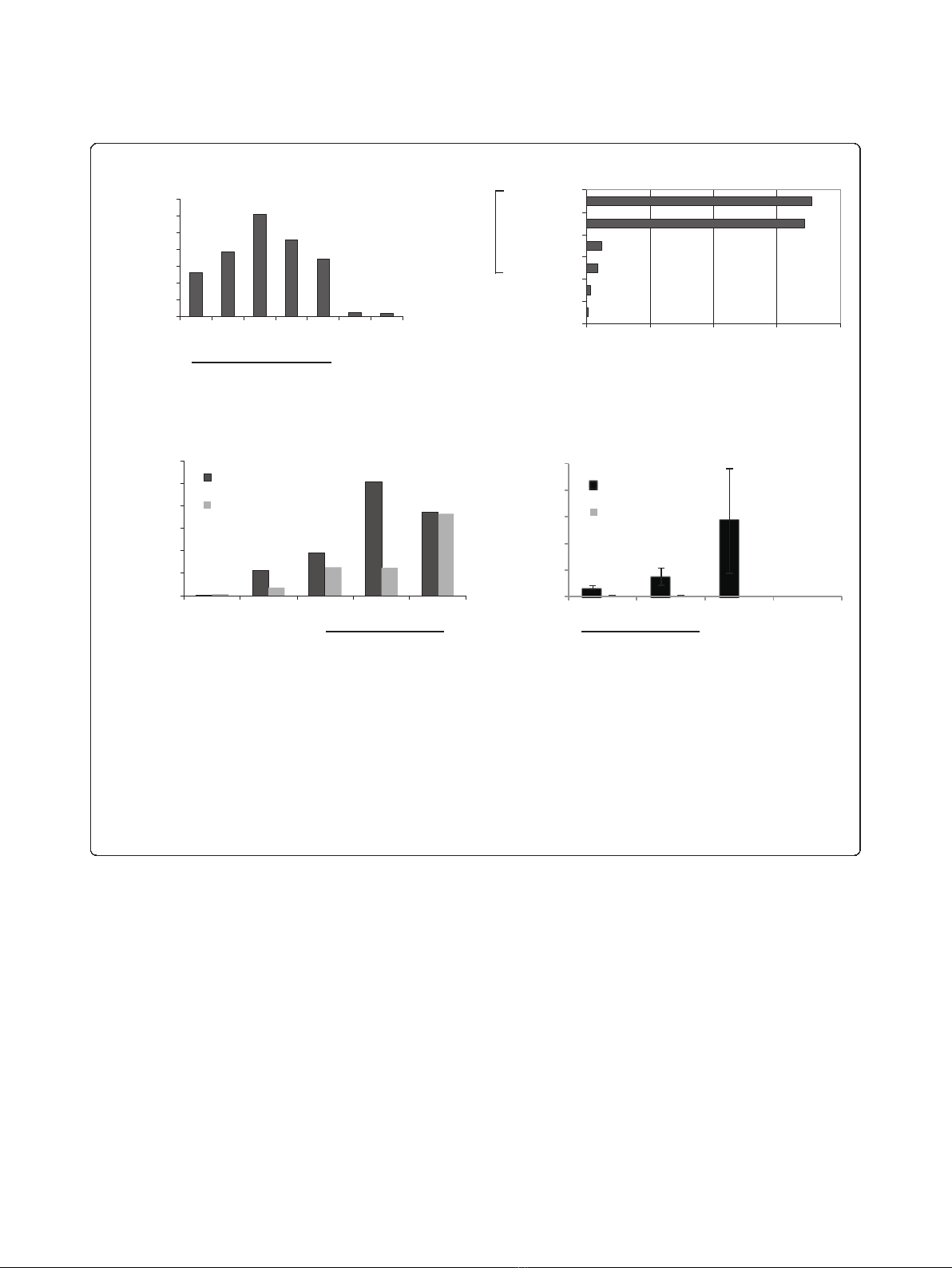
Figure 1 KT64/BBL aAPCs support the expansion of TILs in a CD28-independent manner.(a) TILs cultures established for 3-4 weeks in 600
IU/ml IL-2 were expanded using aAPCs loaded with anti-CD3 and anti-CD28 mAbs at various aAPC to T cell ratios in the continued presence of
IL-2 (100 IU/mL). In this representative experiment (one of three), a 62-fold expansion of TILs was achieved 9 days after a single stimulation with
aAPCs at the 2:1 aAPC to T cell ratio. A 3-fold expansion occurred after continued culture in IL-2. TILs stimulated with aAPCs underwent greater
expansion at all aAPC to TIL ratios compared to continued growth in IL-2 or growth in medium alone. (b) KT64/BBL aAPC-based TIL expansion is
CD28 costimulation-independent but augmented by provision of IL-2 support. Established TIL cultures were expanded for 9 days using aAPC
loaded with anti-CD3 antibody in the presence or absence of clone 9.3 anti-CD28 antibody, in the presence or absence of IL-2 supplement. (c)
CD28 costimulation augments the aAPC-based expansion of peripheral blood T cells, but not autologous TILs. CD3/28 beads do not support TIL
expansion (3:1 bead to T cell ratio). Day 9 cell counts are shown. (d) TILs stimulated with KT64/BBL aAPCs with or without anti-CD28 antibody
do not secrete IL-2 after overnight culture, but peripheral blood lymphocytes do. IL-2 secretion by PBL is increased by provision of CD28
costimulation and supported by CD3/28 bead stimulation. Mean IL-2 (pg/mL) concentration ± SEM from three independent TIL cultures is
shown.
Ye et al.Journal of Translational Medicine 2011, 9:131
http://www.translational-medicine.com/content/9/1/131
Page 5 of 13


![Vaccine và ứng dụng: Bài tiểu luận [chuẩn SEO]](https://cdn.tailieu.vn/images/document/thumbnail/2016/20160519/3008140018/135x160/652005293.jpg)


