MET H O D O LO G Y Open Access
Generation of high-titer viral preparations by
concentration using successive rounds of
ultracentrifugation
Christine V Ichim
1,2
and Richard A Wells
1,2,3,4*
Abstract
Background: Viral vectors provide a method of stably introducing exogenous DNA into cells that are not easily
transfectable allowing for the ectopic expression or silencing of genes for therapeutic or experimental purposes.
However, some cell types, in particular bone marrow cells, dendritic cells and neurons are difficult to transduce
with viral vectors. Successful transduction of such cells requires preparation of highly concentrated viral stocks,
which permit a high virus concentration and multiplicity of infection (MOI) during transduction. Pseudotyping with
the vesicular stomatitis virus G (VSV-G) envelope protein is common practice for both lentiviral and retroviral
vectors. The VSV-G glycoprotein adds physical stability to retroviral particles, allowing concentration of virus by
high-speed ultracentrifugation. Here we describe a method report for concentration of virus from large volumes of
culture supernatant by means of successive rounds of ultracentrifugation into the same ultracentrifuge tube.
Method: Stable retrovirus producer cell lines were generated and large volumes of virus-containing supernatant
were produced. We then tested the transduction ability of virus following varying rounds of concentration by ultra-
centrifugation. In a second series of experiments lentivirus-containing supernatant was produced by transient
transfection of 297T/17 cells and again we tested the transduction ability of virus following multiple rounds of
ultra-centrifugation.
Results: We report being able to centrifuge VSV-G coated retrovirus for as many as four rounds of
ultracentrifugation while observing an additive increase in viral titer. Even after four rounds of ultracentrifugation
we did not reach a plateau in viral titer relative to viral supernatant concentrated to indicate that we had reached
the maximum tolerated centrifugation time, implying that it may be possible to centrifuge VSV-G coated retrovirus
even further should it be necessary to achieve yet higher titers for specific applications. We further report that VSV-
G coated lentiviral particles may also be concentrated by successive rounds of ultracentrifugation (in this case four
rounds) with minimal loss of transduction efficiency.
Conclusion: This method of concentrating virus has allowed us to generate virus of sufficient titers to transduce
bone marrow cells with both retrovirus and lentivirus, including virus carrying shRNA constructs.
Introduction
Viral vectors are commonly used to introduce exogen-
ous genetic material in experimental systems, and have
been used successfully in human gene therapy trials to
treat patients with primary immunodeficiencies such as
X-linked severe combined immunodeficiency (SCID)
[1-3] and adenosine deaminase deficiency [1-3]. Suitable
vectors frequently used in the laboratory and clinical
setting include retroviral and lentiviral vectors. However,
the ability to transduce difficult-to-infect cells such as
primary hematopoietic cells, hematopoietic stem cells,
and neuronal cells with these vectors is dependent on
the ability to produce stocks of high viral titers [4,5].
Retro- and lentivirus is produced by transfecting pro-
ducer cell lines with viral plasmids resulting in the pro-
duction of virions that are released into the supernatant.
Target cells may be transduced using the supernatant or
alternatively by using supernatant that has been
* Correspondence: rwells@sri.utoronto.ca
1
Department of Medical Biophysics, University of Toronto, Toronto, ON M5G
2M9, Canada
Full list of author information is available at the end of the article
Ichim and Wells Journal of Translational Medicine 2011, 9:137
http://www.translational-medicine.com/content/9/1/137
© 2011 Ichim and Wells; licensee BioMed Central Ltd. This is an Open Access article distributed under the terms of the Creative
Commons Attribution License (http://creativecommons.org/licenses/by/2.0), which permits unrestricted use, distribution, and
reproduction in any medium, provided the original work is properly cited.

concentrated to increase the viral titer. Ultracentrifuga-
tion is one method that may be used to concentrate
supernatant containing retroviral and lentiviral vectors
that were pseudotyped with the G envelope glycoprotein
of the vesicular stomatitis virus (VSV-G)[6-8]. In con-
trast to endogenous envelope proteins, VSV-G is a
sturdy glycoprotein that can withstand the stresses of
prolonged ultracentrifugation [7]. Furthermore, trans-
duction with VSV-G coated virions occurs via mem-
brane fusion [9] not by receptor-mediated uptake,
thereby expanding the cellular tropism of the viral parti-
cles [10]
Nevertheless, even after concentration of virus, titers
may still not be high enough for the successful trans-
duction of difficult-to-infect cells such as primary bone
marrow cells. This is especially relevant if the vector is
not amenable to the production of high viral titers, as is
often the case with shRNA vectors [11] One method of
increasing the concentration of virus, in principle, would
be to simply scale up and increase the volume of super-
natant concentrated. However, the amount of viral
supernatant concentrated in currently used protocols is
limited by the capacity of the rotor tube, typically 30
mL. To yield a higher concentration of virus some pro-
tocols allow for a second round of ultracentrifugation
[7] In these cases following one round of centrifugation,
the supernatant is decanted into a waste container and
the viral pellet remains in the bottom of the centrifuge
tube. Another 30 mL of viral supernatant is added to
the previously used ultracentrifuge tube that contains a
viral pellet, and the tube is centrifuged a second time.
Following this second round of centrifugation the super-
natant is decanted and the virus is resuspended
overnight.
Here we report that performing multiple successive
rounds of ultracentrifugation of retrovirus pseudotyped
using the VSV-G envelope protein additively increases
the titer of viral preparations. We have observed that
even after four successive rounds of ultracentrifugation
(6 hours of centrifugation) the transduction efficiency of
the retroviral particles remains uncompromised. We
further observe that this protocol is suitable for concen-
trating shRNA lentiviral particles to a titer sufficient for
transduction of bone marrow cells.
Materials and methods
Cell lines
The 293GPG packaging cell line [12] (kind gift from Dr.
Richard Mulligan) was maintained in 293GPG medium
(Dulbecco’s Modified Eagles Medium (DMEM) with
high glucose, L-glutamine and sodium pyruvate supple-
mented with 10% heat-inactivated FBS, G418, Tetracy-
cline, puromycin and penicillin/streptomycin) as
previously described [12]. NIH/3T3 and 293T/17 cells
were obtained from ATCC and maintained in DMEM
medium with 10% defined bovine calf serum (Hyclone
Cat # SH30073.03) and penicillin/streptomycin.
Creation of stable producer cell lines
293GPG cells were cultured in 15cm plates with 30 mL
of 293GPG medium. 12 hours after removal of antibio-
tics, cells were transiently transfected with 25 μg of plas-
mid DNA using Lipofectamine 2000 (Invitrogen). In this
studyweusedeithertheMMP retroviral vector [13,14]
in which the cDNA for human NR2F6 (EAR-2) was sub-
cloned upstream of an IRES-EGFP cassette [15], and
also the MMP-EGFP control vector. Virus was collected
on days 3 to 7, concentrated by centrifugation at 16,500
RPM for 90 minutes and used to transduce a second
culture of 293GPG cells grown in 293GPG medium.
Transduction of > 95% of cells was confirmed by flow
cytometry. Stable producer cell lines were cultured in
DMEM supplemented with G418, Tetracycline and
puromycin.
Generation of retrovirus
To produce virus, 293GPG cells were grown to conflu-
ence and culture media was replaced with DMEM sup-
plemented with 10% heat-inactivated FBS and penicillin/
streptomycin, free of tetracycline, puromycin and G418.
Medium was changed every 24 hours. Viral supernatant
was collected at 72, 96, 120, 144, and 168 hours. Super-
natant was filtered through a 0.45 μmporesizepoly-
ethersulfone (PES) bottle-top filter (Nalgene, Thermo
Fisher Scientific).
Supernatant from each time point was pooled and
then ultracentrifuged.
Ultracentrifugation
Beckman Ultra-Clear centrifuge tubes (Cat # 344058)
were sterilized for 15 minutes by exposure to UV light
in a biological safety cabinet. For each round of ultra-
centrifugation 30 mL of viral supernatant was centri-
fuged at 16500 rpm (RCF avg: 36026; RCF max: 49092)
for 90 minutes at 4°C in a Beckman SW28 swinging
bucket rotor lined with a Beckman Ultra-Clear centri-
fuge tube. Following centrifugation, medium was care-
fully decanted into a bleach-filled container. To obtain
similar final volumes, for the final round of centrifuga-
tion as the medium was being decanted a P1000 pipette
was used to remove the final drop of medium so that all
tubes would be in similar final volumes. Centrifuge
tubes where then either covered in parafilm and then
stored at 4°C overnight in an up-right position, or
returned to the rotor bucket and loaded with another 30
mL of viral supernatant for another round of ultracen-
trifugation under the conditions described above. Pellets
were kept over-night at 4 degrees. The following day
Ichim and Wells Journal of Translational Medicine 2011, 9:137
http://www.translational-medicine.com/content/9/1/137
Page 2 of 8

pellets were gently resuspended by pipetting 20 times
using a P200 pipette, care being taken to minimize the
creation of foam. Viral stocks from replicate centrifuge
tubes were pooled and the pooled viral stock was
titrated.
Titration
Titers were determined by transducing 1 × 10
6
NIH/
3T3 cells seeded in one well of a 6-well plate in 4 mL of
medium containing 4 μg/mL of polybrene (Sigma). After
5 hours virus was washed off the NIH/3T3 cells and
fresh medium was added. After 48 hours the number of
cells expressing GFP was determined using flow cytome-
tery and viral titers were calculated based on the pro-
portion of transduced cells. Admittedly, this approach
will only give an approximation of the true viral titre as
we have not established that conditions ensure the
transduction of only one viral particle per cell, neither
have we controlled for the possibility that multiple parti-
cles could infect each cell.
Transduction of bone marrow cells
12-week old C57Bl/6 mice were given 5 fluorouracil,
150 μg/g body mass, by intraperitoneal injection and
humanely killed ninety-six hours later. Bone marrow
was collected from femurs and tibiae and cultured in
Iscove’s Modified Dulbecco’s Medium previously condi-
tioned by culturing on OP-9 cells (T Nakano, Japan) for
72 hours, supplemented with fetal bovine serum (5%), c-
Kit ligand conditioned medium (3%), Flt-3 ligand (30
ng/mL), TPO (30 ng/mL), IL-11 (30 ng/mL), Insulin (10
μg/mL), bovine serum albumin (0.5%), conditions that
minimize differentiation but initiate cycling of long-term
repopulating cells.
Following prestimulation, 2.0 × 10
6
cells were seeded
per well of a 24 well plate in 400 μLofbonemarrow
culture medium, plus 4 μg/mL polybrene (Sigma) and
10 mM HEPES (Gibco-Invitrogen). 75-150 μL of retro-
virus was added to the cells to give an MOI of what our
method of titration estimated to be 100. One round of
spin-infection was carried out by centrifugation at 3000
RPM on a Beckman GH 3.8 rotor for 45 minutes at
room temperature. Forty-eight hours after retroviral
transduction GFP-positive cells were assessed by flow
cytometry.
Generation of lentivirus
The packaging vectors pRSV Rev, pMD2.G (VSV-G) and
pMDLg/pRRE, as well as the shRNA vector H1GIP (a
kind gift from John Dick, University Health Network)
were grown in STBL2 competent cells (Invitrogen,
Carlsbad, CA) at 30 degrees. Plasmid DNA was
extracted using the EndoFree Mega kit (Qiagen).
293T/17 cells were passaged 1:4 to 1:6 three times a
week, before reaching 80% confluence. This passaging
schedule was intended to maintain the cells at a density
where they would be in a log state of proliferation, as
well as to maintain them as individual cells (as opposed
to cell aggregates) which would also increase transfec-
tion efficiency. Only early passages of the 293T/17 cells
lines were used for the production of lentivirus, further-
more, batches of cells were not maintained in culture
for more than a month. Care was taken to maintain
293T/17 cells endotoxin free.
293T/17 cells were transfected using the CalPhos
Mammalian Transfection Kit (Clonetech, Palo Alto, CA)
in 15 cm plates. Briefly, 12 × 10
6
cells were plated in a
15 cm dish the day prior to transfection. Two hours
before transfection medium was aspirated and cells were
fed 25 mL of fresh medium. Calcium Phosphate precipi-
tates were prepared in 50 mL conical tubes in master
mixes sufficient for transfecting 6 plates. Each plate
received a solution containing 63.4 μg of DNA (28.26 μg
of the H1 shRNA hairpin vector; 18.3 μgofpMDLg/
pRRE; 9.86 μg of pMD2.G and 7.04 μgofpRSVRev)
and 229.4 μL of 2 M Calcium solution in a total volume
of 3.7 mL. The transfection solution was incubated 20
minutes at room temperature and was then added drop
wise to each plate. Plates were incubated overnight with
transfection precipitate, and washed with PBS the next
morning.
Lentiviral supernatent was collected after 24 and 48
hours. Supernatant was centrifuged in a table-top centri-
fuge for 10 minutes to remove debris and then pooled
andfilteredthrougha0.45μmporesizepolyethersul-
fone (PES) bottle-top filter (Nalgene, Thermo Fisher
Scientific). Ultracentrifugation was conducted as
described above.
Results
Generation of stable 293GPG cell lines
293GPG cells were transformed into stable producer cell
lines by transduction with retrovirus obtained from a
previous round of viral production by transient transfec-
tion. We generated several polyclonal producer cell lines
corresponding to a number of different viral constructs
using the MMP backbone containing an IRES-GPF cas-
sette. Polyclonal producer cells were stable over time in
both expression of GFP (Figure 1A) and protein (Figure
1B). Although these lines produced virus at higher titres
than those achieved by transient transfection of a suita-
ble retroviral vector (MMP vector) (Figure 1C), we were
not able to achieve high rates of transduction of bone
marrow cells (Figure 1D), either using virus generated
by transient transfection (data not shown) or from stable
producer cell lines.
Ichim and Wells Journal of Translational Medicine 2011, 9:137
http://www.translational-medicine.com/content/9/1/137
Page 3 of 8
Concentration of retrovirus using successive rounds of
ultracentrifugation
While it is common protocol to concentrate VSV-G
pseudotyped retrovirus by ultracentrifugation (Figure
2A-C), protocols recommend conducting a single round
of centrifugation, with some giving the user the option
of conducting a second round of centrifugation. Since
our viral titres were not sufficiently high to transduce
bone marrow cells we sought a method of increasing
viral titres. We hypothesized that successive rounds of
ultracentrifugation into the same centrifuge tube would
allow the viral pellet to increase in size having an addi-
tive effect on viral titre.
The appeal of this protocol is that it is conceptually
very simple: one fills a tube with virus containing med-
ium (Figure 2A), the medium is centrifuged (Figure 2B),
the virus is pelleted while the supernatant now devoid
of virus is decanted into an appropriate biohazard waste
receptacle (Figure 2C), the tube containing the pellet is
then re-filled with more virus containing medium (Fig-
ure 2D) and they cycle is repeated for a total of four
rounds of centrifugation. We chose four rounds arbitra-
rily for pragmatic reasons so that the centrifugation pro-
cedure may be finished in an 8-hour day.
To test whether we would be able to increase viral
titres using sequential rounds of ultracentrifugation,
medium from stable 293GPG producer cell lines that
had been induced to produce virus by removal of anti-
biotics was concentrated by ultracentrifugation for a var-
ious numbers of rounds, and the concentrated stocks
titred (Figure 3A and 3B). To reduce variation, superna-
tant used for these experiments taken was from a single
batch of viral supernatant derived from pooling culture
EARͲ2
Actin
B
CD45
GFP
Mock
Infected
Retroviral
Infected
4.4%0.8%
C D
A
4
.
4%
FSC
GFP
99.9%0.01%
293GPG 293GPG-
GFP
99.3%
293GPG-
EAR-2
Transient
transfection
Stable
p
roducer
0.0E+00
2.0E+07
4.0E+07
6.0E+07
8.0E+07
Titer (particles/mL)
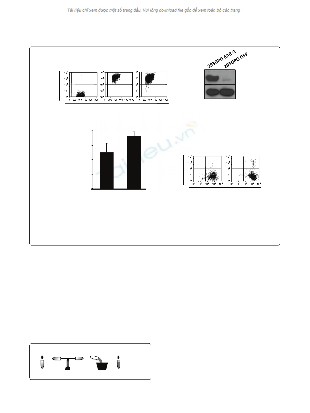
Figure 1 Stable producer cell lines generated by transduction of 293GPG cells.A. 293GPG stable producer cell lines for the GFP-empty
vector control virus and the human EAR-2 -GFP virus are stable in expression of GFP. Flow cytometry performed after two months continuous
culture shows GFP expression in > 99.5% of cells. B. 293GPG-EAR-2 cell lines were stable with respect to protein expression. Immunoblot analysis
performed on transduced cells after two months continuous culture shows strong expression of EAR-2 protein. C. 293GPG stable producer cell
lines were able to produce virus at titers significantly higher than those achieved by transient transfection. Virus was concentrated (one round).
Error bars denote standard deviation. D. Transduction of bone marrow using virus produced from stable producer cell lines (1 round of
ultracentrifugation) is not able to achieve high transduction rates in primary murine bone marrow cells.
SUPERNATANT CENTRIFUGE
WASTE
DECANT SUPERNATANT
REPEAT:
4 rounds of
centrifugation tota
l
A
BCD
Figure 2 Schematic of centrifugation protocol.
Ichim and Wells Journal of Translational Medicine 2011, 9:137
http://www.translational-medicine.com/content/9/1/137
Page 4 of 8
supernatant from numerous plates and filtered into the
same bottle. Therefore, each experimental group was
concentrated from supernatant with identical viral titers.
Following the appropriate number of rounds of centrifu-
gation centrifuge tubes were stored at 4 degrees. Upon
titration we observed that viral titers indeed increased
with each subsequent round of centrifugation (Figure
3A) and showed that this increase is additive, as demon-
strated by the linear relationship in the fold change of
viral titres (Figure 3B).
To test whether such long centrifugation periods had
a detrimental effect on viral titres we compared the
titres of two experimental groups that differed only in
the amount of centrifugation they received (Figure 3C).
Initially all tubes were subjected to three rounds of cen-
trifugation, in which tubes were centrifuged, decanted
and fresh viral containing medium added to the
previous viral pellet. Following three rounds of centrifu-
gation, half the tubes in the rotor were decanted and
stored for at 4°C for titration the next day, while the
other half of tubes were centrifuged an addition round
(without decanting supernatant or addition of fresh viral
medium), after which they too were decanted and stored
at 4°C for titration the next day. Both groups hence con-
tained the same quantity of virus, and differed only in
the amount of centrifugation each received. We did not
observe a significant difference in the titres between
these two experimental groups (Figure 3C) suggesting a
minimal effect of centrifugation on viral titres.
It is a common belief that centrifugation is able to pull
down cellular debris, membrane fragments, and proteins
from the virus containing medium. Conceivably, these
putative byproducts might have a detrimental effect on
any target cell, especially primary cells which are even
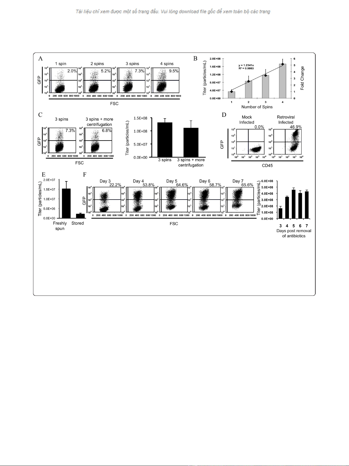
Figure 3 Retrovirus coated with VSV-G may be concentrated using multiple rounds of centrifugation.A. Assessment by flow cytometry
of transduction by retrovirus following concentration using different numbers of rounds of centrifugation. 1 μL of retrovirus was added for each
transduction. B. Titration of concentrated viral stocks. Bars denote the mean viral titer ± standard deviation. Diamonds represent the fold change
in viral titer. The trendline shows a linear relationship between the fold change in viral titer and the number of rounds of centrifugation. C.
Addition of an addition round of centrifugation without addition of unconcentrated supernatant does not result in a decrease in viral titre. D.
Demonstration by flow cytometry of successful transduction of primary mouse bone marrow cells by retroviral particles concentrated using
multiple rounds of centrifugation. E. Viral titers rapidly decrease following storage of virus at 4 degrees C for 7 days. F. Time course of viral titers
obtained following four rounds of centrifugation of supernatant collected on the given day post-induction (removal of antibiotics/tetracycline).5
μL of retrovirus was added for each titration.
Ichim and Wells Journal of Translational Medicine 2011, 9:137
http://www.translational-medicine.com/content/9/1/137
Page 5 of 8


