
BioMed Central
Page 1 of 10
(page number not for citation purposes)
Virology Journal
Open Access
Research
Prevention of genital herpes in a guinea pig model using a
glycoprotein D-specific single chain antibody as a microbicide
Jianmin Chen, Sanat K Davé and Anthony Simmons*
Address: University of Texas Medical Branch, Galveston, Texas, USA
Email: Jianmin Chen - jiachen@utmb.edu; Sanat K Davé - skdave@utmb.edu; Anthony Simmons* - ansimmon@utmb.edu
* Corresponding author
Abstract
Background: Genital herpes (GH) is a recurrent sexually transmitted infection (STI) that causes
significant morbidity and is also the major source of herpes simplex virus (HSV) in cases of neonatal
herpes. Vaccination is a current goal which has had limited success so far in preventing GH and
microbicides offer an attractive alternative. Treatment of primary disease cannot prevent
establishment of latent infections and thus, cannot prevent subsequent recurrent disease. Recently,
many of the molecular events leading to entry of HSV into cells have been elucidated, resulting in
the description of a number of herpesvirus entry mediators (HVEMs) that interact with HSV
glycoprotein D (gD) on the surface of virions. Described here is a strategy for interrupting the
spread of HSV based on interfering with these interactions. The hypothesis addressed in the
current report was that single chain antibody variable fragments (scFv) that interrupt associations
between gD and HVEMs would not only prevent infection in vitro but could also be used as
microbicides to interfere with acquisition GH.
Results and Conclusions: Here we show that a scFv derived from a particular hybridoma, DL11,
not only inhibits infection in vitro but also prevents development of GH in a guinea pig model when
applied intravaginally in an inert vehicle. Comparison of different anti-gD single chain antibodies
supported the hypothesis that the activity of DL11-scFv is based on its ability to disrupt the
associations between gD and the two major receptors for HSV, nectin-1 and HveA. Further, the
results predict that bacterial expression of active single chain antibodies can be optimized to
manufacture inexpensively a useful microbicidal product active against HSV.
Background
GH is generally caused by HSV type 2 (HSV-2), though
HSV type 1 (HSV-1) is increasingly recognized as a signif-
icant cause of primary infections [1]. Throughout the last
few decades there were substantial advances in under-
standing the epidemiology of genital HSV infections. Pri-
mary infection is almost always self-limited but on
healing virus is not eliminated from the host but rather,
viral genomes remain in a latent (dormant) state in sen-
sory neurons innervating initially infected skin and
mucous membranes [2,3]. The significance of latency is
that it is a reservoir of infection that can periodically reac-
tivate, causing virus to travel down nerve fibers to skin or
mucous membranes in the dermatome of primary infec-
tion. This may be manifest clinically as recurrent GH or
more frequently, causes unrecognized shedding of infec-
tious HSV [4-7] which despite being unrecognized is
responsible for the majority of new HSV-2 infections [8].
Published: 23 November 2004
Virology Journal 2004, 1:11 doi:10.1186/1743-422X-1-11
Received: 11 November 2004
Accepted: 23 November 2004
This article is available from: http://www.virologyj.com/content/1/1/11
© 2004 Chen et al; licensee BioMed Central Ltd.
This is an Open Access article distributed under the terms of the Creative Commons Attribution License (http://creativecommons.org/licenses/by/2.0),
which permits unrestricted use, distribution, and reproduction in any medium, provided the original work is properly cited.
Virology Journal 2004, 1:11 http://www.virologyj.com/content/1/1/11
Page 2 of 10
(page number not for citation purposes)
The epidemiology is further complicated by the fact that
many primary infections are asymptomatic or unrecog-
nized, which has the important implication that the first
clinical presentation of GH, often referred to as the initial
episode, may be caused by a recurrence of a prior asymp-
tomatic primary infection [9].
In the latter half of the 20th century, there were great
strides in antiviral therapy for GH but unfortunately, treat-
ing primary disease does not prevent establishment of
infection [10] and thus, cannot prevent subsequent recur-
rent disease. Barrier contraception provides some protec-
tion but its efficacy remains unclear [11] owing to the
complex nature of HSV pathogenesis, in which virus is
shed frequently and asymptomatically from multiple sites
below the waist [5]. Hence condoms are not as effective at
preventing transmission of GH as they are for other sexu-
ally transmitted infections. Vaccination is a current goal
which has had limited success to date. A recent trial of a
glycoprotein D-based sub-unit vaccine protected only
double (HSV-1 and 2) seronegative women but not men
[12]. Further, protection was mainly measured by preven-
tion of primary disease rather than infection. It is known
that treating primary disease does not prevent establish-
ment of latency and consequently, the long term efficacy
of this vaccine against subsequent recurrences remains
unknown.
Thus, the number of strategies for preventing sexual trans-
mission of GH is limited. Recently, there has been consid-
erable interest in topical microbicides as a potentially
attractive alternative to vaccination for prevention of sex-
ually transmitted infections, including GH [13]. Women
are able to control the use of vaginal microbicides and sev-
eral products are currently being used or tested, including
acid buffers and sulphated polymer-based inhibitors or
surfactants [14] like nonoxynol-9 (N-9) [13]. N-9 has
been used as a spermicide for 30 years and was thought to
provide some protection against gonorrhea and chlamy-
dia, a view was recently proven to be erroneous [14]. A
major factor limiting the efficacy and long-term viability
of N-9 and similar chemical compounds as topical agents
is their irritant effects on the vaginal epithelium [15]. Fur-
ther, recent data suggest that N-9, contrary to prior belief,
is not effective at protecting against HIV but rather it was
shown to increase rather than decrease the risk of acquir-
ing HIV in some populations studied, particularly women
at high risk of infection [14].
An evolving strategy that may be useful for preventing spe-
cific sexually transmitted viral infections is blocking of
virus entry into cells or subsequent inhibition cell-to-cell
spread. Many of the molecular events leading to entry of
HSV into cells have now been unraveled, resulting in the
description of two prominent cell-surface herpesvirus
entry mediators (Hve-A and nectin-1, also known as Hve-
C) that interact with HSV glycoprotein D (gD) on the sur-
face of virions [16-20]. In a recent study [21], nectin-1 was
shown to be expressed in the vaginal epithelium of
humans throughout the estrous cycle. In contrast, in mice
nectin-1 was expressed in vaginal epithelium only during
the stage of estrous at which they are susceptible to HSV.
Using a mouse model of GH, pre-incubation of HSV-2
with soluble recombinant nectin-1 was shown to block
entry of virus through vaginal mucosa [21], suggesting the
importance of nectin-1 in mediating entry of HSV into the
female genital tract. Hve-A and nectin bind to conforma-
tionally overlapping regions of gD and we were able
exploit this information together with the results of prior
studies that had mapped the sites on gD recognized by a
panel of monoclonal antibodies [22-26]. Antibody DL11
was of particular interest because it binds to an epitope on
gD that blocks the interactions between gD and both Hve-
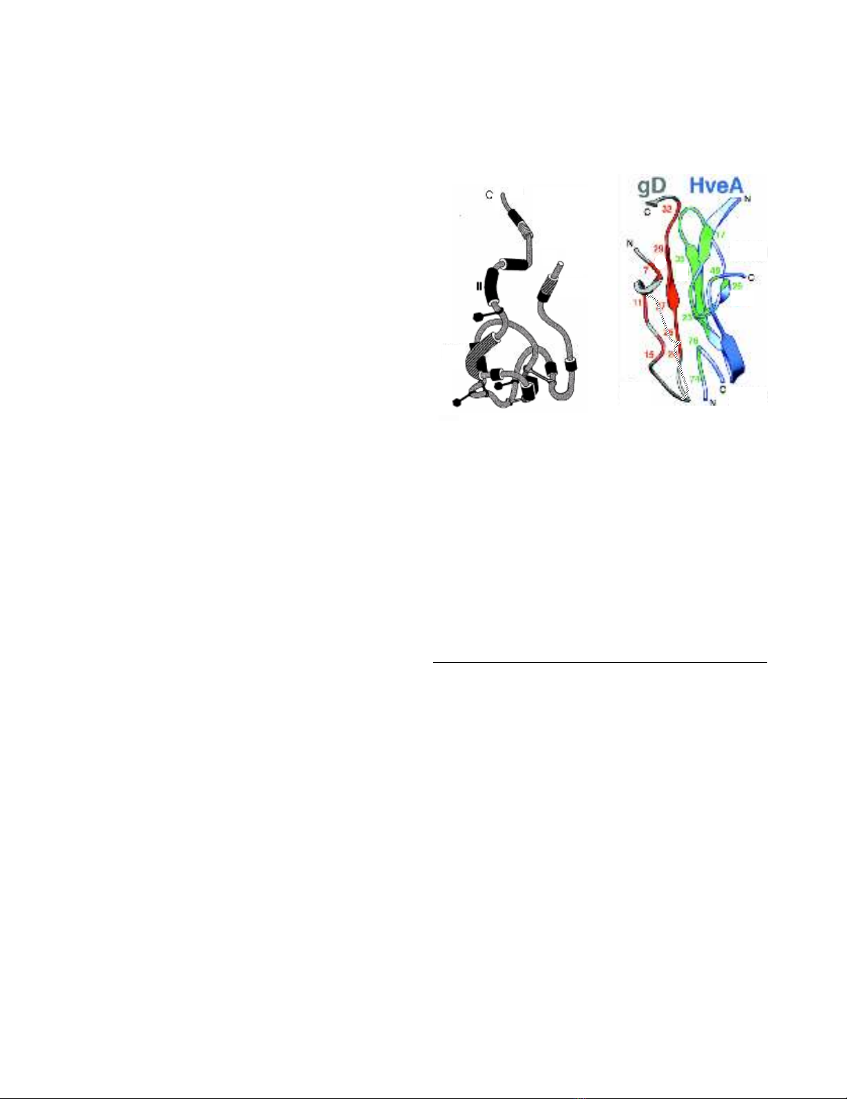
Panel A: Hypothetical model illustrating the antigenic struc-ture of gD and demonstrating the clustering of antigenic sites into seven groups, as defined by locations of amino acids bound by various monoclonal antibodiesFigure 1
Panel A: Hypothetical model illustrating the antigenic struc-
ture of gD and demonstrating the clustering of antigenic sites
into seven groups, as defined by locations of amino acids
bound by various monoclonal antibodies. Disulphide bonds
location defined by braces. Diagram adapted with permission
from Nicola et al, 1998 [22]. Of particular relevance to this
study are the locations of sites VII (amino acid residues 11–
19), which is bound by antibody 1D3, and site Ib, a discontin-
uous epitope that includes residues 222 to 252 that is bound
by antibody DL11. Panel B: Diagram showing interface
between N-terminal amino acids of gD and HveA and the
approximate residues (blue) bound by monoclonal antibody
1D3 and, by inference, 1D3 scFv (adapted with permission
from Connolly et al, 2003 [19].
1A 1B
VII
N
1
Ib
369
Virology Journal 2004, 1:11 http://www.virologyj.com/content/1/1/11
Page 3 of 10
(page number not for citation purposes)
A and nectin-1 [19] (figure 1). We show here that a single
chain antibody variable fragment (scFv) constructed from
DL11 neutralizes HSV infection in vitro, inhibits cell-to-
cell spread of virus and can be used to prevent infection in
a guinea pig model of GH.
Results
Construction and expression of single chain antibodies
against gD
Four from the panel of anti-HSV gD hybridomas available
were selected for scFv construction based on the known
locations of their epitopes [22] (summarized in figure 1)
and knowledge about the neutralization properties of the
antibodies produced by them. Of particular note are the
properties of DL11, which neutralizes both HSV-1 and
HSV-2 in the absence of complement and antibody bind-
ing to its conformational epitope is known to disrupt the
interactions of gD both with Hve-A and nectin-1. Also
1D3 binds to a group VII neutralizing epitope that directly
Table 1: Degenerate PCR primers used for amplification of VL (kappa) and VH (gamma).
Nomenclature Primer sequences used for PCR reactions
Signal sequence/framework primers
Kappa 1 GGTGATATCGTGATRACMCARGATGAACTCTC
Kappa 2 GGTGATATCWTGMTGACCCAAWCTCCACTCTC
Kappa 3 GGTGATATCGTKCTCACYCARTCTCCAGCAAT
Kappa 4 CTGWTGTTCTGGATTCCTG
Kappa 5 GTGCTCTGGATTCGGGAA
Kappa 6 TCAGCTTCYTGCTAATCAGTG
Kappa 7 TGGGTATCTGGTRCSTGTG
Kappa 8 GTTTCMAGGTRCCAGATGT
Kappa 9 TGTTTTCAAGGTRCCAGATGT
Kappa 10 CTSTGGTTGTCTGGTGTTGA
Kappa 11 TGCTKCKCTGGGTTCCAG
C region kappa primer TGGTGGGAAGATGGA
Signal sequence/framework primers
Gamma 1 GAGGTGAAGCTGCAGGAGTCAGGACCTAGCCTGGTG
Gamma 2 AGGTVMAACTGCAGVAGTCWGG
Gamma 3 AGGTVVAGCTGCAGVAGTCWGG
Gamma 4 ACTGCAGGTRTCCACTCC
Gamma 5 RCTACAGGTGTCCACTCC
Gamma 6 GCYACAGMTGTCCACTCC
Gamma 7 ACTGCAGGTGTCCTCTCT
Gamma 8 RCTRCAGGYGTCCACTCT
Gamma 9 CCAAGCTGTGTCCTRTCC
Gamma 10 TGTTGACAGYCVTT CCKGGT
Gamma 11 TAYTTTAAAARGTGTCMAGTGT
Gamma 12 CTYTTAAAAGGKGTCCAGWG
Gamma 13 CYTTTAMATGGTATCCAGTGT
Gamma 14 ATGGCAGCWGCYCAAAG
Gamma 15 CTTTTAAAAGWTGTCCAGKGT
Gamma 16 CTTCCTGATGGCAGTGGTT
C region gamma primer TAACCCTTGACCAGGCATCC
Key to degenerate nucleotides: R = A+G; M = A+C; W = A+T; K = G+T; S = G+C; Y = C+T; H = A+T+C; B = G+T+C; D = G+A+T; N =
A+C+G+T; V = G+A+C
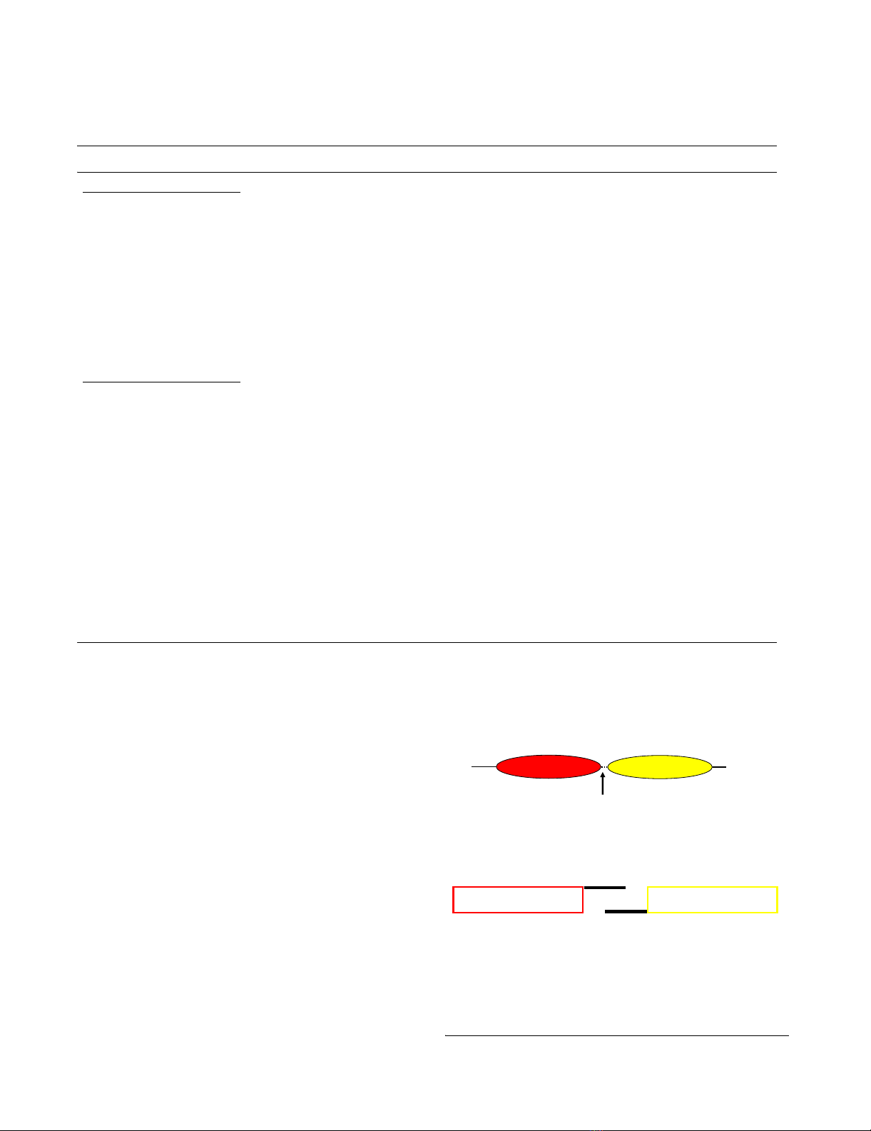
Panel A: Structure of an scFv cassette spliced using a (Gly4Ser)3 hingeFigure 2
Panel A: Structure of an scFv cassette spliced using a
(Gly4Ser)3 hinge. Panel B. Alternative glycine codons were
used in the overlapping region of the hinge to avoid produc-
tion of completely overlapping regions, thereby generating a
sub-optimal (Gly4Ser)2 hinge.
A.
B.
VLVH
VL
VLVH
VH
N
V
L
V
H
(Hinge)
(Gly
4
Ser)
3
COOH
Virology Journal 2004, 1:11 http://www.virologyj.com/content/1/1/11
Page 4 of 10
(page number not for citation purposes)
interferes with the interface between gD and HveA (figure
1B). A fifth scFv cassette, against carcinoembryonic anti-
gen (CEA) was excised from a plasmid encoding an anti-
tumor chimeric T-cell receptor, kindly provided by Hin-
rich Abken (Cologne University, Germany). For produc-
tion of cDNAs, individual VL and VH regions from each
hybridoma were reverse transcribed using primers near
the VH-CH and VL-CL junctions. For PCR cloning these
primers were paired with a panel of degenerate primers
derived from VH or VL signal sequences (Table 1) that were
able to amplify all hybridoma heavy and light chains
tested so far (14/14) irrespective of antibody class or sub-
class (data not shown). PCR products were sequenced
directly to facilitate design of new primer sets allowing, on
re-amplification of hybridoma cDNAs, elimination of
degenerate primer sequences introduced in the first reac-
tion and introduction of 2/3 of a 15 amino acid hinge
region comprising three repeats of four glycine and one
serine residues (Figure 2). VL and VH are not covalently
linked in nature but flexible hinges of this design and
length were shown previously [27] to allow reconstruc-
tion of antibody binding sites when VL and VH are linked
end-to-end (figures 3, 4). Finally, the PCR products con-
taining the overlapping hinge regions were ligated, PCR
amplified and the resultant scFv cassette was TA cloned
into pCR2.1TOPO. To generate the desired single chain
antibodies, the cassettes were subcloned into the bacterial
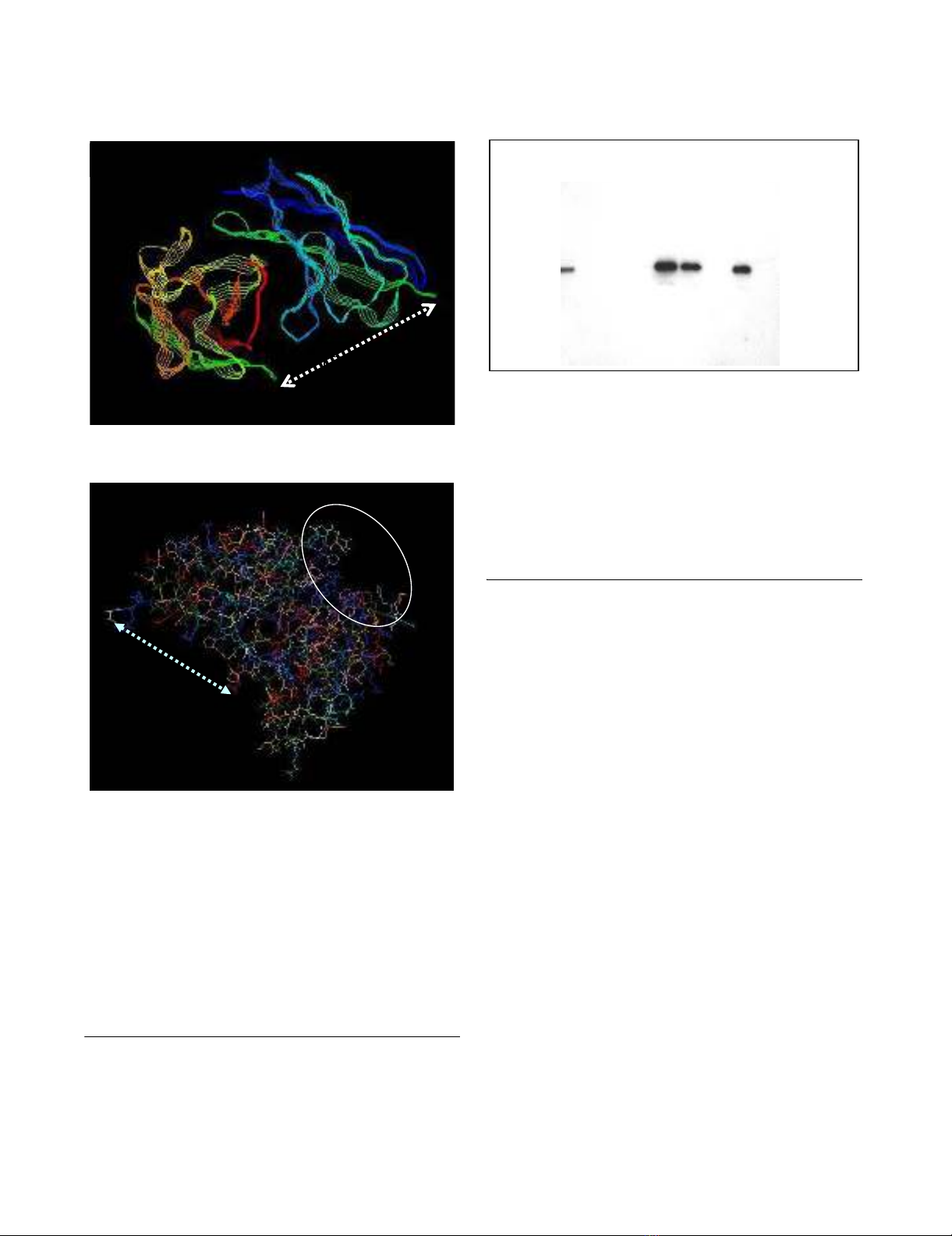
Single plain view of a 3-D model of DL11 scFv, showing its predicted structureFigure 3
Single plain view of a 3-D model of DL11 scFv, showing its
predicted structure. Panel A: Strand view, colored by
group, demonstrates relative orientation of the kappa (top)
and gamma (bottom) chains, which shows the positions of
residues to which the (Gly4Ser)3 hinge is attached. Panel B:
Wireframe image illustrating hinge attachment sites on one
side of the molecule (linked by dashed line) and clustering on
the opposite side (inside the circle) of the complementary
determining regions (CDRs) predicted by the Kabat antibody
database. The clustering of CDRs suggests correct conforma-
tion of the molecule with formation of an antigen binding
site.
Hinge
CDR
cluster
Hinge
B
A
Western blot demonstrating expression of DL11 scFv by E, ColiFigure 4
Western blot demonstrating expression of DL11 scFv by E,
Coli. BL21 cells were transfected with p-TOPO10 containing
the scFv cassette. Bacterial lysates were purified using a
nickel chelation column and the reaction with anti-V5 of total
lysates and various fractions from the column are shown.
Lane 1, unpurified total bacterial lysate; lane 2, nickel column
flow through; lanes 3 and 4, saline washes; lane 6, eluate from
Ni beads; lane 7, bacterial supernatant; lane 8, scFv remaining
on nickel column after elution; lane 9: supernatant from un-
induced bacteria.
34kd
1 2 3 4 5 6 7 8 9
Virology Journal 2004, 1:11 http://www.virologyj.com/content/1/1/11
Page 5 of 10
(page number not for citation purposes)
expression vector pET101-D. An antibody modeling algo-
rithm, verified by the locations of the complementary
determining regions, was used to predict the 3-D struc-
tures of all four of the anti-gD single chain antibodies. The
results were consistent with reconstitution of the original
antigen binding sites (e.g. figure 3, DL11; others not
shown).
Bacterial expression and extraction of anti-gD single chain
antibodies
The single chain antibodies were expressed in E. Coli
strain BL21 using pET101-D (Invitrogen), which attaches
hexa-His and V5 tags to expressed proteins for their isola-
tion and identification. Bacteria were induced with IPTG,
centrifuged and the supernatants tested for the presence of
scFvs by western blotting using anti-His antibody (figure
4). Bacterial pellets were sonicated in phosphate buffered
saline to release inclusion bodies and proteins were sol-
ubulized by addition of 6 M guanidine (BL21). Nickel
bead chelation was used to extract the His-tagged protein.
Western blots of eluates from nickel beads (e.g. DL11 scFv
from DL21; Fig. 4, lanes 6 and 7) identified scFvs that
were released by this procedure. They were generally iso-
lated at concentrations of 500–1000 µg/ml from BL21.
Re-folding and intra-chain disulphide bond formation
were maximized by gradually reducing guanidine concen-
tration by step-wise dialysis from 6 M initially to 3 M, then
2 M, 1 M, 0.5 M and finally 0 M, with addition of L-
arginine and oxidized glutathione (GSSG) in final two
steps [28]. The ability of the single chain antibodies pro-
duced in this way to bind their target antigen was tested by
determining their reaction with plastic bound gD by
ELISA. Binding ratios were calculated in relation to the
background binding of CEA scFv (e.g. DL11-based scFv;
figure 5)
Selected anti-gD single chain antibodies neutralize HSV in
vitro
The hypothesis that selected single chain antibodies can
block infection of cells in vitro by reacting with an epitope
that disrupts the interface between gD and HVEMs was
tested by comparing the activities of the various bacteri-
ally expressed anti-gD scFv in a Vero cell-based HSV-1
plaque reduction assay. A scFv directed against an epitope
on carcinoembryonic antigen was included as an unre-
lated control. The results showed that pre-incubation of
virus with DL11 and 1D3 scFvs inhibited plaque forma-
tion with decreasing efficiency. DL6 scFv showed minimal
but reproducible activity (data not shown), whereas the
other scFvs tested (DL2 and CEA) had no plaque reducing
capability at all (figure 6). Against HSV-2, only DL11
showed neutralizing activity in a similar plaque reduction
assay (data not shown), confirming the type common
nature of its epitope. In addition to inhibition of plaque
formation, pre-incubating HSV-2 with 100 µg/ml DL11
caused an 80% reduction in plaque numbers and a ~50%
reduction (figure 7) in the size of plaques (0.95 ± 0.3 mm
with DL11scFv vs. 1.9 ± 0.4 mm without, respectively).
The same was true for HSV-1 and DL11 (not shown). It
was concluded that DL11scFv could not only block infec-
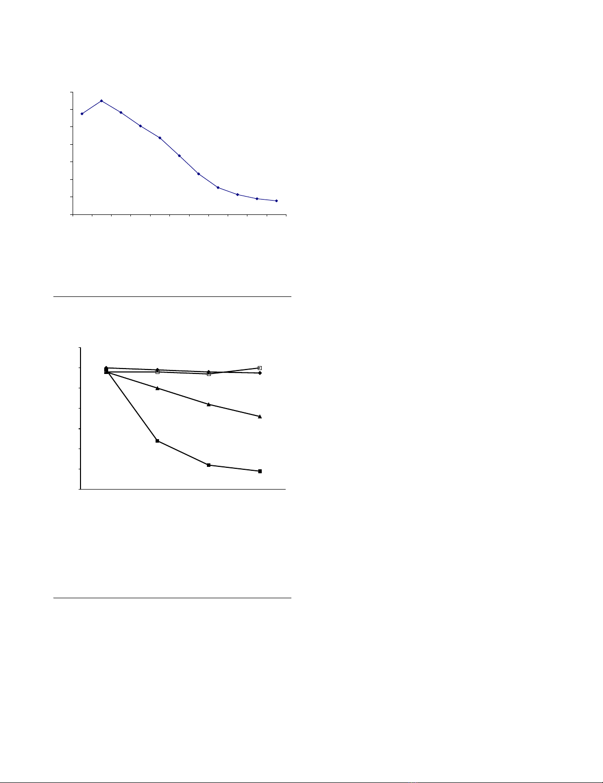
Binding of scFv to plastic bound gDFigure 5
Binding of scFv to plastic bound gD. Binding ratios of DL11
scFv to gD compared with an irrelevant (CEA) scFv at the
same protein concentrations.
Specific reduction of HSV-2 plaque numbers by incubation of virus with anti-gD scFvFigure 6
Specific reduction of HSV-2 plaque numbers by incubation of
virus with anti-gD scFv. Vero cells were pre-incubated with
approximately 120 PFU HSV-2, strain G with single chain
antibodies generated from hybridomas D11 (¦), 1D3 (?),
DL2(?) and an irrelevant CEA-specific construct (?).
0
1
2
3
4
5
6
7
12481632641282565121024
Dilution
DL11/CEA binding ratio
0
20
40
60
80
100
120
140
1234
Number of plaques
81.25Pg/ml 162.5Pg/ml 325Pg/ml 750Pg/ml
Concentration of scFv


![Vaccine và ứng dụng: Bài tiểu luận [chuẩn SEO]](https://cdn.tailieu.vn/images/document/thumbnail/2016/20160519/3008140018/135x160/652005293.jpg)


%20--%3e%3cdefs%3e%3cstyle%3e%20.st0%20{%20fill:%20%23fff;%20}%20.st1%20{%20fill:%20%237800fa;%20}%20%3c/style%3e%3c/defs%3e%3cpath%20class='st1'%20d='M117.78,12.18H43.11c2.9,3.47,4.65,7.94,4.65,12.82,0,5.6-2.3,10.66-6.01,14.29h76.02l7.22-13.56-7.22-13.56Z'/%3e%3cg%3e%3cpath%20class='st0'%20d='M53.58,26.17h-.59v-1.46h.59v-4.96h2.83c1.78,0,2.67.94,2.67,2.82v5.76c0,1.87-.89,2.81-2.67,2.81h-2.83v-4.96ZM55.36,21.37v3.34h1.1v1.46h-1.1v3.34h1.01c.61,0,.91-.37.91-1.1v-5.93c0-.74-.3-1.1-.91-1.1h-1.01Z'/%3e%3cpath%20class='st0'%20d='M65.99,31.14h-1.8l-.31-2.07h-2.19l-.31,2.07h-1.64l1.82-11.39h2.62l1.82,11.39ZM65.28,18.04c-.25.46-.51.77-.75.94-.21.15-.47.22-.79.22-.26,0-.57-.07-.92-.22l-.38-.15c-.14-.05-.26-.07-.37-.07-.3,0-.53.18-.71.54l-.91-.68c.25-.46.51-.77.75-.94.21-.14.48-.21.79-.21.26,0,.57.07.92.21l.38.15c.14.05.26.07.37.07.3,0,.53-.18.71-.54l.91.68ZM61.91,27.52h1.73l-.87-5.76-.87,5.76Z'/%3e%3cpath%20class='st0'%20d='M74.53,26.89v1.52c0,1.91-.89,2.86-2.67,2.86s-2.67-.95-2.67-2.86v-5.93c0-1.91.89-2.86,2.67-2.86s2.67.95,2.67,2.86v1.11h-1.69v-1.22c0-.75-.31-1.12-.93-1.12s-.93.37-.93,1.12v6.15c0,.74.31,1.11.93,1.11s.93-.37.93-1.11v-1.63h1.69Z'/%3e%3cpath%20class='st0'%20d='M81.4,31.14h-1.8l-.31-2.07h-2.19l-.31,2.07h-1.64l1.82-11.39h2.62l1.82,11.39ZM75.9,19.2l1.52-1.91h1.71l1.51,1.91h-1.61l-.76-.95-.75.95h-1.61ZM77.32,27.52h1.73l-.87-5.76-.87,5.76ZM83.1,15.99l-1.76,1.91h-1.26l1.17-1.91h1.86Z'/%3e%3cpath%20class='st0'%20d='M84.86,19.75c1.78,0,2.67.94,2.67,2.82v1.48c0,1.87-.89,2.81-2.67,2.81h-.85v4.28h-1.79v-11.39h2.64ZM84.01,21.37v3.86h.85c.58,0,.87-.36.87-1.08v-1.71c0-.71-.29-1.07-.87-1.07h-.85Z'/%3e%3cpath%20class='st0'%20d='M93.51,19.75c1.78,0,2.67.94,2.67,2.82v1.48c0,1.87-.89,2.81-2.67,2.81h-.85v4.28h-1.79v-11.39h2.64ZM92.66,21.37v3.86h.85c.58,0,.87-.36.87-1.08v-1.71c0-.71-.29-1.07-.87-1.07h-.85Z'/%3e%3cpath%20class='st0'%20d='M98.8,31.14h-1.79v-11.39h1.79v4.88h2.03v-4.88h1.83v11.39h-1.83v-4.88h-2.03v4.88Z'/%3e%3cpath%20class='st0'%20d='M105.36,24.55h2.46v1.62h-2.46v3.34h3.09v1.63h-4.88v-11.39h4.88v1.63h-3.09v3.18ZM108.17,17.29l-1.76,1.91h-1.26l1.17-1.91h1.86Z'/%3e%3cpath%20class='st0'%20d='M112.2,19.75c1.78,0,2.67.94,2.67,2.82v1.48c0,1.87-.89,2.81-2.67,2.81h-.85v4.28h-1.79v-11.39h2.64ZM111.35,21.37v3.86h.85c.58,0,.87-.36.87-1.08v-1.71c0-.71-.29-1.07-.87-1.07h-.85Z'/%3e%3c/g%3e%3ccircle%20class='st1'%20cx='25'%20cy='25'%20r='20'/%3e%3cpath%20class='st0'%20d='M32.78,19.27c2.92,0,4.43,2.55,5.28,5.33l.71,2.17c.14.38-.33.75-.71.75h-5.61c.19-.33.24-.71.09-1.08l-.75-2.45c-.43-1.32-.99-2.64-1.79-3.77.75-.57,1.65-.94,2.78-.94h0ZM25,18.38c3.25,0,4.9,2.78,5.89,5.89l.76,2.45c.14.42-.33.8-.8.8h-11.69c-.42,0-.94-.38-.8-.8l.75-2.45c.99-3.11,2.64-5.89,5.89-5.89h0ZM25,11.35c1.74,0,3.11,1.37,3.11,3.11s-1.37,3.11-3.11,3.11-3.11-1.41-3.11-3.11,1.41-3.11,3.11-3.11h0ZM17.27,19.27c1.08,0,1.98.38,2.73.94-.8,1.13-1.37,2.45-1.74,3.77l-.8,2.45c-.14.38-.05.75.09,1.08h-5.56c-.42,0-.9-.38-.75-.75l.71-2.17c.9-2.78,2.41-5.33,5.33-5.33h0ZM17.27,12.91c1.51,0,2.78,1.27,2.78,2.83s-1.27,2.83-2.78,2.83-2.83-1.27-2.83-2.83,1.27-2.83,2.83-2.83h0ZM32.78,12.91c1.56,0,2.78,1.27,2.78,2.83s-1.23,2.83-2.78,2.83-2.83-1.27-2.83-2.83,1.27-2.83,2.83-2.83h0ZM27.07,28.56v.09c0,.57-.24,1.08-.61,1.46h0v.05c-.38.33-.9.57-1.46.57s-1.08-.24-1.46-.61h0c-.38-.38-.61-.9-.61-1.46v-.09h1.41v.09c0,.19.05.38.19.47v.05c.09.09.28.19.47.19s.38-.09.47-.19v-.05c.14-.09.24-.28.24-.47t-.05-.09h1.41ZM30.99,28.56v.09c0,1.65-.66,3.16-1.74,4.24-1.08,1.08-2.59,1.79-4.24,1.79s-3.16-.71-4.24-1.79l-.05-.05c-1.04-1.08-1.7-2.55-1.7-4.2v-.09h1.41v.09c0,1.27.47,2.4,1.27,3.25h.05c.85.85,1.98,1.37,3.25,1.37s2.4-.52,3.25-1.37c.85-.8,1.37-1.98,1.37-3.25v-.09h1.37ZM34.99,28.56v.09c0,2.78-1.13,5.28-2.92,7.07-1.79,1.79-4.29,2.92-7.07,2.92s-5.23-1.13-7.07-2.92c-1.79-1.79-2.92-4.29-2.92-7.07v-.09h1.41v.09c0,2.4.94,4.53,2.5,6.08,1.56,1.56,3.72,2.5,6.08,2.5s4.52-.94,6.08-2.5c1.56-1.56,2.5-3.68,2.5-6.08v-.09h1.41Z'/%3e%3c/svg%3e)