
RESEARCH Open Access
CD4
+
T cells spontaneously producing human
immunodeficiency virus type I in breast milk from
women with or without antiretroviral drugs
Diane Valea
1,2†
, Edouard Tuaillon
3,4,5†
, Yassine Al Tabaa
3,4
, François Rouet
1
, Pierre-Alain Rubbo
3,4
, Nicolas Meda
2
,
Vincent Foulongne
3,5
, Karine Bollore
3,4
, Nicolas Nagot
3
, Philippe Van de Perre
3,5
and Jean-Pierre Vendrell
3,4,5,6*
Abstract
Background: Transmission of human immunodeficiency virus type 1 (HIV-1) through breast-feeding may involve
both cell-free and cell-associated virus. This latter viral reservoir remains, however, to be fully explored. CD4
+
T cell-
associated virus production in breast milk was therefore investigated.
Methods: The ex vivo spontaneous production of HIV-1 antigen and HIV-1 RNA by CD4
+
T cells was measured in
paired blood and breast milk samples from 15 HIV-1 infected women treated or not with antiretroviral drugs.
Spontaneous antigen secreting cells (HIV-1-AgSCs) from breast milk and blood were enumerated by an ELISpot
assay, and cell-associated HIV-1 RNA was quantified by real-time PCR in supernatants of CD4
+
T cells cultured for
18 hours without addition of polyclonal activators.
Results: Among the CD4
+
T cells present in breast milk, memory cells expressing high levels of cell-surface
activation markers were predominant. Spontaneous HIV-1-AgSCs were detected and enumerated in the breast milk
of all 15 women, with a median number of 13.0 and 9.5 HIV-1- AgSCs/106 CD4
+
T cells in aviremic (n = 7) and
viremic (n = 8) women, respectively. Cell- associated HIV-1 RNA was detected in cell-free supernatants from 4/7
aviremic and 5/8 viremic individuals at median levels of 190 and 245 copies/ml, respectively.
Conclusions: Activated CD4
+
T cells producing HIV-1 are detected in the breast milk of untreated individuals as
well as those receiving highly active antiretroviral therapy. This finding strongly suggests that HIV-1 replication
occurs in latently infected CD4
+
T cells that, upon spontaneous activation, revert to productively infected cells.
These cells might be responsible for a residual breast milk transmission despite maternal highly active antiretroviral
therapy.
Background
Today, while improvements have been made in prophy-
lactic measures to prevent the perinatal transmission of
HIV-1, its transmission through breastfeeding is still the
cause of over half the estimated yearly 420,000 new
pediatric infections worldwide [1]. Indeed, while it is
universally recognized as the optimal source of nutrition
and defense against disease in infants, breast milk is also
a major mode of HIV-1 transmission from mother to
child [2-4]. The mechanisms by which this occurs,
however, remain poorly understood [5]. In breast milk,
HIV-1 may be present in three different forms of poten-
tially unequal transmission risk: (i) free virions measured
as HIV-1 RNA, (ii) integrated provirus measured as
HIV-1 DNA, and (iii) HIV-1 RNA that is released by
activated cells that sustain the virus replication cycle
and is measured as cell-associated HIV-1 RNA. High
levels of free HIV-1 RNA in maternal plasma and in
breast milk are associated with a high risk of breastfeed-
ing transmission [6-11]. A similar association has been
demonstrated with HIV-1 proviral DNA levels in breast
milk, thus suggesting that both cell-free and cell-asso-
ciated HIV-1s are involved in breastfeeding transmission
[9,12-14]. Results of a study performed in Botswana
* Correspondence: jp-vendrell@chu-montpellier.fr
†Contributed equally
3
Faculté de Pharmacie, 15 Avenue Charles Flahault, Montpellier 34060,
France
Full list of author information is available at the end of the article
Valea et al.Retrovirology 2011, 8:34
http://www.retrovirology.com/content/8/1/34
© 2011 Valea et al; licensee BioMed Central Ltd. This is an Open Access article distributed under the terms of the Creative Commons
Attribution License (http://creativecommons.org/licenses/by/2.0), which permits unrestricted use, distribution, and reproduction in
any medium, provided the original work is properly cited.

suggest that up to 9 months postpartum, HIV-1 is
mainly transmitted by cells containing the provirus
while the cell-free virus is more frequently involved later
on [15]. Furthermore, preliminary observations suggest
that some babies breastfed by HIV-1 infected women
taking antiretroviral therapy (ART) get infected despite
undetectable levels of HIV-1 RNA in their mother’s
plasma or breast milk [16,17]. Importantly, the in vitro
infectivity of the cell-associated virus has been found to
be 100 to 1000 times higher than that of cell-free virus
stocks [18]. Taken together, these observations strongly
suggest that cell-associated virus is frequently involved
in the transmission of HIV-1 by breastfeeding. HIV-1
persists in a latent form in resting CD4
+
T cells, even in
patients receiving antiretroviral treatment (ART) and in
whom the viral load is undetectable. These latently
infected cells constitute a viral reservoir, which may be
regarded as a cell type or anatomical site in which a
functional form of the virus persists with increased sta-
bility compared to the pool of actively replicating virus
[19]. A recent study shows that cell-free and, to a much
lesser extent, cell-associated HIV-1 RNA levels in breast
milk are suppressed by antiretroviral regimens used to
prevent mother to child transmission, whereas no signif-
icant reduction in latently HIV-1 infected resting CD4
+
T cells is observed [20].
We recently demonstrated that breast milk contains
such resting CD4
+
T lymphocytes and that these cells
are capable of producing viral antigens (Ags) and virions
after in vitro polyclonal-cell activation. In addition, these
CD4
+
T lymphocytes showed a greater capacity to pro-
duce viral particles than their circulating blood counter-
parts [21]. Moreover, it has also been demonstrated that
CD4
+
T cells from most viremic HIV-1 infected
patients, spontaneously secrete HIV-1 virions as a con-
sequence of an ongoing viral replication in the absence
of ART or a residual HIV-1 replication under ART
[22,23]. Thus, we hypothesized that breast milk contains
CD4
+
T cells able to spontaneously produce HIV-1 pro-
teins, RNA. and infectious particles.
In this study, we (i) characterized activated CD4
+
T
cells in breast milk, (ii) enumerated CD4
+
T cells sponta-
neously producing HIV-1 antigens (HIV-1-AgSCs), and
(iii) measured cell-associated HIV-1 RNA in cell-free
supernatants in infected women treated or not with anti-
retroviral drugs. The human milk-derived activated CD4
+
T cells that spontaneously produced HIV-1 were barely
affected by maternal antiretroviral therapy and might
therefore be responsible for residual HIV-1 transmission.
Results
Study subjects
Women’s characteristics, antiretroviral treatments and
breast milk sample collection conditions are described
in Table 1. According to national policy guidelines, 9
women received perinatal prophylactic treatment to pre-
vent mother to child transmission of HIV-1, consisting
of zidovudine given from between the 34th and 36th
weeks of pregnancy until delivery plus a single dose of
nevirapine during labor/delivery. The remaining 6
women were eligible for ART during pregnancy and
received zidovudine, lamivudine and ritonavir-boosted
lopinavir. The mean duration of ART until delivery was
36.4 days. Among the 15 women, the mean CD4
+
T cell
count was 519 cells/mm3 and the mean plasma HIV-1
RNA level 13,105 copies/ml. Seven women, 5 treated
with ART (nos. 1, 3, 9, 12 and 13) and two with the
short perinatal prophylactic treatment (nos. 6 and 11),
had undetectable plasma HIV-1 RNA load. The remain-
ing seven women who received the short perinatal pro-
phylactic treatment (nos. 2, 4, 5, 7, 10, 14 and 15) had a
detectable plasma HIV-1 RNA load, and the one
remaining woman receiving ART (no. 8) showed detect-
able viraemia. HIV-1 RNA was detected in the breast
milk of five (35%) women; (mean 140 HIV-1 RNA
copies/ml, range < 145-4,062 HIV-1 RNA copies/ml),
four of whom had stopped ART at time of sampling
and showed detectable HIV-1 plasma viral load.
Characterization of CD4
+
T cells in breast milk
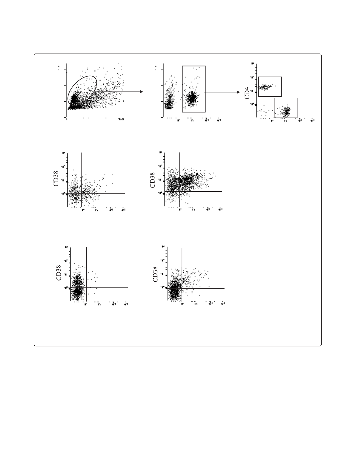
As shown in one representative case (patient no. 10), we
characterized the CD3
+
, CD4
+
and CD8
+
T cells as well as
CD4
+
and CD8
+
T cells expressing HLA-DR and CD38
receptors in breast milk and blood by flow cytometry
prior to CD4
+
Tcellenrichment(Figure1A,B,C).The
CD4
+
T cells in the breast milk of 15 women represented
on average 22.2% of the total T cell count, and the CD3
+
CD8
+
T cells represented 60.1%. A similar distribution
was found in blood samples. The majority of CD4
+
and
CD8
+
T cells in milk did not express the CD45RA recep-
tors characteristic of naive T cells (mean 92.4% and 79%,
respectively). The percentage of CD4
+
and CD8
+
T cells
not expressing CD45RA was significantly lower in blood
(mean 64.3% and 45.3%, respectively, P< 0.001). These
results imply that the majority of T cells found in the
milk are mainly memory T cells. This observation was
confirmed by the high level of cell-surface CD45RO
receptor expression on these cells (data not shown). In
addition,asshowninTable2,breastmilkCD4
+
and
CD8
+
T cells expressed higher levels of activation mar-
kers when compared with blood CD4
+
and CD8
+
Tcells.
Thus, breast milk from HIV-1 infected women contains
predominantly activated memory CD4
+
T cells.
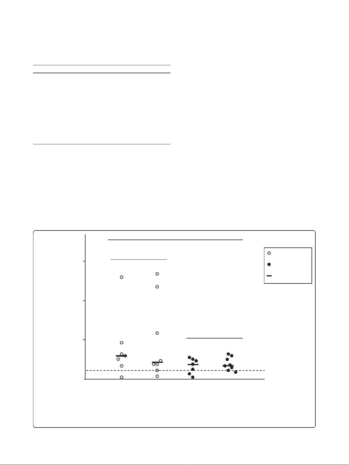
Enumeration of HIV-1-AgSCs in breast milk and blood
derived CD4
+
T cells
To evaluate the ability of the CD4
+
T lymphocytes to
spontaneously secrete HIV-1 Ag and viral particles, freshly
Valea et al.Retrovirology 2011, 8:34
http://www.retrovirology.com/content/8/1/34
Page 2 of 12

purified CD4
+
T cells from paired breast milk and blood
samples were directly tested using our ELISpot assay.
HIV-1-AgSCs were detected in breast milk cells from all
women. As shown in Figure 2, the median number of
HIV-1-AgSCs was similar in aviremic (n= 7) and viremic
(n= 8) subjects, 13.0 HIV-1-AgSCs/10
6
CD4
+
T cells
[Interquartile Range (IQR) 9.5-16.5 HIV-1- AgSCs/10
6
CD4
+
T cells] and 9.5 HIV-1-AgSCs/10
6
CD4
+
Tcells
(IQR 8.1-29.4 HIV-1-AgSCs/10
6
CD4
+
T cells), respec-
tively (P > 0.05). HIV-1-AgSCs were also detected in the
blood of viremic and aviremic women, median, 8.1/10
6
CD4
+
T cells (IQR, 4.0-9.5/10
6
CD4
+
T cells) and 6.25/10
6
CD4
+
T cells (IQR, 5.4-10.3/10
6
CD4
+
T cells, respec-
tively), the numbers of which showed no significant differ-
ence between the two groups (P> 0.05).
Detection of cell-associated HIV-1 RNA in supernatants
from breast milk- and blood-derived CD4
+
T cell cultures
HIV-1 RNA was also quantified in the culture supernatant
following 18 hours culturing of breast milk- and blood-
derived CD4
+
T cells. As shown in Figure 3, concerning
the breast milk samples, breast milk cell-associated HIV-1
RNA was detectable in 10 of the 15 subjects (66.7%), the
HIV-1 RNA levels were similar in women with detectable
or undetectable plasma viral load: median, 245 RNA
copies/ml (IQR, 113-12,300 RNA copies/ml) and 190
RNA copies/ml (IQR, 30-261 copies/ml), respectively. No
correlation was observed between the number of HIV-1
RNA copies detected in the supernatants and the number
of HIV-1-AgSCs. These data suggest that the presence of
cells spontaneously producing HIV-1 RNA in breast milk
is independent of plasma HIV-1 RNA levels. In blood
samples, cell-associated HIV-1 RNA was detected in 14/15
individuals (93.3%) with a median level of 2,261 RNA
copies/ml (IQR, 1,629-5,190 RNA copies/ml) in aviremic
women (range 583-119,981) and 13,855 (IQR, 40,051-
111,390 RNA copies/ml) in viremic women. Unexpectedly,
although a similar number of HIV-1-AgSCs was found in
the breast milk of aviremic and viremic women, the cell-
associated HIV-1 RNA copies were significantly higher in
the women with detectable viral load (P<0.01).CD4
+
T
cell-associated HIV-1 RNA levels were significantly higher
Table 1 Characteristics of HIV-1 infected women
Patients
no.
Initiation of
antiretroviral treatment
(days before delivery)
Duration of lactation until
sampling (days)
Antiretroviral
regimen
Treatment at time
of sampling
CD4
+
T cell
counts/mm
3
HIV-1 RNA level
(copies/ml)
plasma Breast
milk
1 15 54 ART
a
Ongoing NT ND
b
NT
2 18 65 Short-course
prophylaxis
c
Withdrawal since 65
days
400 1776 ND
3 34 33 ART Ongoing 762 ND ND
4 35 11 Short-course
prophylaxis
Withdrawal since 11
days
521 12,878 ND
5 38 14 Short-course
prophylaxis
Withdrawal since 14
days
270 83,547 ND
6 26 55 Short-course
prophylaxis
Withdrawal since 55
days
646 ND ND
7 47 57 Short-course
prophylaxis
Withdrawal since 57
days
658 6,790 ND
8 32 50 ART Ongoing 305 34,937 4,062
9 17 29 ART Ongoing 416 ND ND
10 65 91 Short-course
prophylaxis
Withdrawal since 91
days
628 50,036 772
11 58 77 Short-course
prophylaxis
Withdrawal since 77
days
618 ND 190
12 15 52 ART Ongoing 444 ND ND
13 69 21 ART Ongoing 533 ND ND
14 46 9 Short-course
prophylaxis
Withdrawal since 9
days
688 1,049 145
15 31 15 Short-course
prophylaxis
Withdrawal since 15
days
384 4,526 308
a
ART, antiretroviral therapy.
b
Threshold: 300 copies/ml plasma and 60 copies/ml for breast milk.
c
Short-course perinatal prophylaxis (zidovudine until delivery and a single-dose of nevirapine during labor).
NT: not tested.
ND: not detected, < threshold.
Valea et al.Retrovirology 2011, 8:34
http://www.retrovirology.com/content/8/1/34
Page 3 of 12
in blood than in breast milk (P<0.01).Insubjectswith
undetectable HIV-1 viral load in plasma and breast milk
(n = 5), both cell-associated HIV-1 RNA and HIV-1-
AgSCs were detected in the breast milk, suggesting that
the antiretroviral treatment was not fully effective at sup-
pressing spontaneous virus production in breast milk.
In vitro infection of CD4
+
T cells using breast milk- and
blood-cell culture supernatants
The infectivity of the virus secreted in breast milk- and
blood- cell culture supernatants was assessed by
infection of in vitro activated CD4
+
T cells provided by
healthy blood donors. As shown in Figure 4, a decrease
in HIV-1 RNA levels, followed by a sustained rebound
of HIV-1 RNA, was observed in three blood-derived
supernatants and two breast milk-derived supernatants,
demonstrating the infectiousness of the virus. Successful
in vitro infections were obtained using samples from
women not receiving ART. The resulting supernatant
fluids exhibited a viral load of over 10,000 copies/ml
after 18 hours of CD4
+
T cell incubation. Within the
first few days of in vitro infection, we observed a
26.2% 59.3%
2.2% 12.3%
Breast milk CD4+T cells
Structure
Size
Size
CD3 CD8
HLA-DR HLA-DR
HLA-DR HLA-DR
Blood CD8+T cells
A
&BBreast milk CD8+T cells
Blood CD4+T cells
Figure 1 Representative dot plots from breast milk and blood samples of an HIV-1-infected woman (no 8) (A) Gating strategy to
explore breast milk CD4
+
T cells and CD8
+
T cells.(B) Analysis of CD38 and HLA-DR cell-surface expression on breast milk CD4
+
T cells (left)
and CD8
+
T cells (right). (C) CD38 and HLA-DR cell surface expression on blood CD4
+
T cells (left) and CD8
+
T cells (right) using the same gating
strategy. The percentage of cells positive for both HLA-DR and CD38 staining is given in the upper quadrant of each dot plot.
Valea et al.Retrovirology 2011, 8:34
http://www.retrovirology.com/content/8/1/34
Page 4 of 12
decrease in HIV-1 viral load in the breast milk derived
supernatant. This may be related to the membrane fixa-
tion and entry of the HIV-1 into the target cells before
completion of the virus cycle. The decline in viral load
appears less visible during the first few days of target
cell culture with blood-derived compared to breast
milk-derived supernatant. This may be related to the
higher HIV-1 viral load in blood supernatant for the
same number of target CD4
+
cells.
Quantification of HIV-1 DNA in breast milk- and blood-
derived CD4
+
T cells
HIV-1-proviral DNA was measured in 12 of the 15
breast milk samples. The median HIV-1 DNA level was
3,178 DNA copies/10
6
CD4
+
T cells (IQR, 460-23,646
DNA copies/10
6
CD4
+
T cells) and showed no signifi-
cant difference between aviremic- and viremic-women.
HIV-1 DNA was also detected in the circulating CD4
+
T cells of the same 12 subjects, median 23,310 copies/
10
6
CD4
+
T cells (IQR, 1,875-117,886 copies/10
6
CD4
+
T cells), again with no significant difference between
aviremic versus viremic subjects.
Discussion
To investigate the cells potentially involved in HIV-1
postnatal transmission through breastfeeding, freshly
purified breast milk CD4
+
T cells were enumerated and
characterized for their capacity to spontaneously pro-
duce HIV-1 Ag, using a sensitive HIV-1 Ag ELISpot
assay. In parallel, after an overnight cell-culture step,
Table 2 Cell-surface marker expression on breast milk
and blood T lymphocytes
Cell-surface marker Breast milk Blood P
CD3
+
CD4
+
22.2 (4.1-62.3)
a
29.2 (10.6-46.0) NS
b
CD3
+
CD8
+
60.1 (18.7-83.4) 56.3 (39.1-82.7) NS
CD4
+
CD45RA
-
92.4 (64.2-98.1) 64.3 (43.4-88.1) < 0.001
CD8
+
CD45RA
-
79.0 (69.6-99.3) 45.4 (25.3-72.5) 0.003
CD4
+
HLA-DR
+
42.6 (19.2-87.5) 12.0 (1.0-18.1) 0.004
CD4
+
CD38
+
39.2 (22.1-72.8) 51.3 (24.5-81.2) NS
CD4
+
CD38
+
HLA-DR
+
23.3 (12.6-46.6) 8.1 (0.3-15.3) 0.01
CD8
+
HLA-DR
+
76.4 (24.5-89.2) 20.6 (11.5-45.9) < 0.001
CD8
+
CD38
+
92.5 (45.4-98.2) 54.2 (27.2-99.8) < 0.001
CD8
+
CD38
+
HLA-DR
+
72.3 (16.3-95.6) 11.7 (9.3-43.2) < 0.001
a
mean (range).
b
NS, not significant
0
20
40
60
Breast milk
Blood
Median
HIV-1 Ag-secreting cells/106CD4+T cells
a
v
ir
e
mi
cv
ir
e
mi
c
a
v
ir
e
mi
cv
ir
e
mi
c
P>
0
.
0
5
P> 0.05
P> 0.05
Plasma HIV-1 RNA
Figure 2 Detection of ex vivo HIV-1 Ag secreting CD4
+
T lymphocytes in breast milk and blood.HIV-1infectedCD4
+
T cells able to
spontaneously produce HIV-1 Ag were enumerated by an ELISpot assay aimed at detecting p24 secretion. Spontaneous HIV-1-AgSCs were
detected in breast milk cell samples from all the women tested. Dotted line indicates the lower limit of quantification of the test (3 HIV-1-AgSCs/
10
6
CD4
+
T cells). The number of HIV-1-AgSCs showed no significant difference between individuals in whom plasma HIV-1 RNA was detectable
or not nor was any difference found between breast milk and blood compartments (Mann Whitney U test, P > 0.05).
Valea et al.Retrovirology 2011, 8:34
http://www.retrovirology.com/content/8/1/34
Page 5 of 12


%20--%3e%3cdefs%3e%3cstyle%3e%20.st0%20{%20fill:%20%23fff;%20}%20.st1%20{%20fill:%20%237800fa;%20}%20%3c/style%3e%3c/defs%3e%3cpath%20class='st1'%20d='M117.78,12.18H43.11c2.9,3.47,4.65,7.94,4.65,12.82,0,5.6-2.3,10.66-6.01,14.29h76.02l7.22-13.56-7.22-13.56Z'/%3e%3cg%3e%3cpath%20class='st0'%20d='M53.58,26.17h-.59v-1.46h.59v-4.96h2.83c1.78,0,2.67.94,2.67,2.82v5.76c0,1.87-.89,2.81-2.67,2.81h-2.83v-4.96ZM55.36,21.37v3.34h1.1v1.46h-1.1v3.34h1.01c.61,0,.91-.37.91-1.1v-5.93c0-.74-.3-1.1-.91-1.1h-1.01Z'/%3e%3cpath%20class='st0'%20d='M65.99,31.14h-1.8l-.31-2.07h-2.19l-.31,2.07h-1.64l1.82-11.39h2.62l1.82,11.39ZM65.28,18.04c-.25.46-.51.77-.75.94-.21.15-.47.22-.79.22-.26,0-.57-.07-.92-.22l-.38-.15c-.14-.05-.26-.07-.37-.07-.3,0-.53.18-.71.54l-.91-.68c.25-.46.51-.77.75-.94.21-.14.48-.21.79-.21.26,0,.57.07.92.21l.38.15c.14.05.26.07.37.07.3,0,.53-.18.71-.54l.91.68ZM61.91,27.52h1.73l-.87-5.76-.87,5.76Z'/%3e%3cpath%20class='st0'%20d='M74.53,26.89v1.52c0,1.91-.89,2.86-2.67,2.86s-2.67-.95-2.67-2.86v-5.93c0-1.91.89-2.86,2.67-2.86s2.67.95,2.67,2.86v1.11h-1.69v-1.22c0-.75-.31-1.12-.93-1.12s-.93.37-.93,1.12v6.15c0,.74.31,1.11.93,1.11s.93-.37.93-1.11v-1.63h1.69Z'/%3e%3cpath%20class='st0'%20d='M81.4,31.14h-1.8l-.31-2.07h-2.19l-.31,2.07h-1.64l1.82-11.39h2.62l1.82,11.39ZM75.9,19.2l1.52-1.91h1.71l1.51,1.91h-1.61l-.76-.95-.75.95h-1.61ZM77.32,27.52h1.73l-.87-5.76-.87,5.76ZM83.1,15.99l-1.76,1.91h-1.26l1.17-1.91h1.86Z'/%3e%3cpath%20class='st0'%20d='M84.86,19.75c1.78,0,2.67.94,2.67,2.82v1.48c0,1.87-.89,2.81-2.67,2.81h-.85v4.28h-1.79v-11.39h2.64ZM84.01,21.37v3.86h.85c.58,0,.87-.36.87-1.08v-1.71c0-.71-.29-1.07-.87-1.07h-.85Z'/%3e%3cpath%20class='st0'%20d='M93.51,19.75c1.78,0,2.67.94,2.67,2.82v1.48c0,1.87-.89,2.81-2.67,2.81h-.85v4.28h-1.79v-11.39h2.64ZM92.66,21.37v3.86h.85c.58,0,.87-.36.87-1.08v-1.71c0-.71-.29-1.07-.87-1.07h-.85Z'/%3e%3cpath%20class='st0'%20d='M98.8,31.14h-1.79v-11.39h1.79v4.88h2.03v-4.88h1.83v11.39h-1.83v-4.88h-2.03v4.88Z'/%3e%3cpath%20class='st0'%20d='M105.36,24.55h2.46v1.62h-2.46v3.34h3.09v1.63h-4.88v-11.39h4.88v1.63h-3.09v3.18ZM108.17,17.29l-1.76,1.91h-1.26l1.17-1.91h1.86Z'/%3e%3cpath%20class='st0'%20d='M112.2,19.75c1.78,0,2.67.94,2.67,2.82v1.48c0,1.87-.89,2.81-2.67,2.81h-.85v4.28h-1.79v-11.39h2.64ZM111.35,21.37v3.86h.85c.58,0,.87-.36.87-1.08v-1.71c0-.71-.29-1.07-.87-1.07h-.85Z'/%3e%3c/g%3e%3ccircle%20class='st1'%20cx='25'%20cy='25'%20r='20'/%3e%3cpath%20class='st0'%20d='M32.78,19.27c2.92,0,4.43,2.55,5.28,5.33l.71,2.17c.14.38-.33.75-.71.75h-5.61c.19-.33.24-.71.09-1.08l-.75-2.45c-.43-1.32-.99-2.64-1.79-3.77.75-.57,1.65-.94,2.78-.94h0ZM25,18.38c3.25,0,4.9,2.78,5.89,5.89l.76,2.45c.14.42-.33.8-.8.8h-11.69c-.42,0-.94-.38-.8-.8l.75-2.45c.99-3.11,2.64-5.89,5.89-5.89h0ZM25,11.35c1.74,0,3.11,1.37,3.11,3.11s-1.37,3.11-3.11,3.11-3.11-1.41-3.11-3.11,1.41-3.11,3.11-3.11h0ZM17.27,19.27c1.08,0,1.98.38,2.73.94-.8,1.13-1.37,2.45-1.74,3.77l-.8,2.45c-.14.38-.05.75.09,1.08h-5.56c-.42,0-.9-.38-.75-.75l.71-2.17c.9-2.78,2.41-5.33,5.33-5.33h0ZM17.27,12.91c1.51,0,2.78,1.27,2.78,2.83s-1.27,2.83-2.78,2.83-2.83-1.27-2.83-2.83,1.27-2.83,2.83-2.83h0ZM32.78,12.91c1.56,0,2.78,1.27,2.78,2.83s-1.23,2.83-2.78,2.83-2.83-1.27-2.83-2.83,1.27-2.83,2.83-2.83h0ZM27.07,28.56v.09c0,.57-.24,1.08-.61,1.46h0v.05c-.38.33-.9.57-1.46.57s-1.08-.24-1.46-.61h0c-.38-.38-.61-.9-.61-1.46v-.09h1.41v.09c0,.19.05.38.19.47v.05c.09.09.28.19.47.19s.38-.09.47-.19v-.05c.14-.09.24-.28.24-.47t-.05-.09h1.41ZM30.99,28.56v.09c0,1.65-.66,3.16-1.74,4.24-1.08,1.08-2.59,1.79-4.24,1.79s-3.16-.71-4.24-1.79l-.05-.05c-1.04-1.08-1.7-2.55-1.7-4.2v-.09h1.41v.09c0,1.27.47,2.4,1.27,3.25h.05c.85.85,1.98,1.37,3.25,1.37s2.4-.52,3.25-1.37c.85-.8,1.37-1.98,1.37-3.25v-.09h1.37ZM34.99,28.56v.09c0,2.78-1.13,5.28-2.92,7.07-1.79,1.79-4.29,2.92-7.07,2.92s-5.23-1.13-7.07-2.92c-1.79-1.79-2.92-4.29-2.92-7.07v-.09h1.41v.09c0,2.4.94,4.53,2.5,6.08,1.56,1.56,3.72,2.5,6.08,2.5s4.52-.94,6.08-2.5c1.56-1.56,2.5-3.68,2.5-6.08v-.09h1.41Z'/%3e%3c/svg%3e)