
BioMed Central
Page 1 of 13
(page number not for citation purposes)
Retrovirology
Open Access
Research
Evolution of antibody landscape and viral envelope escape in an
HIV-1 CRF02_AG infected patient with 4E10-like antibodies
Tessa Dieltjens*1, Leo Heyndrickx1, Betty Willems1, Elin Gray2, Lies Van
Nieuwenhove3, Katrijn Grupping1, Guido Vanham1,4 and Wouter Janssens1
Address: 1Department of Microbiology, Unit of Virology, Institute of Tropical Medicine, Antwerp, Belgium, 2National Institute for Communicable
Diseases, Johannesburg, South Africa, 3Department of Parasitology, Unit of Parasite Diagnostics, Institute of Tropical Medicine, Antwerp, Belgium
and 4Department of Biomedical Sciences, University of Antwerp, Antwerp and Faculty of Medicine and Pharmacy, Free University of Brussels,
Belgium
Email: Tessa Dieltjens* - tdieltjens@itg.be; Leo Heyndrickx - lheyndrickx@itg.be; Betty Willems - bwillems@itg.be; Elin Gray - eling@nicd.ac.za;
Lies Van Nieuwenhove - lvnieuwenhove@itg.be; Katrijn Grupping - kgrupping@itg.be; Guido Vanham - gvanham@itg.be;
Wouter Janssens - wouterjanssens@live.be
* Corresponding author
Abstract
Background: A minority of HIV-1 infected individuals develop broad cross-neutralizing (BCN)
plasma antibodies that are capable of neutralizing a spectrum of virus variants belonging to different
HIV-1 clades. The aim of this study was to identify the targeted epitopes of an individual with BCN
plasma antibodies, referred to as ITM4, using peptide phage display. This study also aimed to use
the selected mimotopes as tools to unravel the evolution of the antibody landscape and the viral
envelope escape which may provide us with new insights for vaccine design.
Results: This study led us to identify ITM4 plasma antibodies directed to the 4E10 epitope located
in the gp41 membrane-proximal external region (MPER). Analysis of antibody specificities revealed
unusual immunogenic properties of the ITM4 viral envelope, as not only the V3 loop and the gp41
MPER but also the C1 and lentivirus lytic peptide 2 (LLP2) region seem to be targets of the immune
system. The 4E10-like antibodies are consistently elicited during the 6-year follow up period. HIV-
1 ITM4 pseudoviruses showed an increasing resistance over time to MPER monoclonal antibodies
4E10 and 2F5, although no changes are found in the critical positions of the epitope. Neutralization
of COT6.15 (subtype C; 4E10-sensitive) pseudoviruses with alanine substitutions in the MPER
region indicated an overlapping specificity of the 4E10 monoclonal antibody and the ITM4 follow
up plasma. Moreover the 4E10-like antibodies of ITM4 contribute to the BCN capacity of the
plasma.
Conclusions: Using ITM4 BCN plasma and peptide phage display technology, we have identified
a patient with 4E10-like BCN antibodies. Our results indicate that the elicited 4E10-like antibodies
play a role in virus neutralization. The viral RNA was isolated at different time points and the ITM4
envelope sequence analysis of both early (4E10-sensitive) and late (4E10-resistant) viruses suggest
that other regions in the envelope, outside the MPER region, contribute to the accessibility and
sensitivity of the 4E10 epitope. Including ITM4 specific HIV-1 Env properties in vaccine strategies
may be a promising approach.
Published: 14 December 2009
Retrovirology 2009, 6:113 doi:10.1186/1742-4690-6-113
Received: 1 September 2009
Accepted: 14 December 2009
This article is available from: http://www.retrovirology.com/content/6/1/113
© 2009 Dieltjens et al; licensee BioMed Central Ltd.
This is an Open Access article distributed under the terms of the Creative Commons Attribution License (http://creativecommons.org/licenses/by/2.0),
which permits unrestricted use, distribution, and reproduction in any medium, provided the original work is properly cited.

Retrovirology 2009, 6:113 http://www.retrovirology.com/content/6/1/113
Page 2 of 13
(page number not for citation purposes)
Background
During the course of Human Immunodeficiency Virus 1
(HIV-1) infection, a huge variety of HIV-1 variants,
termed 'quasispecies' are generated. This is driven by a
high mutation rate and a high turnover rate of HIV-1 in
vivo, as well as by selective immune responses. In response
to the high degree of antigenic polymorphism, HIV-1
infected patients develop a strong and persistent immune
response characterized by CD8+ cytotoxic T-lymphocyte
activity and the production of HIV-1 specific antibodies.
Antibodies with neutralizing capacities against primary
isolates emerge after seroconversion relatively late, and
their neutralization spectrum broadens over time [1,2].
Broad cross neutralizing (BCN) antibodies that target con-
served regions on diverse HIV-1 clades are generated in a
minority of the infected patients during natural infection.
Nevertheless some BCN monoclonal antibodies that neu-
tralize HIV-1 in vitro have been identified and include IgG
b12 (directed against the CD4 binding site), 2G12 (anti-
gp120 carbohydrate), 2F5 (anti-gp41) and 4E10 (anti-
gp41). Out of this small panel of BCN monoclonal anti-
bodies, 4E10 has the most broadly neutralizing activity
described to date [3]. Studies applying passive immuniza-
tion with these monoclonal antibodies show protection
against in vivo challenges with SHIV in rhesus macaques
[4-7]. In humans, the passive transfer of neutralizing
monoclonal antibodies 2G12, 2F5, and 4E10 resulted in
a delay of HIV-1 rebound after cessation of antiretroviral
therapy [8]. As such, it is hoped that by inducing a suffi-
ciently high BCN antibody concentration in addition to
antiviral CD8+ lymphocyte immunologic responses
through vaccination, an individual might be protected
against HIV-1 by any natural transmission route. One of
the current challenges remains to generate immunogens
that are capable of inducing a high titer of neutralizing
antibodies. However, using envelope (Env) proteins pre-
senting these neutralizing epitopes has not yet resulted in
eliciting BCN antibody responses as measured by com-
monly used neutralization assays [9,10]. The optimal
presentation of the corresponding neutralization epitopes
may be restricted to the conformational Env context of
particular virus variants that induce these antibodies in
natural infection [11-14].
In the present study we aimed at unravelling the antigenic
landscape of the HIV-1 Env of ITM4, a CRF02_AG infected
patient with BCN antibodies, using M13 phage display
peptide libraries. Peptide phage display is a simple meth-
odology for screening interactions between antibodies
and their epitopes with the major advantage that both lin-
ear and conformational B-cell epitopes can be identified
without pre-existing notions about the nature of the inter-
action. Previously, several groups used this technology to
successfully map the epitope specificities of serum neu-
tralizing antibodies of HIV-infected individuals We subse-
quently tested several mimotopes as immunogens [15-
17]. We explored the targets of neutralizing antibodies
present in the patient's plasma and investigated the evolu-
tion of the humoral immune responses during disease
progression. The results of the panning revealed the pres-
ence of 4E10-like antibodies. Studies by Yuste et al. [18]
suggest that 4E10 and 2F5-like neutralizing specificities
are rare in HIV-1 infected individuals. Furthermore, the
initial isolation of the monoclonal antibodies (Mabs) 2F5
and 4E10 was done without reference to the original
blood donors; so the viruses of the respective donors have
never been isolated and identified. Therefore, studies ana-
lyzing the virus envelope evolution in patients with 2F5 or
4E10-like antibodies are of great interest. Recently, an
HIV-1 infected patient with 2F5-like antibodies was dis-
covered and analyzed in detail by Shen et al. [19]. In addi-
tion to this finding, we report here on a patient with 4E10-
like antibodies, which we refer to as ITM4. We describe
four functional envelope clones isolated at different time
points during disease progression. The correlation
between viral escape and the presence or appearance of
several antibodies was explored. The contribution of the
4E10-like antibody in the broad cross neutralizing activity
of the plasma was further examined.
Results
Identification of patient ITM4
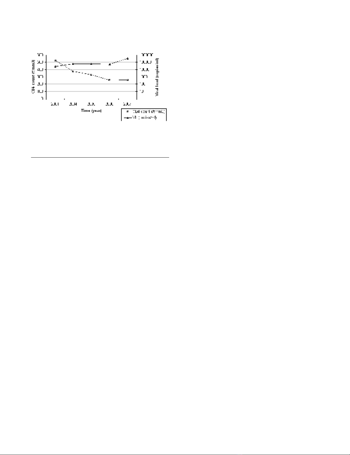
ITM4 is a male HIV-1 Circulating Recombinant Form
CRF02_AG infected individual, who has been infected by
heterosexual transmission. He first consulted the Institute
of Tropical Medicine in 2001. Between 2001 and 2007,
his viremia increased from 42,000 copies/ml (sample
ITM4_01.1) to 330,000 copies/ml (sample ITM4_07.2),
and his CD4 T cell counts decreased from 550 per mm3
(sample ITM4_01.1) to 250 per mm3 (sample ITM4_07.2)
(Fig. 1). During this follow up period, patient ITM4 never
received anti-retroviral therapy. ITM4 was selected for the
unique capacity of his plasma, taken in 2005 (further
referred to as ITM4_05) to neutralize a broad spectrum of
primary virus isolates from subtypes A (3/4), B (2/4), C
(4/4), D (4/4), CRF01_AE (4/4) and CRF02_AG (5/5) in
a primary virus/PBMC neutralization assay (Table 1)[20].
The BCN capacity of his plasma was confirmed for a sam-
ple taken in 2007 in a pseudovirus/TZM-bl assay (Table
1). The panel of pseudoviruses tested was neutralized with
ID50s ranging between 33 and >640, with the subtypes C
and D Envs being the most sensitive and subtypes B Envs
being more neutralization resistant.
Peptide phage selection and localization
In order to map the antibody responses directed against
the HIV-1 envelop in patient ITM4, peptide phage display
technology was applied. A random 12-mer phage library
was panned against a pool of ITM4 plasma samples. After
3 selection rounds, peptide sequences were deduced for
Retrovirology 2009, 6:113 http://www.retrovirology.com/content/6/1/113
Page 3 of 13
(page number not for citation purposes)
phage displaying positive reactivity in ELISA with ITM4
plasma, as well as low or no reactivity with an HIV nega-
tive plasma pool. The generated peptide sequences were
aligned and ranked according to homology, resulting in
four groups of peptide sequences with a commonly simi-
lar motif (Table 2). Phage clones presenting a peptide
with a NWFNLTQTLMPR motif were predominantly doc-
umented (n = 18); twelve peptide phages represented the
KxWWxA motif. Furthermore, mimotopes with a SLxxLRL
motif (n = 7) and a KxxxIGPHxxY motif (n = 3) were iden-
tified. Peptide sequences were compared with the linear
Env sequences of ITM4 to localize each of the mimotope
groups. The NWFNLTQTLMPR peptide shares key amino
acid residues of the 4E10 epitope [WFx(I/L)(T/S)xx(L/
I)W] located in the membrane-proximal external region
(MPER) of gp41 [21,22]. Mimotopes with the KxxxIG-
PHxxY motif showed homology to the crown of the V3
loop of the ITM4 gp160 sequences (Table 2). The KxW-
WxA motif shared linear homology to C1 sequences of
ITM4 isolates of 2007 (Fig. 2). A last group of mimotopes
sharing the SLxxLRL motif is predicted to bind antibodies
directed to the lentivirus lytic peptide 2 (LLP2) region of
gp41.
Recognition of the ITM4 phage mimotopes by other HIV-1
infected individuals
In a capture ELISA, eighty random plasma samples of
HIV-1 positive individuals were screened for antibody
cross reactivity to the selected ITM4 peptide phage groups.
The highest cross-reactivity was seen for the mimotope
representing the immunodominant part of the V3 region,
ten (12.5%) of the tested HIV1 plasma had antibodies
that bound this mimotope (Table 2). The mimotope
localized in the gp41 MPER is more exclusive; it was only
recognized by one plasma sample (later referred as
CrossR1). This is in accordance with previous publica-
tions indicating that antibodies against the MPER are
found in a minority of HIV infected individuals [18,23-
25]. The random plasma samples also showed a very weak
cross reactivity (1/80) towards the LLP2 mimotope, while
none of the plasma samples that were tested recognized
the C1 mimotope. This result indicated that this epitope
is unique for the virus circulating in ITM4.
Evolution of the antibody development in ITM4 follow up
plasma samples
Next, an ELISA was performed to determine the reactivity
of the different phage groups with the individual plasma
follow up samples of ITM4 (2001-2007) (Fig. 3). Reactiv-
ity patterns of the peptide phage groups with ITM4 follow
up plasma were not uniform. Each phage group had a
unique reaction pattern: (1) Antibodies elicited against
the MPER (NWFNLTQTLMPR) mimotope were already
present in 2001 and showed a comparable high reactivity
in all the follow up samples, whereas (2) the antibodies
against the C1 (KxWWxA) mimotope were absent in most
of the samples and only appeared in the plasma samples
taken in 2007. The V3-specific antibodies (3) showed a
gradual decreasing reactivity over time; in contrast (4) an
increase of binding antibodies is seen for the LLP2 (SLxx-
LRL) mimotope. These data demonstrate that the anti-
body development in ITM4 is a continuously dynamic
event.
Specificity of the MPER mimotope
To further explore the characteristics of the antibodies
binding to the NWFNLTQTLMPR mimotope, additional
ELISA experiments were performed. First, the ability of the
monoclonal antibody 4E10 to bind this phage peptide
was analyzed. We observed a high signal (OD = 3.0) when
4E10 was added to the mimotope, indicating the ability of
the NWFNLTQTLMPR peptide to bind 4E10-like antibod-
ies (Fig. 4). In a second part of the experiment, different
peptides overlapping the 4E10 region were used in a com-
petition ELISA. The results clearly demonstrated that pep-
tide 6376 obtained from the NIH AIDS Reagent Program,
SLWNWFDITNWLWYI, presenting the 4E10 epitope,
strongly competed with the peptide phage for 4E10-bind-
ing (Fig. 4). A similar observation was made for the
plasma from ITM4 (Fig. 4), the same peptide occupied the
binding places for the antibodies binding the NWF-
NLTQTLMPR mimotope as for the monoclonal antibody
4E10. Taken together, the results above suggest that 4E10-
like antibodies are present in our subject of interest.
Autoreactive antibodies
As shown by Haynes et al. [26] and Scherer et al. [27], Mab
4E10 has an affinity for the autoantigen cardiolipine (CL),
due to the epitope position which is recognized in the
context of the viral membrane. In our study, the cross-
reactivity to CL of five broadly cross neutralizing plasma
samples was analyzed, including both patients with anti-
CD4+ T-cell numbers and viral loads detected in follow up plasma samples of ITM4 over a period of 6 yearsFigure 1
CD4+ T-cell numbers and viral loads detected in fol-
low up plasma samples of ITM4 over a period of 6
years.

Retrovirology 2009, 6:113 http://www.retrovirology.com/content/6/1/113
Page 4 of 13
(page number not for citation purposes)
bodies against the NWFNLTQTLMPR mimotope: ITM4
and CrossR1. The presence of anti-CL antibodies was
measured in an ELISA, and plasma samples were ranked
according to their reactivity, with >20 GPL units catego-
rized as positive; 10-20 GPL units as weakly positive, and
< 10 GPL units as negative. Plasma ITM4_07 showed the
highest reactivity (34 GPL units) and thus scored strongly
positive. The second highest score (16 GPL units) was
obtained with plasma from patient CrossR1. Two other
BCN plasma were weakly positive (12 and 11 GPL units
respectively), one BCN plasma scored negative (8 GPL
units) (data not shown). Additionally, we analyzed the
presence of other auto-antibodies (anti dsDNA, anti Ro/
Ssa, and anti Jo1 antibodies) in the selected plasma sam-
ples. None of the samples showed positive reactivity with
any of these auto-antigens (data not shown).
We further noted that the clinical status of patient ITM4
was regularly followed between 2001 and 2007 by the
team of medical doctors at the clinic of the Institute of
Table 1: Neutralization profile of patient ITM4 in a primary virus/PBMC assay (left) and a pseudovirus/TZM-bl assay (right)
PBMC/Primary Virus Assay Plasma Sample ITM4_05 Pseudovirus/TZM bl Assay Plasma Sample ITM4_07
Subtype Virus % NeutralizationaPseudovirus ID50b
A VI 191 66 PV 92RW009 222
92UG037 100 PV PIC 32281 54
VI 820 99
VI 1031 100
B 89.6 0 PV SF162 322
93US076 69 PV JRFL 88
93US077 97 PV AC10 62
93US143 100 PV CAAN 33
CVI 829 80 PV VI 829 180
VI 882 99 PV VI 882 316
VI 1358 99 PV VI 1358 208
VI 1144 100 PV 92BR025 155
PV Du174 333
DVI 656 97 PV UG024 >640
VI 693 100
VI 824 91
VI 865 93
CRF01 VI 1249 98 PV CM244 93
CA 10 97
VI 1888 96
92TH022 99
CRF02 VI 1090 94 PV VI 1090 221
CA18 88 PV CA18 63
VI 2680 99
VI 1380 98
VI 2727 93
a % neutralization obtained with 1:20 plasma dilution, ≥80% reduction in virus titer is indicated in bold.
b Plasma dilution causing 50% reduction of relative light units compared to the virus control, ID50 ≥50 is indicated in bold.
Table 2: Overview of the selected mimotope groups
Mimotope AA Motif Location in the ITM4 Env Sequence Times Selected Cross Reactivity a
NWFNLTQTLMPR gp41 MPER region 18 1/80 (1.3%)
KxWWxA gp120 C1 region 12 0/80 (0.0%)
SLxxLRL gp41 LLP2 region 7 1/80 (1.3%)
KxxxIGPHxxY gp120 V3 region 3 10/80 (12.5%)
a Number of HIV-1+ plasma samples cross-reacting with the mimotope group.
Retrovirology 2009, 6:113 http://www.retrovirology.com/content/6/1/113
Page 5 of 13
(page number not for citation purposes)
Tropical Medicine (Antwerp, Belgium). The fact that no
symptoms of auto-immune disease were reported in this
follow up period, suggests that the ITM4 antibodies react-
ing with the CL autoantigen are non-pathogenic, induced
by the HIV-infection, and are not present due to an
autoimmune disorder [28].
Genotypic Analysis of the MPER of ITM4
MPER sequences of 4 clones per follow-up sample (n = 7)
were generated and analyzed (Fig. 5). For clones of sam-
ples ITM4_01.1 and ITM4_01.2 taken in 2001, two
diverse 2F5 epitope variants were documented: ALDKWA
and ALNKWA, having a D664N substitution. Clones of
samples from 2004 and later time points displayed wild
type ALDKWA and/or A667 mutant sequences (Fig. 5).
ALDKWA represents the consensus subtype A 2F5 epitope,
whereby the DKW motif is crucial for binding 2F5
[29,30]. The D664N substitution resulting in ALNKWA,
has been described as a 2F5 escape variant with a lower
but still relatively high infectivity in vitro and displaying
resistance to 2F5 neutralization [31].
The 4E10 epitope of clones of ITM4 follow-up samples
did not present mutations in key amino acids [WFx(I/
L)(T/S)xx(L/I)W]. The subtype A 4E10 epitope NWFDIT-
NWLW was conserved in clones of samples ITM4_01.1
and ITM4_01.2 taken in 2001. Clones of 2004 and sam-
ples of later time points displayed D674 mutant
sequences. One N677K mutation is seen in a clone iso-
lated from the 2005 sample. Both mutations at position
674 and 677 do not confer resistance to 4E10 [21], but
substitutions at these positions may have an impact on
4E10 neutralization sensitivity [32].
Neutralization sensitivity of functional ITM4 clones
To investigate the autologous neutralizing activity, we
cloned and pseudotyped full length envelope genes of
functional clones from 4 different time points (2001,
2004, 2007.1, 2007.2) and examined the sensitivity of the
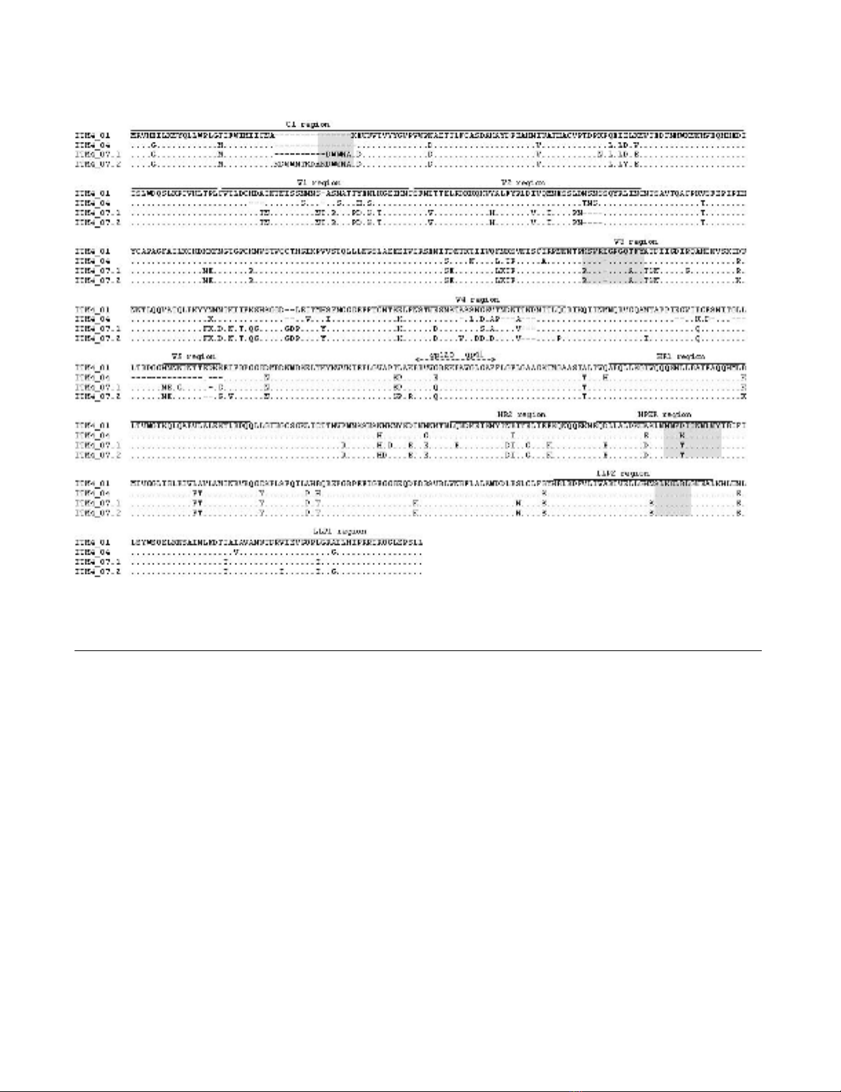
Env amino acid alignment of ITM4 follow-up pseudovirusesFigure 2
Env amino acid alignment of ITM4 follow-up pseudoviruses. Dots are included for alignment purposes. Mimotope
localizations are highlighted in grey.


![Báo cáo seminar chuyên ngành Công nghệ hóa học và thực phẩm [Mới nhất]](https://cdn.tailieu.vn/images/document/thumbnail/2025/20250711/hienkelvinzoi@gmail.com/135x160/47051752458701.jpg)


%20--%3e%3cdefs%3e%3cstyle%3e%20.st0%20{%20fill:%20%23fff;%20}%20.st1%20{%20fill:%20%237800fa;%20}%20%3c/style%3e%3c/defs%3e%3cpath%20class='st1'%20d='M117.78,12.18H43.11c2.9,3.47,4.65,7.94,4.65,12.82,0,5.6-2.3,10.66-6.01,14.29h76.02l7.22-13.56-7.22-13.56Z'/%3e%3cg%3e%3cpath%20class='st0'%20d='M53.58,26.17h-.59v-1.46h.59v-4.96h2.83c1.78,0,2.67.94,2.67,2.82v5.76c0,1.87-.89,2.81-2.67,2.81h-2.83v-4.96ZM55.36,21.37v3.34h1.1v1.46h-1.1v3.34h1.01c.61,0,.91-.37.91-1.1v-5.93c0-.74-.3-1.1-.91-1.1h-1.01Z'/%3e%3cpath%20class='st0'%20d='M65.99,31.14h-1.8l-.31-2.07h-2.19l-.31,2.07h-1.64l1.82-11.39h2.62l1.82,11.39ZM65.28,18.04c-.25.46-.51.77-.75.94-.21.15-.47.22-.79.22-.26,0-.57-.07-.92-.22l-.38-.15c-.14-.05-.26-.07-.37-.07-.3,0-.53.18-.71.54l-.91-.68c.25-.46.51-.77.75-.94.21-.14.48-.21.79-.21.26,0,.57.07.92.21l.38.15c.14.05.26.07.37.07.3,0,.53-.18.71-.54l.91.68ZM61.91,27.52h1.73l-.87-5.76-.87,5.76Z'/%3e%3cpath%20class='st0'%20d='M74.53,26.89v1.52c0,1.91-.89,2.86-2.67,2.86s-2.67-.95-2.67-2.86v-5.93c0-1.91.89-2.86,2.67-2.86s2.67.95,2.67,2.86v1.11h-1.69v-1.22c0-.75-.31-1.12-.93-1.12s-.93.37-.93,1.12v6.15c0,.74.31,1.11.93,1.11s.93-.37.93-1.11v-1.63h1.69Z'/%3e%3cpath%20class='st0'%20d='M81.4,31.14h-1.8l-.31-2.07h-2.19l-.31,2.07h-1.64l1.82-11.39h2.62l1.82,11.39ZM75.9,19.2l1.52-1.91h1.71l1.51,1.91h-1.61l-.76-.95-.75.95h-1.61ZM77.32,27.52h1.73l-.87-5.76-.87,5.76ZM83.1,15.99l-1.76,1.91h-1.26l1.17-1.91h1.86Z'/%3e%3cpath%20class='st0'%20d='M84.86,19.75c1.78,0,2.67.94,2.67,2.82v1.48c0,1.87-.89,2.81-2.67,2.81h-.85v4.28h-1.79v-11.39h2.64ZM84.01,21.37v3.86h.85c.58,0,.87-.36.87-1.08v-1.71c0-.71-.29-1.07-.87-1.07h-.85Z'/%3e%3cpath%20class='st0'%20d='M93.51,19.75c1.78,0,2.67.94,2.67,2.82v1.48c0,1.87-.89,2.81-2.67,2.81h-.85v4.28h-1.79v-11.39h2.64ZM92.66,21.37v3.86h.85c.58,0,.87-.36.87-1.08v-1.71c0-.71-.29-1.07-.87-1.07h-.85Z'/%3e%3cpath%20class='st0'%20d='M98.8,31.14h-1.79v-11.39h1.79v4.88h2.03v-4.88h1.83v11.39h-1.83v-4.88h-2.03v4.88Z'/%3e%3cpath%20class='st0'%20d='M105.36,24.55h2.46v1.62h-2.46v3.34h3.09v1.63h-4.88v-11.39h4.88v1.63h-3.09v3.18ZM108.17,17.29l-1.76,1.91h-1.26l1.17-1.91h1.86Z'/%3e%3cpath%20class='st0'%20d='M112.2,19.75c1.78,0,2.67.94,2.67,2.82v1.48c0,1.87-.89,2.81-2.67,2.81h-.85v4.28h-1.79v-11.39h2.64ZM111.35,21.37v3.86h.85c.58,0,.87-.36.87-1.08v-1.71c0-.71-.29-1.07-.87-1.07h-.85Z'/%3e%3c/g%3e%3ccircle%20class='st1'%20cx='25'%20cy='25'%20r='20'/%3e%3cpath%20class='st0'%20d='M32.78,19.27c2.92,0,4.43,2.55,5.28,5.33l.71,2.17c.14.38-.33.75-.71.75h-5.61c.19-.33.24-.71.09-1.08l-.75-2.45c-.43-1.32-.99-2.64-1.79-3.77.75-.57,1.65-.94,2.78-.94h0ZM25,18.38c3.25,0,4.9,2.78,5.89,5.89l.76,2.45c.14.42-.33.8-.8.8h-11.69c-.42,0-.94-.38-.8-.8l.75-2.45c.99-3.11,2.64-5.89,5.89-5.89h0ZM25,11.35c1.74,0,3.11,1.37,3.11,3.11s-1.37,3.11-3.11,3.11-3.11-1.41-3.11-3.11,1.41-3.11,3.11-3.11h0ZM17.27,19.27c1.08,0,1.98.38,2.73.94-.8,1.13-1.37,2.45-1.74,3.77l-.8,2.45c-.14.38-.05.75.09,1.08h-5.56c-.42,0-.9-.38-.75-.75l.71-2.17c.9-2.78,2.41-5.33,5.33-5.33h0ZM17.27,12.91c1.51,0,2.78,1.27,2.78,2.83s-1.27,2.83-2.78,2.83-2.83-1.27-2.83-2.83,1.27-2.83,2.83-2.83h0ZM32.78,12.91c1.56,0,2.78,1.27,2.78,2.83s-1.23,2.83-2.78,2.83-2.83-1.27-2.83-2.83,1.27-2.83,2.83-2.83h0ZM27.07,28.56v.09c0,.57-.24,1.08-.61,1.46h0v.05c-.38.33-.9.57-1.46.57s-1.08-.24-1.46-.61h0c-.38-.38-.61-.9-.61-1.46v-.09h1.41v.09c0,.19.05.38.19.47v.05c.09.09.28.19.47.19s.38-.09.47-.19v-.05c.14-.09.24-.28.24-.47t-.05-.09h1.41ZM30.99,28.56v.09c0,1.65-.66,3.16-1.74,4.24-1.08,1.08-2.59,1.79-4.24,1.79s-3.16-.71-4.24-1.79l-.05-.05c-1.04-1.08-1.7-2.55-1.7-4.2v-.09h1.41v.09c0,1.27.47,2.4,1.27,3.25h.05c.85.85,1.98,1.37,3.25,1.37s2.4-.52,3.25-1.37c.85-.8,1.37-1.98,1.37-3.25v-.09h1.37ZM34.99,28.56v.09c0,2.78-1.13,5.28-2.92,7.07-1.79,1.79-4.29,2.92-7.07,2.92s-5.23-1.13-7.07-2.92c-1.79-1.79-2.92-4.29-2.92-7.07v-.09h1.41v.09c0,2.4.94,4.53,2.5,6.08,1.56,1.56,3.72,2.5,6.08,2.5s4.52-.94,6.08-2.5c1.56-1.56,2.5-3.68,2.5-6.08v-.09h1.41Z'/%3e%3c/svg%3e)