
Open Access
Available online http://ccforum.com/content/13/3/R64
Page 1 of 9
(page number not for citation purposes)
Vol 13 No 3
Research
Time course of angiopoietin-2 release during experimental human
endotoxemia and sepsis
Philipp Kümpers1*, Matijs van Meurs2,3*, Sascha David1, Grietje Molema3, Johan Bijzet3,
Alexander Lukasz1, Frank Biertz4, Hermann Haller1 and Jan G Zijlstra2
1Department of Nephrology & Hypertension, Hannover Medical School, Carl-Neuberg-Straße 1, 30625 Hannover, Germany
2Department of Critical Care, University Medical Center Groningen, Hanzeplein 1, 9713 GZ, Groningen, The Netherlands
3Department of Pathology and Medical Biology, University Medical Center Groningen, Hanzeplein 19713 GZ Groningen, The Netherlands
4Department of Biometrics, Hannover Medical School, Carl-Neuberg-Straße 1, 30625 Hannover, Germany
* Contributed equally
Corresponding author: Philipp Kümpers, kuempers.philipp@mh-hannover.de
Received: 12 Feb 2009 Revisions requested: 3 Apr 2009 Revisions received: 21 Apr 2009 Accepted: 5 May 2009 Published: 5 May 2009
Critical Care 2009, 13:R64 (doi:10.1186/cc7866)
This article is online at: http://ccforum.com/content/13/3/R64
© 2009 Kümpers et al.; licensee BioMed Central Ltd.
This is an open access article distributed under the terms of the Creative Commons Attribution License (http://creativecommons.org/licenses/by/2.0),
which permits unrestricted use, distribution, and reproduction in any medium, provided the original work is properly cited.
Abstract
Introduction Endothelial activation leading to vascular barrier
breakdown denotes a devastating event in sepsis. Angiopoietin
(Ang)-2, a circulating antagonistic ligand of the endothelial
specific Tie2 receptor, is rapidly released from Weibel-Palade
and has been identified as a non-redundant gatekeeper of
endothelial activation. We aimed to study: the time course of
Ang-2 release during human experimental endotoxemia; the
association of Ang-2 with soluble adhesion molecules and
inflammatory cytokines; and the early time course of Ang-2
release during sepsis in critically ill patients.
Methods In 22 healthy volunteers during a 24-hour period after
a single intravenous injection of lipopolysaccharide (LPS; 4 ng/
kg) the following measurement were taken by immuno
luminometric assay (ILMA), ELISA, and bead-based multiplex
technology: circulating Ang-1, Ang-2, soluble Tie2 receptor, the
inflammatory molecules TNF-alpha, IL-6, IL-8 and C-reactive
protein, and the soluble endothelial adhesion molecules inter-
cellular adhesion molecule-1 (ICAM-1), E-selectin, and P-
selectin. A single oral dose of placebo or the p38 mitogen
activated protein (MAP) kinase inhibitor drug, RWJ-67657, was
administered 30 minutes before the endotoxin infusion. In
addition, the course of circulating Ang-2 was analyzed in 21
septic patients at intensive care unit (ICU) admission and after
24 and 72 hours, respectively.
Results During endotoxemia, circulating Ang-2 levels were
significantly elevated, reaching peak levels 4.5 hours after LPS
infusion. Ang-2 exhibited a kinetic profile similar to early pro-
inflammatory cytokines TNF-alpha, IL-6, and IL-8. Ang-2 levels
peaked prior to soluble endothelial-specific adhesion molecules.
Finally, Ang-2 correlated with TNF-alpha levels (r = 0.61, P =
0.003), soluble E-selectin levels (r = 0.64, P < 0.002), and the
heart rate/mean arterial pressure index (r = 0.75, P < 0.0001).
In septic patients, Ang-2 increased in non-survivors only, and
was significantly higher compared with survivors at baseline, 24
hours, and 72 hours.
Conclusions LPS is a triggering factor for Ang-2 release in men.
Circulating Ang-2 appears in the systemic circulation during
experimental human endotoxemia in a distinctive temporal
sequence and correlates with TNF-alpha and E-selectin levels.
In addition, not only higher baseline Ang-2 concentrations, but
also a persistent increase in Ang-2 during the early course
identifies septic patients with unfavorable outcome.
Introduction
Microvascular capillary leakage resulting in tissue edema,
vasodilation refractory to vasopressors, and increased recruit-
ment of leukocytes denote key features of sepsis-related
endothelial-cell activation. During the course of severe sepsis
and septic shock, widespread endothelial cell activation con-
ALI: acute lung injury; Ang: angiopoietin; ANOVA: analysis of variance; ARDS: acute respiratory distress syndrome; AUC: area under the curve; CI:
confidence interval; CRP: C-reactive protein; ELISA: enzyme linked immuno sorbent assay; ICAM-1: inter-cellular adhesion molecule-1; ICU: intensive
care unit; IL: interleukin; ILMA: immunoluminometric sandwich assay; LPS: lipopolysaccharide; MAP: mitogen activated protein; OR: odds ratio; ROC:
receiver operator characteristics; TNF-alpha: tumor necrosis factor-alpha; VCAM-1: vascular cell adhesion molecule-1; WPB: Weibel-Palade body.

Critical Care Vol 13 No 3 Kümpers et al.
Page 2 of 9
(page number not for citation purposes)
tributes to the initiation and progression of multi-organ failure
[1]. Recently, Angiopoietin (Ang)-2 has emerged as a key reg-
ulator of endothelial cell activation [2]. In critically ill patients,
Ang-2 increases endothelial permeability and is considered a
key molecule in the pathogenesis of acute lung injury (ALI) and
acute respiratory distress syndrome (ARDS) [3,4].
Ang-1 and Ang-2 are antagonistic ligands, which bind to the
extracellular domain of the Tie2 receptor, which is almost
exclusively expressed by endothelial cells [5,6]. Binding of the
agonist Ang-1 to the endothelial Tie2 receptor maintains ves-
sel integrity, inhibits vascular leakage, suppresses inflamma-
tory gene expression, and prevents recruitment and
transmigration of leukocytes [7,8]. In contrast, binding of Ang-
2 to the Tie2 receptor disrupts protective Ang-1/Tie2 signaling
and facilitates endothelial inflammation in a dose-dependent
fashion [9].
In vitro, Ang-2 simultaneously mediates disassembly of cell–
cell and cell–matrix contacts, and causes active endothelial
cell contraction in a Rho kinase-dependent fashion, followed
by massive plasma leakage and loss of vasomotor tone [3,10].
Furthermore, Ang-2 facilitates up-regulation of inter-cellular
adhesion molecule-1 (ICAM-1), vascular cell adhesion mole-
cule -1 (VCAM-1), and E-selectin [3,7,10,11].
In vivo, Ang-2-deficient mice do not exhibit any vascular inflam-
matory responses in experimental sepsis, and vessels in Ang-
1-overexpressing mice are resistant to leakage to inflammatory
stimuli [12,13]. As a Weibel-Palade body-stored molecule
(WPB), Ang-2 is rapidly released upon endothelial stimulation
and is regarded the dynamic regulator within the Ang/Tie sys-
tem [7,12]. Consistently, exceptionally high levels of circulat-
ing Ang-2 have been detected in critically ill patients with
sepsis and sepsis-related organ dysfunction [14-16].
Beyond its role as a mediator, Ang-2 has been identified as a
promising strong marker of endothelial activation in various
diseases [17-19]. In critically ill septic patients, we recently
showed that admission levels of circulating Ang-2 correlates
with surrogate markers of tissue hypoxia, disease severity, and
is a strong and independent predictor of mortality [20]. How-
ever, the exact time course of Ang-2 release during sepsis and
the role of inflammatory cytokines thereof remain elusive. Fur-
thermore, the tempting sequential concept [7] of Ang-2 as a
primer for excess endothelial adhesion molecule (e.g. ICAM-1,
VCAM-1, and E-selectin) expression in sepsis has not been
investigated in human sepsis.
To address these issues, we wanted to study the time course
of Ang-2 release, and the association of Ang-2 with soluble
adhesion molecules and inflammatory cytokines in a graded
and well-defined human endotoxemia model. Therefore, we re-
measured circulating Ang-2, cytokines, and adhesion mole-
cules in blood samples from a placebo-controlled interven-
tional trial on pharmacologic p38 mitogen-activated protein
(MAP) kinase inhibition during experimental human endotox-
emia [21]. Furthermore, we analyzed circulating Ang-2 in sep-
tic patients during a 72 hour time course after admission to the
intensive care unit (ICU).
Materials and methods
Subjects
Twenty-one healthy male subjects, mean age 29 (range 19 to
44) years, were admitted to the research unit of our ICU (Med-
ical Department) at University Medical Center of Groningen,
Groningen, The Netherlands. The local Medical Ethics Com-
mittee approved the study and written informed consent was
obtained from the subjects. A radial arterial catheter was
placed for blood sampling. Thirty minutes before the infusion
of lipopolysaccharide (LPS), the volunteers received a single
oral dose of RWJ-67657 (4-(4-(Fluorophenyl)-1-(3-phenylpro-
pyl)-5-(4-pyrindinyl)-1H-imidazol-2-yl)-3-butyn-1-ol), supplied
in an oral pharmaceutical formulation (R.W. Johnson Pharma-
ceutical Research Institute, Bassersdorf, Switzerland). Three
dose levels were tested, placebo-controlled: placebo (n = 6),
350 mg (n = 5), 700 mg (n = 6), and 1400 mg (n = 4). At time
point t = 0, LPS (Escherichia coli, batch EC-6, US Pharmaco-
peia, Twinbrook Parkway, Rockville, MD, USA) was adminis-
tered as a one minute infusion at a dose of 4 ng/kg body
weight (10.000 LPS units/mg). Blood samples were drawn at
several time points between pre-medication (t = 0) and 24
hours after administration of LPS. Samples were placed on
ice, centrifuged, stored at -80°C, and analyzed in a blinded
fashion [21].
Patients
The time course of Ang-2 release during the early course of
human sepsis was studied in 21 ICU patients (Internal Medi-
cine Department) recruited at Hannover Medical School (terti-
ary care university hospital), Hannover, Germany. Patient
characteristics are shown in Table 1. Enrollment was per-
formed after obtaining written informed consent from the
patient or his/her legal representatives. If the patient was
recovering and able to communicate, he/she was informed of
the study purpose and consent was required to further main-
tain status as a study participant. Twenty-eight day survival
was the primary outcome studied and was calculated from the
day of ICU admission to day of death from any cause. Patients
who did not die within the follow-up were censored at the date
of last contact. The study was carried out in accordance with
the declaration of Helsinki and was approved by the institu-
tional review board. Serum samples were obtained at baseline
(admission), 24 hours, and 72 hours, placed on ice, centri-
fuged, stored at -80°C, and analyzed in a blinded fashion.
Quantification of circulating angiopoietin-1 and 2, and
soluble Tie2
Ang-1 and Ang-2 were measured by in-house immuno lumino-
metric assay (ILMA), and ELISA as previously described

Available online http://ccforum.com/content/13/3/R64
Page 3 of 9
(page number not for citation purposes)
[17,18,20]. Soluble Tie2 was measured by commercially avail-
able ELISA kit (R&D Systems, Oxford, UK) according to the
manufacturer's instructions.
Quantification of soluble endothelial-adhesion
molecules and cytokines
Soluble ICAM-1, E-selectin, and P-selectin were measured
using Fluorokine® MultiAnalyte Profiling kits and a Luminex®
Bioanalyzer (R&D Systems, Oxford, UK) according to the man-
ufacturers' instructions. TNF-alpha, IL-6, IL-8, and c-reactive
protein (CRP) were determined using Medigenix Easia kits
from BioSource (BioSource, Nivelles, Belgium) and reported
previously [22].
Statistical analysis
The modified Kolmogorov-Smirnov test was used to test for a
normal distribution of continuous variables. In the human endo-
toxemia model, a non-parametric analysis of variance
(ANOVA) (Friedman's test) with Dunn's test for multiple com-
parison (two-sided) was used to demonstrate statistical
changes in Ang-2, cytokines, and adhesion molecules. Corre-
lations of Ang-2 with TNF-alpha, E-selectin, and the heart rate/
mean arterial pressure index were calculated with Pearsons's
correlation and linear regression analysis after log-transforma-
tion. Data are presented as mean ± standard error of the mean
unless otherwise stated.
In patients, differences between survivors and non-survivors at
baseline and during follow-up were compared by non-para-
metric two-sided Mann Whitney U test. Receiver operator
characteristic (ROC) procedures identified optimal cut-off val-
ues for Ang-2 to differentiate between survivors and non-sur-
vivors. Contingency table-derived data and likelihood ratios
were calculated using the StatPages website [23]. Two-sided
P < 0.05 were considered statistically significant for all statis-
tical procedures used. All statistical analyses were performed
using the SPSS package (SPSS Inc., Chicago, IL, USA) and
the GraphPad Prism software (GraphPad Prism Software Inc.
San Diego, CA, USA).
Results
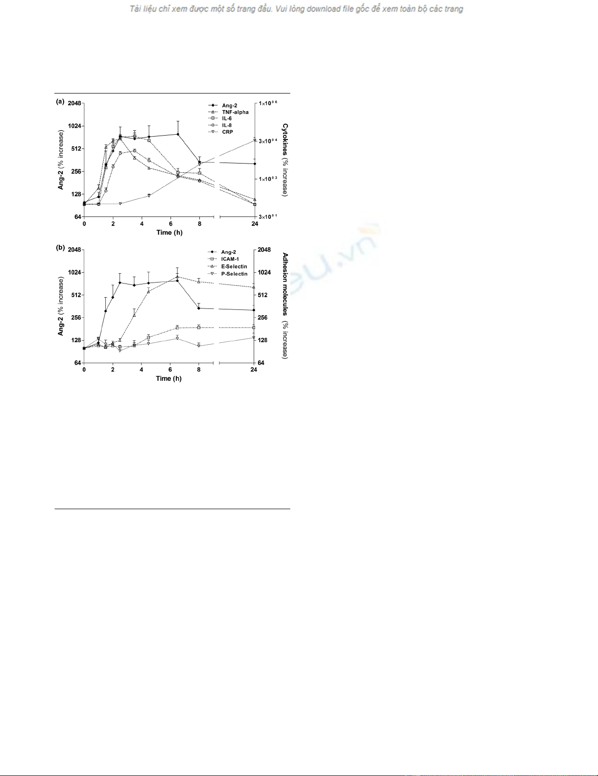
Angiopoietin-2 is released in a distinctive pattern after
endotoxin challenge in healthy volunteers
Normal Ang-2 concentrations (0.57 ± 0.20 ng/mL) were
present at baseline in healthy volunteers (Table 2). Ang-2 lev-
els started to increase at two hours, were significantly elevated
from 2.5 hours until 6.5 hours (P < 0.01), reaching peak levels
(2.42 ± 0.54 ng/mL) 4.5 hours after LPS infusion (P < 0.0001;
Figure 1; n = 6, placebo group).
In our cohort of healthy volunteers, neither endogenous sTie2,
nor circulating Ang-1 concentrations changed during 24
hours after endotoxin challenge (Table 2).
Angiopoietin-2 release runs in parallel with early pro-
inflammatory cytokines and precedes endothelial
inflammation after endotoxin challenge
Plasma levels of TNF-alpha were already significantly elevated
at 1.5 hours (P < 0.01) compared with baseline, and 30 min-
utes earlier compared with Ang-2 and IL-6 (Figure 1a). IL-8
appeared in the circulation about 30 minutes later than Ang-2
and IL-6. Elevated Ang-2 levels declined more slowly than that
of TNF-alpha, IL-6, and IL-8.
Soluble E-selectin appeared in the circulation later than Ang-
2 and E-selectin levels were elevated from 4.5 hours until 24
hours (all P < 0.0001). Similarly, ICAM-1 levels were elevated
from 6.5 hours until 24 hours after LPS infusion (all P <
0.0001; Figure 1b). However, P-selectin did not increase after
endotoxin challenge in the present study (P = 0.151).
Angiopoietin-2 release after endotoxin challenge is
attenuated by p38 MAP kinase inhibition
Our previous studies have shown that inhibition of the intrac-
ellular p38 MAP kinase attenuated inflammatory responses
during human endotoxemia [21]. Thus, we hypothesized that
p38 MAP kinase inhibition would also have an impact on Ang-
2 release. In addition to LPS-treated subjects that received
Table 1
Characteristics of septic ICU patients on admission
Characteristics Value
Patients, number 21
Male 8 (38.1%)
Female 13 (61.9%)
Age, years, median (min to max) 57 (36 to 72)
Reason for medical ICU admission
Pneumonia 12 (57.1%)
Peritonitis 4 (19.0%)
Urinary tract infection 2 (9.5%)
Systemic mycosis 2 (9.5%)
Endocarditis 1 (4.8%)
Mediastinitis 1 (4.8%)
Mean arterial pressure (mmHg) 78 (58 to 108)
Heart rate (beats/minute) 95 (53 to 125)
Vasopressor support, number 12 (57.1%)
Mechanically ventilated, number 19 (90.5%)
Fraction of inspired oxygen (%) 40 (25 to 95)
APACHE II score 22 (12 to 48)
SOFA score 10 (3 to 19)
Mortality, number 11 (52.4%)
APACHE = Acute Physiology and Chronic Health Evaluation; ICU =
intensive care unit; SOFA = Sequential Organ Failure Assessment.
Critical Care Vol 13 No 3 Kümpers et al.
Page 4 of 9
(page number not for citation purposes)
placebo (n = 6), circulating Ang-2 was determined in LPS-
treated subjects that were randomized to different doses of an
oral p38 MAP kinase inhibitor [21]. In contrast to the placebo
group (LPS without p38 MAP kinase inhibitor), no statistically
significant Ang-2 release occurred in any of the three interven-
tional groups (i.e. 350 mg, 700 mg, or 1400 mg of RWJ-
67657). However, when the areas under the curves (AUC)
during the time course were calculated, a dose dependent
effect of RWJ-67657 on Ang-2 release was present.
The AUC of absolute Ang-2 values (ng/ml) were 39.8, 31.0,
32.1, and 17.8 in the placebo and the three interventional
groups, respectively. Correspondingly, the AUC of percent-
age increase in Ang-2 from baseline were 9850, 4765, 3435,
and 2567 in the placebo and the three interventional groups,
respectively.
Circulating angiopoietin-2 correlates with TNF-alpha
levels, soluble E-selectin levels, and the heart rate/mean
arterial pressure index
TNF-alpha levels correlated well with Ang-2 at 3.5 (r = 0.44, P
= 0.04), 4.5 hours (r = 0.54, P = 0.012), 6.5 hours (r = 0.61,
P = 0.003), and 8 hours (r = 0.49, P = 0.024; Figure 2a). Like-
wise, levels of soluble E-selectin were closely associated with
Ang-2 at 4.5 hours (r = 0.5, P = 0.005), 6.5 hours (r = 0.64,
P = 0.0013), and 24 hours (r = 0.69, P < 0.0004; Figure 2b),
when all subjects in the endotoxin model were analyzed (n =
21). Finally, we analyzed the increase in heart rate/mean arte-
rial pressure index as a dynamic surrogate marker of hemody-
namic compromise. Indeed, a close correlation was found
between the increase in circulating Ang-2 and the increase in
heart rate/mean arterial pressure index at 4.5 hours (r = 0.6, P
= 0.003), 6.5 hours (r = 0.58, P = 0.006), and 8 hours (r =
0.75, P < 0.0001; Figure 2c), when all subjects were analyzed
(n = 21).
Excess Ang-2 on admission and increasing Ang-2 level
during the early course indicate unfavorable 28-day
survival in septic patients
First, circulating Ang-2 on admission was 9.8 ± 3.2 ng/ml in
septic patients (n = 21). Regarding the kinetics of Ang-2 dur-
ing follow-up, mean Ang-2 levels remained unchanged at 24
hours (14.3 ± 4.0 ng/ml) and 72 hours (18.2 ± 6.0 ng/ml)
when all patients were analyzed (non-parametric repeated
measures ANOVA (Friedman's test); P = 0.146; Figure 3).
Second, when analyzed separately, non-survivors (n = 11) had
higher Ang-2 levels compared with survivors (n = 10) on
admission (9.7 ± 1.6 ng/ml vs. 4.7 ± 1.3 ng/ml; P = 0.032),
after 24 hours (13.3 ± 3.2 ng/ml vs. 5.0 ± 1.3 ng/ml; P =
0.027) and 72 hours (21.5 ± 6.0 ng/ml vs. 4.3 ± 1.6 ng/ml; P
= 0.008). In non-survivors, Ang-2 levels were significantly
increased after 72 h (9.7 ± 1.6 ng/ml vs. 21.5 ± 6.0 ng/ml; P
= 0.019). In contrast, no increase in Ang-2 level was detected
in survivors during follow-up (4.7 ± 1.3 ng/ml vs. 4.3 ± 1.6 ng/
ml; P = 0.83; Figure 3).
Finally, we calculated sensitivity, specificity, and predictive val-
ues by 2 × 2 tables including all patients (n = 21) to compare
the predictive value between absolute Ang-2 at baseline,
absolute Ang-2 at 72 hours, and the decrease/increase of
Ang-2 between baseline and during 72 hours. At baseline
(admission), a ROC-optimized Ang-2 cut-off value more than
5.9 ng/ml best identified non-survivors with 90% specificity
and 81% sensitivity. The positive predictive value was 90%
and the negative predictive value 81%. In patients with Ang-2
values more than 5.9 ng/ml, the odds ratio (OR) was 40.5
(95% confidence interval (CI) = 3.7 to 398.1) for death during
28-day follow-up (Fisher exact test P = 0.002). Essentially the
same results were obtained at 72 hours when a ROC-opti-
mized Ang-2 cut-off value of more 5.0 ng/ml was used (Fisher
exact test P = 0.002). In a similar fashion, albeit with a lower
statistical significance, the Ang-2 time course (as a categorical
Figure 1
Time course of Ang-2, cytokines, and adhesion molecules after LPS challenge in healthy subjectsTime course of Ang-2, cytokines, and adhesion molecules after LPS
challenge in healthy subjects. (a) Concentrations of circulating angi-
opoietin (Ang)-2 compared with plasma levels of TNF-alpha. IL-6, IL-8,
and C-reactive protein (CRP) after lipopolysaccharide (LPS) challenge
in six healthy volunteers. (b) Concentrations of circulating Ang-2 com-
pared with plasma levels of endothelial adhesion molecules E-selectin,
P-selectin, and inter-cellular adhesion molecule-1 (ICAM-1) after LPS
challenge in six healthy volunteers. Non-parametric analysis of variance
(Friedman's test) with Dunn's test for multiple comparison (two-sided)
was used to demonstrate statistical changes in Ang-2, cytokines, and
adhesion molecules (y-axes denote percentage increase; baseline =
100%).
Available online http://ccforum.com/content/13/3/R64
Page 5 of 9
(page number not for citation purposes)
Table 2
Time course after LPS challenge in healthy subjects
Time course after LPS challenge
Variables Pre-dose 1 hour 1.5 hours 2 hours 2.5 hours 3.5 hours 4.5 hours 6.5 hours 8 hours 24 hours P value
Systolic BP
(mmHg)
140 ± 14 139 ± 11 152 ± 11 158 ± 18 151 ± 18 142 ± 22 121 ± 18 107 ± 11 104 ± 11 131 ± 11 < 0.0001
Diastolic BP
(mmHg)
74 ± 10 74 ± 7 80 ± 7 77 ± 12 65 ± 12 61 ± 14 54 ± 11 55 ± 8 55 ± 7 66 ± 7 < 0.0001
MAP (mmHg) 96 ± 11 96 ± 9 104 ± 7 104 ± 13 94 ± 14 88 ± 16 77 ± 13 72 ± 8 71 ± 7 88 ± 7 < 0.0001
Heart rate
(beats/minute)
61 ± 16 59 ± 11 78 ± 19 78 ± 18 92 ± 12 98 ± 8 101 ± 8 97 ± 11 96 ± 13 81 ± 16 < 0.0001
HR/MAP index 0.64 ± 0.11 0.62 ± 0.11 0.75 ± 0.17 0.76 ± 0.22 1.01 ± 0.22 1.15 ± 0.26 1.36 ± 0.32 1.36 ± 0.27 1.4 ± 0.22 0.92 ± 0.16 < 0.0001
Body
temperature
(°C)
35.4 ± 0.38 36.2 ± 0.54 36.4 ± 0.87 37.0 ± 1.04 37.6 ± 1.11 38.5 ± 0.66 38.9 ± 0.53 38.14 ± 0.28 37.9 ± 0.19 36.2 ± 0.29 < 0.0001
White blood
count (103/μl)
5.5 ± 0.7--------10.9 ± 1.60.03
C-reactive
protein (mg/l)
1.1 ± 2.4 0.9 ± 1.8 - - 1.5 ± 2.4 2.2 ± 2.5 1.1 ± 2.3 1.8 ± 2.8 4.8 ± 2.0 60.0 ± 21.5 < 0.0001
Ang-1 (ng/ml) 67.0 ± 20.7 58.2 ± 24.4 - - 61.2 ± 25.0 54.3 ± 19.5 - 64.9 ± 29.1 60.3 ± 31.4 52.3 ± 21.6 0.053
Ang-2 (ng/ml) 0.57 ± 0.50 0.63 ± 0.20 1.04 ± 0.65 1.63 ± 0.89 2.33 ± 0.69 2.35 ± 1.06 2.42 ± 1.32 2.23 ± 1.18 1.61 ± 1.07 1.51 ± 1.03 < 0.0001
Tie2 (ng/ml) 1.34 ± 0.31 1.23 ± 0.29 1.33 ± 0.32 1.53 ± 0.52 1.23 ± 0.20 1.3 ± 0.16 1.31 ± 0.35 1.4 ± 0.3 1.25 ± 0.32 1.43 ± 0.42 0.085
A non-parametric repeated-measures analysis of variance (Friedman's test) was used to test for significant changes of variables during the time course after LPS challenge (placebo group; n = 6). ANG =
angiopoietin; BP = blood pressure; HR = heart rate; LPS = lipopolysaccharide; MAP = mean arterial pressure.


![Báo cáo seminar chuyên ngành Công nghệ hóa học và thực phẩm [Mới nhất]](https://cdn.tailieu.vn/images/document/thumbnail/2025/20250711/hienkelvinzoi@gmail.com/135x160/47051752458701.jpg)


%20--%3e%3cdefs%3e%3cstyle%3e%20.st0%20{%20fill:%20%23fff;%20}%20.st1%20{%20fill:%20%237800fa;%20}%20%3c/style%3e%3c/defs%3e%3cpath%20class='st1'%20d='M117.78,12.18H43.11c2.9,3.47,4.65,7.94,4.65,12.82,0,5.6-2.3,10.66-6.01,14.29h76.02l7.22-13.56-7.22-13.56Z'/%3e%3cg%3e%3cpath%20class='st0'%20d='M53.58,26.17h-.59v-1.46h.59v-4.96h2.83c1.78,0,2.67.94,2.67,2.82v5.76c0,1.87-.89,2.81-2.67,2.81h-2.83v-4.96ZM55.36,21.37v3.34h1.1v1.46h-1.1v3.34h1.01c.61,0,.91-.37.91-1.1v-5.93c0-.74-.3-1.1-.91-1.1h-1.01Z'/%3e%3cpath%20class='st0'%20d='M65.99,31.14h-1.8l-.31-2.07h-2.19l-.31,2.07h-1.64l1.82-11.39h2.62l1.82,11.39ZM65.28,18.04c-.25.46-.51.77-.75.94-.21.15-.47.22-.79.22-.26,0-.57-.07-.92-.22l-.38-.15c-.14-.05-.26-.07-.37-.07-.3,0-.53.18-.71.54l-.91-.68c.25-.46.51-.77.75-.94.21-.14.48-.21.79-.21.26,0,.57.07.92.21l.38.15c.14.05.26.07.37.07.3,0,.53-.18.71-.54l.91.68ZM61.91,27.52h1.73l-.87-5.76-.87,5.76Z'/%3e%3cpath%20class='st0'%20d='M74.53,26.89v1.52c0,1.91-.89,2.86-2.67,2.86s-2.67-.95-2.67-2.86v-5.93c0-1.91.89-2.86,2.67-2.86s2.67.95,2.67,2.86v1.11h-1.69v-1.22c0-.75-.31-1.12-.93-1.12s-.93.37-.93,1.12v6.15c0,.74.31,1.11.93,1.11s.93-.37.93-1.11v-1.63h1.69Z'/%3e%3cpath%20class='st0'%20d='M81.4,31.14h-1.8l-.31-2.07h-2.19l-.31,2.07h-1.64l1.82-11.39h2.62l1.82,11.39ZM75.9,19.2l1.52-1.91h1.71l1.51,1.91h-1.61l-.76-.95-.75.95h-1.61ZM77.32,27.52h1.73l-.87-5.76-.87,5.76ZM83.1,15.99l-1.76,1.91h-1.26l1.17-1.91h1.86Z'/%3e%3cpath%20class='st0'%20d='M84.86,19.75c1.78,0,2.67.94,2.67,2.82v1.48c0,1.87-.89,2.81-2.67,2.81h-.85v4.28h-1.79v-11.39h2.64ZM84.01,21.37v3.86h.85c.58,0,.87-.36.87-1.08v-1.71c0-.71-.29-1.07-.87-1.07h-.85Z'/%3e%3cpath%20class='st0'%20d='M93.51,19.75c1.78,0,2.67.94,2.67,2.82v1.48c0,1.87-.89,2.81-2.67,2.81h-.85v4.28h-1.79v-11.39h2.64ZM92.66,21.37v3.86h.85c.58,0,.87-.36.87-1.08v-1.71c0-.71-.29-1.07-.87-1.07h-.85Z'/%3e%3cpath%20class='st0'%20d='M98.8,31.14h-1.79v-11.39h1.79v4.88h2.03v-4.88h1.83v11.39h-1.83v-4.88h-2.03v4.88Z'/%3e%3cpath%20class='st0'%20d='M105.36,24.55h2.46v1.62h-2.46v3.34h3.09v1.63h-4.88v-11.39h4.88v1.63h-3.09v3.18ZM108.17,17.29l-1.76,1.91h-1.26l1.17-1.91h1.86Z'/%3e%3cpath%20class='st0'%20d='M112.2,19.75c1.78,0,2.67.94,2.67,2.82v1.48c0,1.87-.89,2.81-2.67,2.81h-.85v4.28h-1.79v-11.39h2.64ZM111.35,21.37v3.86h.85c.58,0,.87-.36.87-1.08v-1.71c0-.71-.29-1.07-.87-1.07h-.85Z'/%3e%3c/g%3e%3ccircle%20class='st1'%20cx='25'%20cy='25'%20r='20'/%3e%3cpath%20class='st0'%20d='M32.78,19.27c2.92,0,4.43,2.55,5.28,5.33l.71,2.17c.14.38-.33.75-.71.75h-5.61c.19-.33.24-.71.09-1.08l-.75-2.45c-.43-1.32-.99-2.64-1.79-3.77.75-.57,1.65-.94,2.78-.94h0ZM25,18.38c3.25,0,4.9,2.78,5.89,5.89l.76,2.45c.14.42-.33.8-.8.8h-11.69c-.42,0-.94-.38-.8-.8l.75-2.45c.99-3.11,2.64-5.89,5.89-5.89h0ZM25,11.35c1.74,0,3.11,1.37,3.11,3.11s-1.37,3.11-3.11,3.11-3.11-1.41-3.11-3.11,1.41-3.11,3.11-3.11h0ZM17.27,19.27c1.08,0,1.98.38,2.73.94-.8,1.13-1.37,2.45-1.74,3.77l-.8,2.45c-.14.38-.05.75.09,1.08h-5.56c-.42,0-.9-.38-.75-.75l.71-2.17c.9-2.78,2.41-5.33,5.33-5.33h0ZM17.27,12.91c1.51,0,2.78,1.27,2.78,2.83s-1.27,2.83-2.78,2.83-2.83-1.27-2.83-2.83,1.27-2.83,2.83-2.83h0ZM32.78,12.91c1.56,0,2.78,1.27,2.78,2.83s-1.23,2.83-2.78,2.83-2.83-1.27-2.83-2.83,1.27-2.83,2.83-2.83h0ZM27.07,28.56v.09c0,.57-.24,1.08-.61,1.46h0v.05c-.38.33-.9.57-1.46.57s-1.08-.24-1.46-.61h0c-.38-.38-.61-.9-.61-1.46v-.09h1.41v.09c0,.19.05.38.19.47v.05c.09.09.28.19.47.19s.38-.09.47-.19v-.05c.14-.09.24-.28.24-.47t-.05-.09h1.41ZM30.99,28.56v.09c0,1.65-.66,3.16-1.74,4.24-1.08,1.08-2.59,1.79-4.24,1.79s-3.16-.71-4.24-1.79l-.05-.05c-1.04-1.08-1.7-2.55-1.7-4.2v-.09h1.41v.09c0,1.27.47,2.4,1.27,3.25h.05c.85.85,1.98,1.37,3.25,1.37s2.4-.52,3.25-1.37c.85-.8,1.37-1.98,1.37-3.25v-.09h1.37ZM34.99,28.56v.09c0,2.78-1.13,5.28-2.92,7.07-1.79,1.79-4.29,2.92-7.07,2.92s-5.23-1.13-7.07-2.92c-1.79-1.79-2.92-4.29-2.92-7.07v-.09h1.41v.09c0,2.4.94,4.53,2.5,6.08,1.56,1.56,3.72,2.5,6.08,2.5s4.52-.94,6.08-2.5c1.56-1.56,2.5-3.68,2.5-6.08v-.09h1.41Z'/%3e%3c/svg%3e)