RESEA R C H Open Access
Vpu-dependent block to incorporation of GaLV
Env into lentiviral vectors
Ilias Christodoulopoulos, Magali E Droniou-Bonzom, Jill E Oldenburg, Paula M Cannon
*
Abstract
Background: The gibbon ape leukemia virus (GaLV) Env protein mediates entry into a wide range of human cells
and is frequently used to pseudotype retroviral vectors. However, an incompatibility exists between GaLV Env and
lentiviral vectors that results in decreased steady-state levels of the mature GaLV Env in cells and prevents its
incorporation into lentiviral vector particles.
Results: We identified the HIV-1 Vpu protein as the major cause of the depletion in GaLV Env levels that occurs
when lentiviral vector components are present. This activity of Vpu targeted the mature (cleaved) form of the GaLV
Env that exists within or beyond the trans-Golgi. The activity required two conserved phospho-serines in the
cytoplasmic tail of Vpu that are known to recruit bTrCP, a substrate adaptor for an SCF E3 ubiquitin ligase
complex, and could be blocked by mutation of lysine 618 in the GaLV Env tail. Moreover, the Vpu-mediated
decrease of GaLV Env levels was inhibited by the lysosomal inhibitor, bafilomycin A1. Interestingly, this activity of
Vpu was only observed in the presence of other lentiviral vector components.
Conclusions: Similar to the mechanism whereby Vpu targets BST-2/tetherin for degradation, these findings
implicate b-TrCP-mediated ubiquitination and the endo-lysosomal pathway in the degradation of the GaLV Env by
lentiviral vector components. Possibly, the cytoplasmic tail of the GaLV Env contains features that mimic bona fide
targets of Vpu, important to HIV-1 replication. Furthermore, the lack of effect of Vpu on GaLV Env in the absence of
other HIV-1 proteins, suggests that a more complex interaction may exist between Vpu and its target proteins,
with the additional involvement of one or more component(s) of the HIV-1 replication machinery.
Background
Both human immunodeficiency virus type 1 (HIV-1)-
based lentiviral and murine leukemia virus (MuLV)-
based retroviral vectors are used clinically in human
gene therapies. However, lentiviral vectors offer an
advantage over the more widely used retroviral vectors
in their ability to transduce non-dividing cells in a range
of organs [[1-3], reviewed in [4]]. A key feature of both
lentiviral and retroviral vectors is their ability to incor-
porate heterologous fusion proteins [reviewed in [5]], in
particular the broadly-tropic vesicular stomatitis virus
(VSV) G protein [6,7], in a process known as pseudotyp-
ing. This allows user-defined host targeting of these vec-
tors, depending on their downstream purpose.
The Gammaretrovirus gibbon ape leukemia virus
(GaLV) Env protein, which has been shown to use a
sodium-dependent phosphate transporter protein (Pit-1)
as its receptor [8,9], is frequently used to pseudotype
retroviral vectors, due to its broad host range and high
efficiency at transducing certain human cell types
[10,11]. Previously we and others reported that although
the GaLV Env could efficientlypseudotyperetroviral
vectors, it was not able to pseudotype lentiviral vectors.
This was in marked contrast to the closely related
amphotropic MuLV Env protein which could efficiently
pseudotype both vector particles [12,13]. This phenom-
enon could be efficiently reversed by either substituting
the cytoplasmic tail of GaLV Env with that from MuLV
Env [12,13], deleting the GaLV Env R-peptide (carboxy-
terminal half of the cytoplasmic domain) [13], or substi-
tuting key residues in the vicinity of the R-peptide clea-
vage site [13,14]. Furthermore, we observed that co-
expression of lentiviral vector components led to
* Correspondence: pcannon@usc.edu
Department of Molecular Microbiology and Immunology, Keck School of
Medicine, 2011 Zonal Avenue, University of Southern California, Los Angeles
90033, California, USA
Christodoulopoulos et al.Retrovirology 2010, 7:4
http://www.retrovirology.com/content/7/1/4
© 2010 Christodoulopoulos et al; licensee BioMed Central Ltd. This is an Open Access article distributed under the terms of the
Creative Commons Attribution License (http://creativecommons.org/licenses/by/2.0), which permits unrestricted use, distribution, and
reproduction in any medium, provided the original work is properly cited.

decreased levels of GaLV Env in cells when compared to
the expression levels observed in the presence of retro-
viral elements [13], suggesting a basis for this
incompatibility
In the present study, we investigated the contribution
of specific lentiviral vector components to the observed
decrease in GaLV Env intracellular levels. Our results
identified a major role for the HIV-1 Vpu protein
which, interestingly, only occurred in the presence of
other lentiviral packaging components. Similarities with
the mechanism whereby Vpu degrades CD4 [15-20] and
BST-2/tetherin [21-23] indicate that the GaLV Env pro-
tein may also contain features that make it a target for
degradation by Vpu.
Results
GaLV TM subunit levels are decreased by lentiviral
packaging constructs expressing HIV-1 accessory proteins
We have previously shown that levels of the GaLV Env
trans-membrane (TM) subunit in cell lysates are
strongly reduced in the presence of the lentiviral packa-
ging plasmid pCMV∆R8.2 (R8.2) and that GaLV Env is
unable to pseudotype lentiviral vector particles [13]. In
contrast, we found that GaLV TM levels were unaf-
fected by expression of the retroviral packaging plasmid
pCgp, and that the GaLV Env was able to efficiently
pseudotype retroviral vectors [13]. The GaLV Env pro-
tein is processed from a polypeptide precursor protein,
Pr85, into SU and TM subunits that remain non-cova-
lently linked in the viral particle [24]. The anti-TM anti-
body used in these studies did not detect Pr85, and in
the absence of an anti-SU antibody, we were unable to
distinguish between an effect of R8.2 co-expression on
GaLV Pr85 stability, Pr85 processing to SU and TM, or
TM stability.
In order to identify the components of the lentiviral
vector system responsible for these effects, we analyzed
GaLV TM levels when lentiviral vectors were generated
by the co-transfection of GaLV Env, a lentiviral vector
transfer plasmid (pHR’-CMVLacZ), and the minimal
lentiviral packaging construct, pCMV∆R8.91 (R8.91).
R8.91 expresses HIV-1 Gag-Pol, Tat and Rev proteins,
but does not express any of the HIV-1 accessory pro-
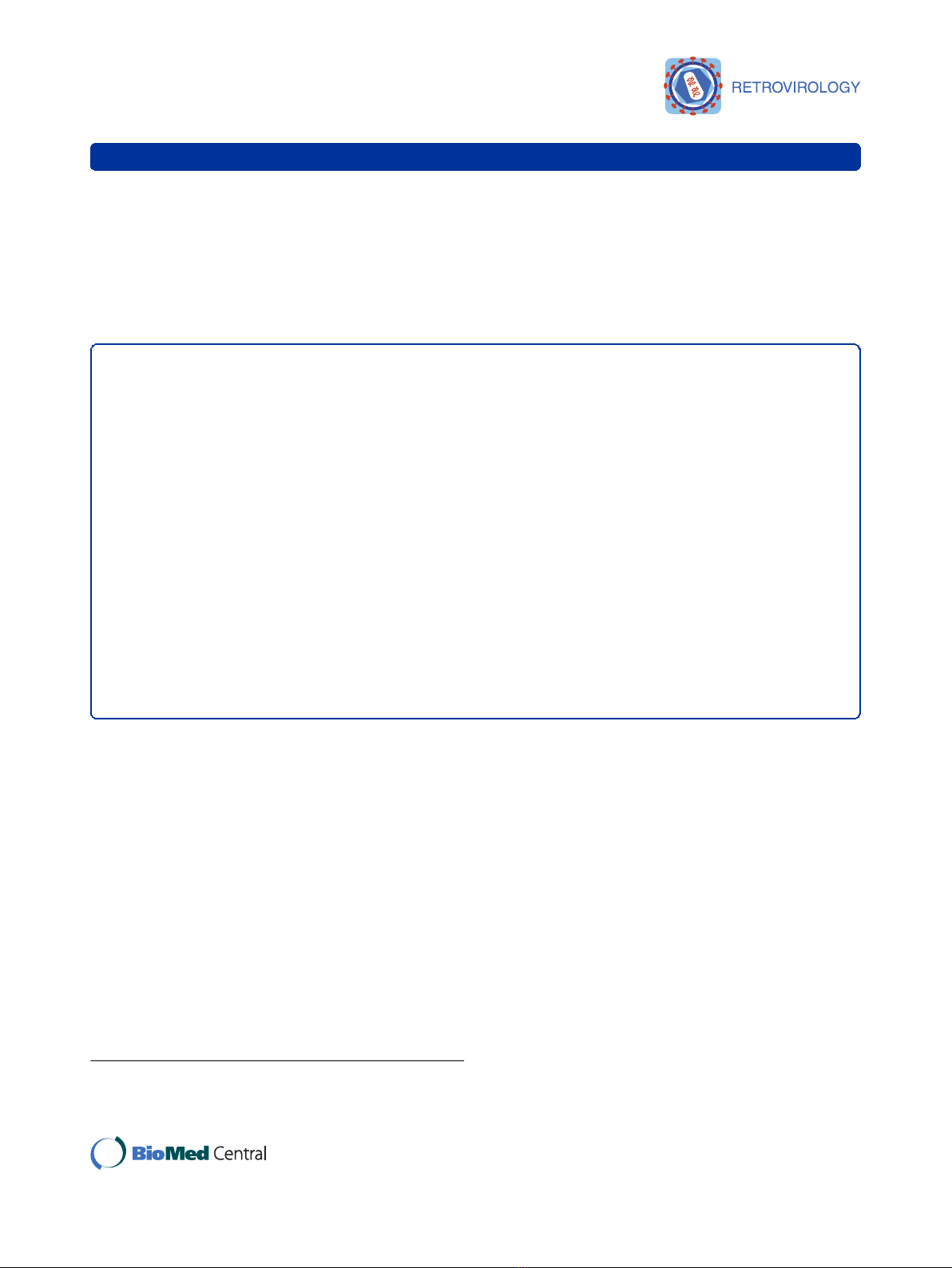
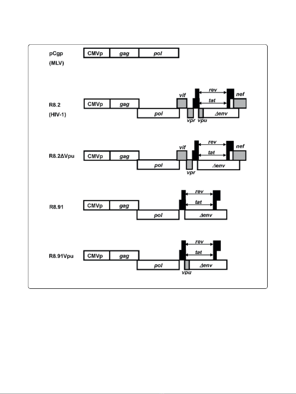
teins (Vpu, Vif, Vpr or Nef) [25] (Figure 1). In contrast
to the results observed in the presence of R8.2, which
decreased the steady-state GaLV TM levels in trans-
fected cells by approximately 60%, we found that co-
expression of R8.91 had no effect on steady state GaLV
TM levels in cell lysates (Figure 2A). This indicates that
one or more accessory proteins of HIV-1 play a key role
in the observed decrease in GaLV TM levels.
Interestingly, despite the lack of effect of R8.91 on
intracellular GaLV TM levels, analysis of vector particles
harvested from culture supernatants revealed a further
defect in the incorporation of GaLV Env into R8.91 gen-
erated vector particles (Figure 2A). Although some TM
protein could be detected in this fraction, it was present
at considerably lower levels than in retroviral particles
produced under the same conditions; and no mature, R-
peptide cleaved form of the protein was apparent.
Removal of the R peptide from the cytoplasmic tail of
GaLV and MLV Env proteins by retroviral proteases
activates their fusogenic potential and is necessary for
full infectivity [26-28]. As expected, the GaLV Env pseu-
dotyped vectors generated using R8.91 had very low
titers (Figure 2B). As we have previously reported [13],
no GaLV TM was detected in R8.2 derived lentiviral
vector particles, and these vectors gave no titer. Taken
together, these results suggest that two areas of incom-
patibility exist between the GaLV Env and lentiviral vec-
tors. First, the expression of one or more HIV-1
accessory proteins in R8.2 reduces the intracellular
steady-state levels of mature (cleaved) GaLV Env, while
an additional block exists that reduces the incorporation
and subsequent R-peptide processing of GaLV Env in
lentiviral particles, even in the absence of any HIV-1
accessory proteins.
Decrease in GaLV TM levels is mostly mediated by the
HIV-1 Vpu protein
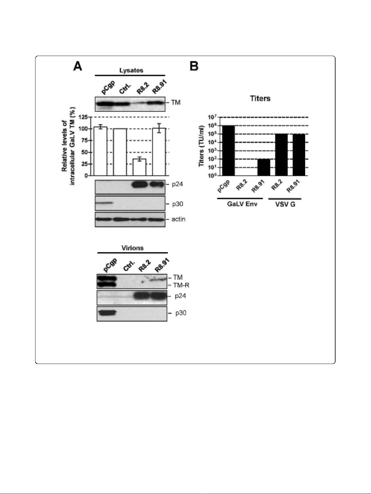
In order to identify which of the HIV-1 accessory pro-
teins expressed from R8.2 was responsible for the
decrease in cellular levels of GaLV TM, we generated
derivatives of R8.2 that were deleted for either Vpu, Nef,
or a combination of the Vif and Vpr proteins, and co-
expressed these plasmids with the lentiviral transfer vec-
tor and the GaLV Env. We found that the loss of Vpu
in construct R8.2∆Vpu (Figure 1) had the greatest effect,
resulting in only a 12% decrease in steady-state GaLV
TM levels compared to the 60% inhibition that resulted
from transfection of R8.2. In contrast, the absence of
the Nef, Vpr or Vif proteins did not significantly stabi-
lize the GaLV TM levels (Figure 3), although we did
observe a consistently small enhancement in TM levels
when Nef was deleted; but, overall, this was not statisti-
cally significant.
To confirm that Vpu was largely responsible for the
effects on TM, we next added back the Vpu ORF into
plasmid R8.91 (Figure 1). Expression of R8.91Vpu led to
a marked decrease in GaLV TM levels, although not
quite as complete as observed with R8.2. This finding
also suggested a minor contribution to Env degradation
from an additional HIV-1 accessory protein(s).
Vpu alone is not sufficient to decrease GaLV TM levels
The ability of the Vpu protein to decrease steady state
levels of GaLV TM was reminiscent of its known role
in removing at least two cellular proteins, CD4 and
BST-2/tetherin, from the cell surface [29,30]. There-
fore, we next examined whether the effect on GaLV
Christodoulopoulos et al.Retrovirology 2010, 7:4
http://www.retrovirology.com/content/7/1/4
Page 2 of 12
TM levels could be caused by expression of Vpu alone.
Surprisingly, we observed that the co-transfection of
GaLV Env and a Vpu expression plasmid (C∆EVpu)
had no effect on GaLV TM levels (Figure 4). We noted
that the level of expression of Vpu resulting from plas-
mid C∆EVpu was lower than from R8.2. However, we
consider that this difference in Vpu expression levels is
not likely to be the reason for the differences observed.
This interpretation was based on the finding that the
co-expression of C∆EVpu with R8.2∆Vpu caused
equivalent reductions in TM levels as occurred with
R8.2, despite the lower level of Vpu protein in the
cells. In addition, we also investigated whether trans-
fecting increasing amounts of the C∆EVpu plasmid,
with a corresponding increase in the levels of Vpu,
could reduce GaLV TM levels in the absence of a
packaging plasmid, but we found that this was not the
case (data not shown).
We next examined the consequences of co-expression
of R8.91 and C∆EVpu, and found that this also resulted
in a decrease in TM levels, in contrast to the results
obtained with R8.91 or C∆EVpu alone. However, the
Figure 1 Schematic representation of retroviral (pCgp) and lentiviral (R8.2, R8.2∆Vpu, R8.91 and R8.91Vpu) packaging constructs.All
lentiviral vectors express Gag, Pol, Tat and Rev; inclusion of Vif, Nef, Vpu and Vpr is as indicated (grey boxes).
Christodoulopoulos et al.Retrovirology 2010, 7:4
http://www.retrovirology.com/content/7/1/4
Page 3 of 12
more complete effect observed when the full set of HIV-
1 genes was present, in the transfection of either R8.2,
or R8.2∆Vpu plus C∆EVpu, suggests that an additional
role is played by one or more of the other HIV-1 acces-
sory proteins.
Mutation of conserved serine residues in Vpu prevents
the decrease in GaLV TM levels
Vpu targets CD4 for proteasomal degradation though
recruitment of the SCF-E3 ubiquitin ligase complex
[18,20]. Vpu binds to both CD4 and the b-TrCP subunit
of the complex, with the interaction with b-TrCP
requiring a conserved motif in Vpu’s cytoplasmic tail
that contains two phosphoserines (DS
52
GXXS
56
) [18,20].
Substitution of these serine residues with asparagine
residues blocks CD4 degradation [15,16]. Recruitment of
b-TrCP by Vpu has also been shown to play a role in
counteracting the host budding restriction factor, BST-
2/tetherin, and promoting either proteasomal [23] or
lysosomal [21,22] degradation. We therefore introduced
serine to asparagine substitutions into the packaging
constructs R8.2 and R8.9.1Vpu, and analyzed their
effects on GaLV TM levels. In both cases we observed
Figure 2 GaLV Env levels and titers of pseudotyped vectors.A. Western blot of a representative experiment showing levels of GaLV TM, HIV-
1 p24, MLV p30 and actin in cell lysates and pelleted supernatants from 293T cells co-transfected with plasmids expressing GaLV Env, together
with pCgp, R8.2, R8.91 or a control (Ctrl.) plasmid, as indicated. Also shown is a quantitative analysis of the steady-state levels of GaLV TM in cell
lysates, made relative to the levels in the presence of control plasmid. Results are mean of three independent experiments. B. Titers of GaLV Env
and VSV G pseudotyped retroviral (pCgp) and lentiviral (R8.2 and R8.91) vectors, as indicated, expressed as transducing units per ml (TU/ml). *
indicates no detectable titer.
Christodoulopoulos et al.Retrovirology 2010, 7:4
http://www.retrovirology.com/content/7/1/4
Page 4 of 12
that loss of the serines prevented the Vpu mediated
decrease in GaLV TM levels (Figure 5). This suggests a
role for the SCF-E3 ubiquitin ligase complex and pro-
tein degradation pathways in the reduction of GaLV
TM levels.
Substitution K618R in the GaLV tail confers resistance to
R8.2
We have previously shown that replacement of the cyto-
plasmic tail of GaLV Env with the equivalent domain
from the MuLV Env prevents the decrease in TM levels
resulting from expression of R8.2 [13]. In addition, we
observed that this effect could be achieved by two GaLV
to MuLV substitutions in the tail, K618Q and I619A
[13]. Schubert et al. [19] previously reported that inhibi-
tion of the degradation of CD4 by Vpu occurred follow-
ing the substitution of four lysines to arginines in CD4’s
cytosolic domain, suggesting ubiquitination as a
mechanism. We therefore tested whether the loss of
K618 alone was sufficient to preserve levels of GaLV
TM by generating mutants K618Q and K618R. We
foundthattheK618RsubstitutionrenderedtheGaLV
Env fully resistant to the R8.2-mediated decrease, similar
to the results obtained when the whole of the GaLV Env
tail was replaced with that of MuLV in construct GM
(TR). In contrast, the K618Q substitution gave an inter-
mediate phenotype (Figure 6).
The SU subunit, but not the Pr85 precursor of GaLV Env,
is affected by R8.2
The GaLV Env protein is synthesized as an 85 kDa pre-
cursor (Pr85). This precursor protein is glycosylated in
the ER and Golgi compartments into a short-lived 95
kDaintermediatethatispresentinthemedialGolgi
[31], and the protein is finally cleaved in the trans-Golgi
network by a cellular furin protease into the mature SU
(70kDa)andTM(15kDa)subunits.SUandTM
remain non-covalently linked and are transported to the
cell surface through the host vesicular transport system
[24]. In an attempt to further understand the effect of
R8.2 on GaLV Env, we investigated the fate of both the
Pr85 and SU proteins in the presence of lentiviral vector
components using a labeled version of the protein con-
taining a FLAG tag at the N-terminus of SU (FGaLV
Env) (Figure 7A). In addition, since the glycosylated
forms of Pr85 and SU are not easily resolved by stan-
dard electrophoresis, we treated cells lysates with Pep-
tide-N-glycosidase F (PNGaseF) in order to distinguish
between the two Env proteins, as previously described
[31,32]. Compared to control cells, we found that the
SU subunit was barely detectable in the presence of
R8.2, and was also significantly reduced by either R8.91
or R8.2∆Vpu, while none of the constructs had any
obvious effect on the Pr85 levels (Figure 7B).
Figure 3 Representative Western blot and quantitative analysis of signal density on blots from three independent experiments
analyzing GaLV TM levels in lysates of 293T cells co-transfected with GaLV Env, pHR’and indicated derivatives of R8.2 and R8.91.
Immunological detection of cellular HIV-1 p24 and actin levels were included as controls.
Christodoulopoulos et al.Retrovirology 2010, 7:4
http://www.retrovirology.com/content/7/1/4
Page 5 of 12


![Báo cáo seminar chuyên ngành Công nghệ hóa học và thực phẩm [Mới nhất]](https://cdn.tailieu.vn/images/document/thumbnail/2025/20250711/hienkelvinzoi@gmail.com/135x160/47051752458701.jpg)


%20--%3e%3cdefs%3e%3cstyle%3e%20.st0%20{%20fill:%20%23fff;%20}%20.st1%20{%20fill:%20%237800fa;%20}%20%3c/style%3e%3c/defs%3e%3cpath%20class='st1'%20d='M117.78,12.18H43.11c2.9,3.47,4.65,7.94,4.65,12.82,0,5.6-2.3,10.66-6.01,14.29h76.02l7.22-13.56-7.22-13.56Z'/%3e%3cg%3e%3cpath%20class='st0'%20d='M53.58,26.17h-.59v-1.46h.59v-4.96h2.83c1.78,0,2.67.94,2.67,2.82v5.76c0,1.87-.89,2.81-2.67,2.81h-2.83v-4.96ZM55.36,21.37v3.34h1.1v1.46h-1.1v3.34h1.01c.61,0,.91-.37.91-1.1v-5.93c0-.74-.3-1.1-.91-1.1h-1.01Z'/%3e%3cpath%20class='st0'%20d='M65.99,31.14h-1.8l-.31-2.07h-2.19l-.31,2.07h-1.64l1.82-11.39h2.62l1.82,11.39ZM65.28,18.04c-.25.46-.51.77-.75.94-.21.15-.47.22-.79.22-.26,0-.57-.07-.92-.22l-.38-.15c-.14-.05-.26-.07-.37-.07-.3,0-.53.18-.71.54l-.91-.68c.25-.46.51-.77.75-.94.21-.14.48-.21.79-.21.26,0,.57.07.92.21l.38.15c.14.05.26.07.37.07.3,0,.53-.18.71-.54l.91.68ZM61.91,27.52h1.73l-.87-5.76-.87,5.76Z'/%3e%3cpath%20class='st0'%20d='M74.53,26.89v1.52c0,1.91-.89,2.86-2.67,2.86s-2.67-.95-2.67-2.86v-5.93c0-1.91.89-2.86,2.67-2.86s2.67.95,2.67,2.86v1.11h-1.69v-1.22c0-.75-.31-1.12-.93-1.12s-.93.37-.93,1.12v6.15c0,.74.31,1.11.93,1.11s.93-.37.93-1.11v-1.63h1.69Z'/%3e%3cpath%20class='st0'%20d='M81.4,31.14h-1.8l-.31-2.07h-2.19l-.31,2.07h-1.64l1.82-11.39h2.62l1.82,11.39ZM75.9,19.2l1.52-1.91h1.71l1.51,1.91h-1.61l-.76-.95-.75.95h-1.61ZM77.32,27.52h1.73l-.87-5.76-.87,5.76ZM83.1,15.99l-1.76,1.91h-1.26l1.17-1.91h1.86Z'/%3e%3cpath%20class='st0'%20d='M84.86,19.75c1.78,0,2.67.94,2.67,2.82v1.48c0,1.87-.89,2.81-2.67,2.81h-.85v4.28h-1.79v-11.39h2.64ZM84.01,21.37v3.86h.85c.58,0,.87-.36.87-1.08v-1.71c0-.71-.29-1.07-.87-1.07h-.85Z'/%3e%3cpath%20class='st0'%20d='M93.51,19.75c1.78,0,2.67.94,2.67,2.82v1.48c0,1.87-.89,2.81-2.67,2.81h-.85v4.28h-1.79v-11.39h2.64ZM92.66,21.37v3.86h.85c.58,0,.87-.36.87-1.08v-1.71c0-.71-.29-1.07-.87-1.07h-.85Z'/%3e%3cpath%20class='st0'%20d='M98.8,31.14h-1.79v-11.39h1.79v4.88h2.03v-4.88h1.83v11.39h-1.83v-4.88h-2.03v4.88Z'/%3e%3cpath%20class='st0'%20d='M105.36,24.55h2.46v1.62h-2.46v3.34h3.09v1.63h-4.88v-11.39h4.88v1.63h-3.09v3.18ZM108.17,17.29l-1.76,1.91h-1.26l1.17-1.91h1.86Z'/%3e%3cpath%20class='st0'%20d='M112.2,19.75c1.78,0,2.67.94,2.67,2.82v1.48c0,1.87-.89,2.81-2.67,2.81h-.85v4.28h-1.79v-11.39h2.64ZM111.35,21.37v3.86h.85c.58,0,.87-.36.87-1.08v-1.71c0-.71-.29-1.07-.87-1.07h-.85Z'/%3e%3c/g%3e%3ccircle%20class='st1'%20cx='25'%20cy='25'%20r='20'/%3e%3cpath%20class='st0'%20d='M32.78,19.27c2.92,0,4.43,2.55,5.28,5.33l.71,2.17c.14.38-.33.75-.71.75h-5.61c.19-.33.24-.71.09-1.08l-.75-2.45c-.43-1.32-.99-2.64-1.79-3.77.75-.57,1.65-.94,2.78-.94h0ZM25,18.38c3.25,0,4.9,2.78,5.89,5.89l.76,2.45c.14.42-.33.8-.8.8h-11.69c-.42,0-.94-.38-.8-.8l.75-2.45c.99-3.11,2.64-5.89,5.89-5.89h0ZM25,11.35c1.74,0,3.11,1.37,3.11,3.11s-1.37,3.11-3.11,3.11-3.11-1.41-3.11-3.11,1.41-3.11,3.11-3.11h0ZM17.27,19.27c1.08,0,1.98.38,2.73.94-.8,1.13-1.37,2.45-1.74,3.77l-.8,2.45c-.14.38-.05.75.09,1.08h-5.56c-.42,0-.9-.38-.75-.75l.71-2.17c.9-2.78,2.41-5.33,5.33-5.33h0ZM17.27,12.91c1.51,0,2.78,1.27,2.78,2.83s-1.27,2.83-2.78,2.83-2.83-1.27-2.83-2.83,1.27-2.83,2.83-2.83h0ZM32.78,12.91c1.56,0,2.78,1.27,2.78,2.83s-1.23,2.83-2.78,2.83-2.83-1.27-2.83-2.83,1.27-2.83,2.83-2.83h0ZM27.07,28.56v.09c0,.57-.24,1.08-.61,1.46h0v.05c-.38.33-.9.57-1.46.57s-1.08-.24-1.46-.61h0c-.38-.38-.61-.9-.61-1.46v-.09h1.41v.09c0,.19.05.38.19.47v.05c.09.09.28.19.47.19s.38-.09.47-.19v-.05c.14-.09.24-.28.24-.47t-.05-.09h1.41ZM30.99,28.56v.09c0,1.65-.66,3.16-1.74,4.24-1.08,1.08-2.59,1.79-4.24,1.79s-3.16-.71-4.24-1.79l-.05-.05c-1.04-1.08-1.7-2.55-1.7-4.2v-.09h1.41v.09c0,1.27.47,2.4,1.27,3.25h.05c.85.85,1.98,1.37,3.25,1.37s2.4-.52,3.25-1.37c.85-.8,1.37-1.98,1.37-3.25v-.09h1.37ZM34.99,28.56v.09c0,2.78-1.13,5.28-2.92,7.07-1.79,1.79-4.29,2.92-7.07,2.92s-5.23-1.13-7.07-2.92c-1.79-1.79-2.92-4.29-2.92-7.07v-.09h1.41v.09c0,2.4.94,4.53,2.5,6.08,1.56,1.56,3.72,2.5,6.08,2.5s4.52-.94,6.08-2.5c1.56-1.56,2.5-3.68,2.5-6.08v-.09h1.41Z'/%3e%3c/svg%3e)