Fatty acids increase the circulating levels of oxidative
stress factors in mice with diet-induced obesity via redox
changes of albumin
Mayumi Yamato
1
, Takeshi Shiba
1
, Masayoshi Yoshida
2
, Tomomi Ide
2
, Naoko Seri
1
,
Wataru Kudou
1
, Shintaro Kinugawa
3
and Hiroyuki Tsutsui
3
1 Department REDOX Medicinal Science, Faculty of Pharmaceutical Sciences, Kyushu University, Fukuoka, Japan
2 Department of Cardiovascular Medicine, Graduate School of Medical Sciences, Kyushu University, Fukuoka, Japan
3 Department of Cardiovascular Medicine, Hokkaido University Graduate School of Medicine, Sapporo, Japan
Metabolic syndrome is characterized by a constella-
tion of multiple risk factors, such as dyslipidemia,
hyperglycemia, hypertension, and abdominal obesity.
Several studies have focused on the role of oxidative
stress in metabolic syndrome [1–6]. For example, oxi-
dative stress markers, such as thiobarbituric acid
reactive substances (TBARS), an index of lipid perox-
idation, 8-hydroxy-2¢-deoxyguanosine, a biomarker of
oxidative DNA damage, and oxidative modification
of low-density lipoprotein, increased in the plasma of
a rat model of metabolic syndrome [6]. Plasma con-
centrations of free fatty acids are also increased in
metabolic syndrome, and might be involved in the
pathogenesis of skeletal muscle insulin resistance [7,8].
Increased fatty acids resulted in cellular damage via
the induction of oxidative stress [9,10]. In skeletal
muscle cells, palmitate-induced mitochondrial DNA
damage and cytotoxicity were caused by the overpro-
duction of peroxynitrite [9]. Zhang et al. demonstra-
ted that fatty acids enhanced monocyte adhesion to
endothelial cells in vitro and that the process was
mediated through the increased generation of reactive
oxygen species and the enhanced expression of the
integrin CD11b [10].
Keywords
ESR; albumin; fatty acid; obesity; oxidative
stress
Correspondence
H. Tsutsui, Department of Cardiovascular
Medicine, Hokkaido University Graduate
School of Medicine, Kita-15, Nishi-7, Kita-ku,
Sapporo 060-8638, Japan
Fax: +81 11 706 7874
Tel: +81 11 706 6973
E-mail: htsutsui@med.hokudai.ac.jp
(Received 11 April 2007, revised 29 May
2007, accepted 31 May 2007)
doi:10.1111/j.1742-4658.2007.05914.x
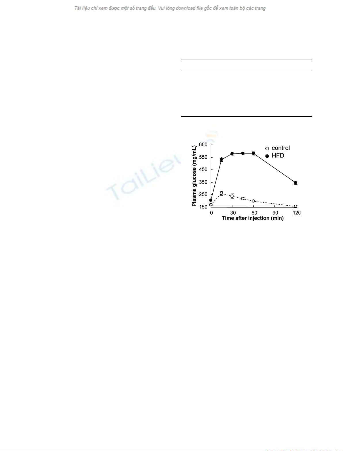
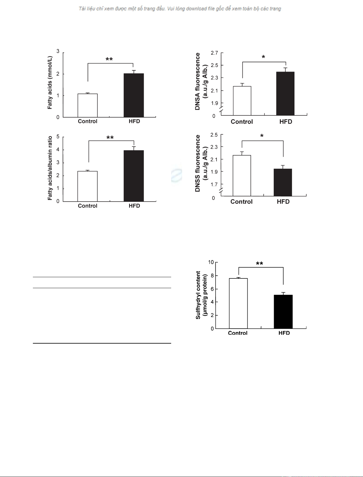
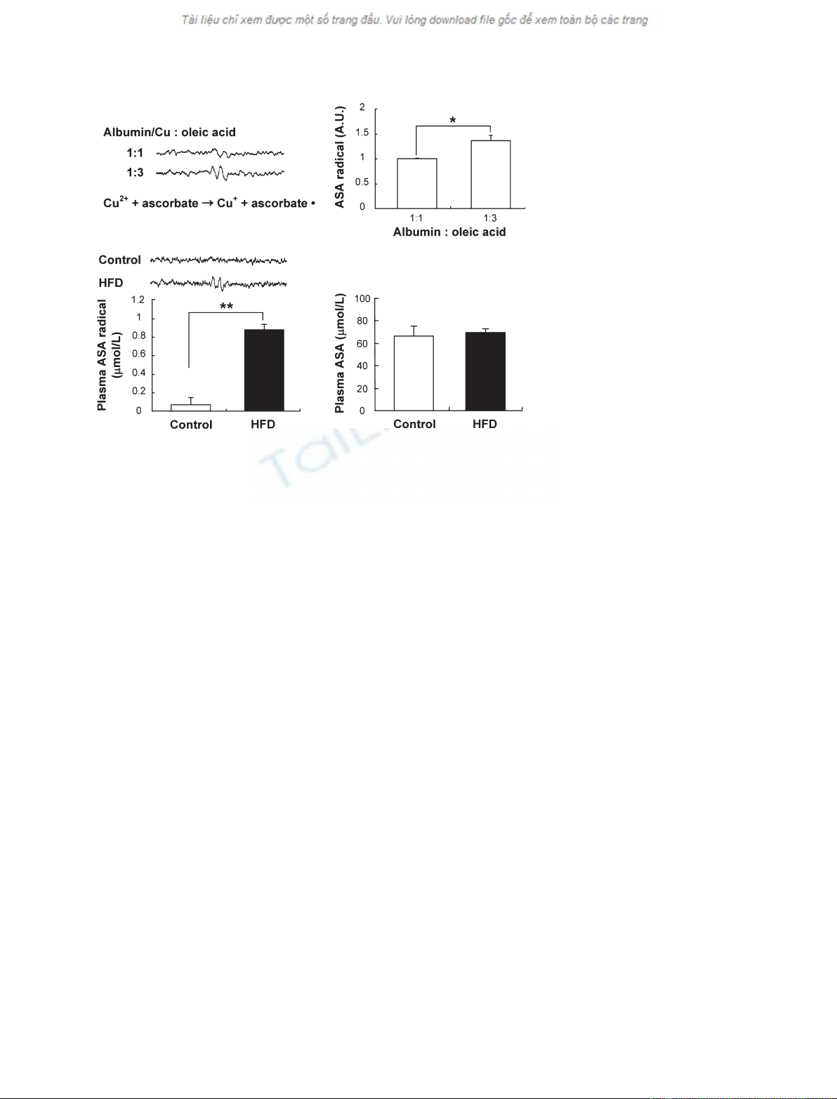
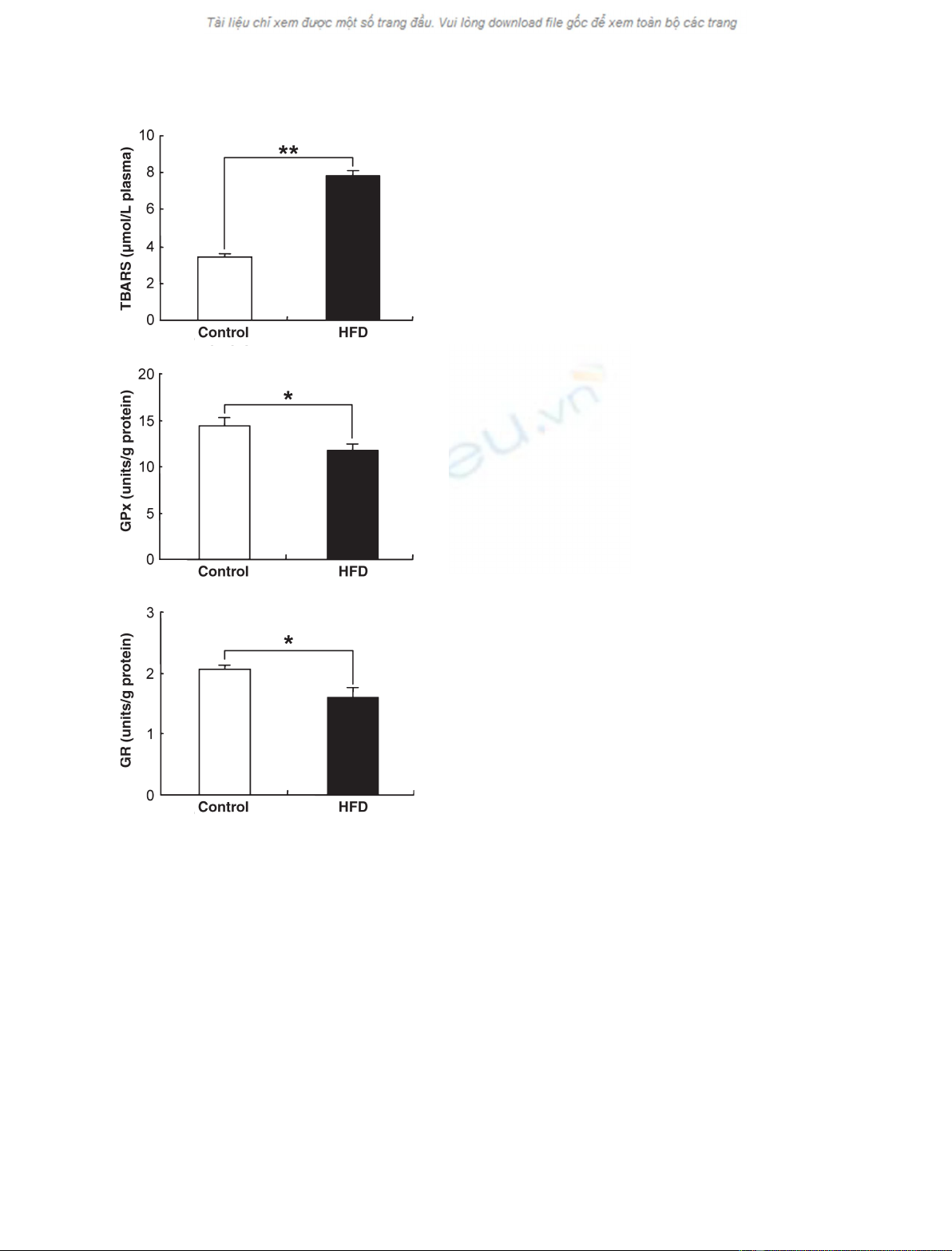
Plasma concentrations of free fatty acids are increased in metabolic syn-
drome, and the increased fatty acids may cause cellular damage via the
induction of oxidative stress. The present study was designed to determine
whether the increase in fatty acids can modify the free sulfhydryl group in
position 34 of albumin (Cys34) and enhance the redox-cycling activity of
the copper–albumin complex in high-fat diet-induced obese mice. The mice
were fed with commercial normal diet or high-fat diet and water ad libitum
for 3 months. The high-fat diet-fed mice developed obesity, hyperlipemia,
and hyperglycemia. The plasma fatty acid albumin ratio also significantly
increased in high-fat diet-fed mice. The increased fatty acid albumin ratio
was associated with conformational changes in albumin and the oxidation
of sulfhydryl groups. Moreover, an ascorbic acid radical, an index of
redox-cycling activity of the copper–albumin complex, was detected only in
the plasma from obese mice, whereas the plasma concentrations of ascorbic
acid were not altered. Plasma thiobarbituric acid reactive substances were
significantly increased in the high-fat diet group. These results indicate that
the increased plasma fatty acids in the high-fat diet group resulted in the
activated redox cycling of the copper–albumin complex and excessive lipid
peroxidation.
Abbreviations
ASA, ascorbic acid; DNSA, dansyl amide; DNSS, dansyl sarcosine; GPx, glutathione peroxidase; GR, glutathione reductase; HFD, high-fat
diet; Nbs
2
, 5,5¢-dithiobis(2-nitrobenzoic acid); TBARS, thiobarbituric acid reactive substances.
FEBS Journal 274 (2007) 3855–3863 ª2007 The Authors Journal compilation ª2007 FEBS 3855
Oxidative stress results from an imbalance between
oxidant production and antioxidant defense mecha-
nisms. In plasma, albumin functions as an extracellular
antioxidant [11,12]. Albumin is composed of mercapt-
albumin (reduced form) and nonmercaptalbumin
(oxidized form), i.e. a protein redox couple. Mercaptal-
bumin has one free sulfhydryl group in position 34
(Cys34) that is known to contribute significantly to the
antioxidant capacity of plasma [13]. Albumin also acts
as the major plasma carrier of various ligands such as
free fatty acids and copper [14–19]. Fatty acids bind to
albumin in long, hydrophobic pockets capped by polar
side chains, many of which are basic [16]. The N-ter-
minal tripeptide (Cu
2+
Ni
2+
-binding motif) of albu-
min is the high-affinity site for copper [15,17].
Previous studies have demonstrated that fatty acids
affect the redox status of albumin in vitro [17,19]. The
binding of fatty acids to albumin increased the reactiv-
ity of Cys34, and facilitated the oxidation of Cys34
[19]. Furthermore, Gryzunov et al. clarified that the
oxidized Cys34 increased the redox-cycling activity of
the copper–albumin complex by binding of fatty acids
[17]. They used ascorbic acid (ASA) radicals to mon-
itor the redox-cycling activity of the copper–albumin
complex with ESR spectroscopy. The one-electron oxi-
dation–reduction reactions of copper are also known
to be cytotoxic, because they can catalyze the produc-
tion of free radical intermediates, which can result in
cellular damage. These reactions may cause lipid per-
oxidation in the plasma, resulting in the development
of atherosclerosis and metabolic disorder. However,
little is known about fatty acid-induced oxidative stress
in vivo.
The present study was designed to determine whe-
ther the increase in fatty acids can modify Cys34 of
albumin and enhance the redox-cycling activity of
the copper–albumin complex in high-fat diet (HFD)-
induced obese mice. We detected ASA radicals in
the plasma by using ESR spectroscopy as an index
of redox-cycling activity of the copper–albumin com-
plex.
Results
Animal characteristics
The HFD-fed mice developed obesity, hyperlipidemia,
and hyperglycemia (Table 1), which are characteristic
for metabolic syndrome. Increased body weight was
associated with an increase in visceral adipose tissue
(data not shown). Glucose tolerance test results were
also abnormal in the HFD group as compared with
the normal diet control group (Fig. 1).
Plasma fatty acids and conformational changes
in albumin
The concentration of plasma fatty acids and the fatty
acid albumin ratio are shown in Fig. 2. There was a
significant increase in the concentration of fatty acids
in the plasma from HFD-fed mice. The fatty
acid albumin ratio in the plasma from the HFD group
was 3.9, and that of the control group was 2.3
(Fig. 2B). The concentrations of representative fatty
acids are shown in Table 2. Most kinds of fatty acid,
such as oleic acid, showed increased concentrations,
although the concentrations of linoleic acid and lino-
lenic acid decreased.
The increased fatty acid albumin ratio should cause
conformational changes in albumin. As expected, the
fluorescence values of dansyl amide (DNSA) were
increased and those of dansyl sarcosine (DNSS) were
decreased in the plasma from HFD-fed mice (Fig. 3).
The concentration of sulfhydryl groups was also signi-
ficantly lower in the HFD group than in the control
Table 1. Animal characteristics. Each value represents the mean ±
SEM. The data were obtained from five animals. HDL, high-density
lipoprotein; LDL, low-density lipoprotein.
Control group HFD group
Body weight (g) 29.9 ± 0.6 46.8 ± 0.7**
Total cholesterol (mgÆdL
)1
) 79.0 ± 3.3 194.2 ± 10.3**
HDL cholesterol (mgÆdL
)1
) 47.5 ± 3.9 109.6 ± 8.6**
LDL cholesterol (mgÆdL
)1
) 21.7 ± 3.7 64.2 ± 7.5**
Triglyceride (mgÆdL
)1
) 49.0 ± 2.3 101.8 ± 9.9**
Phospholipid (mgÆdL
)1
) 78.7 ± 3.3 183.4 ± 7.9**
Glucose (mgÆdL
)1
) 160.5 ± 3.2 195.2 ± 10.6*
*P<0.05 and ** P<0.005 versus control group.
Fig. 1. Plasma glucose during the glucose tolerance test. The
plasma glucose level was significantly higher after the glucose load
in the HFD group than in the control group. Values are means ±
SEM. The data were obtained from five animals at each time point.
Fatty acid-induced oxidative stress in obesity M. Yamato et al.
3856 FEBS Journal 274 (2007) 3855–3863 ª2007 The Authors Journal compilation ª2007 FEBS
group (Fig. 4), indicating that the increased fatty acid
bound plasma albumin, and then facilitated the oxida-
tion of Cys34 in the plasma from HFD-fed mice.
Redox-cycling activity of albumin in mice
The results of the in vitro validation study for the
redox-cycling activity of the copper–albumin complex
are shown in Fig. 5A. The redox-cycling activity of the
copper–albumin complex was enhanced by fatty acids
(Fig. 5A).
We detected the typical doublet signal of ASA radi-
cals with 0.182 mT of hyperfine splitting in the plasma
obtained from HFD-fed mice, and the signal intensity
of the ASA radical in the plasma of the HFD group
A
B
Fig. 2. Plasma fatty acids (A) and fatty acid albumin ratio (B). There
were significant increases in the concentration of fatty acids and
the fatty acid albumin ratio in the plasma from HFD-fed mice. Val-
ues are means ± SEM. **P<0.005 versus control group. The data
were obtained from five animals.
Table 2. Representative fatty acids in the plasma from mice. Each
value represents the mean ± SEM. The data were obtained from
five animals.
Fatty acid (lM) Control group HFD group
Myristic acid 8.8 ± 1.6 18.9 ± 1.7*
Palmitic acid 444.7 ± 30.5 572.0 ± 59.2
Palmitoleic acid 159.5 ± 12.9 179.0 ± 11.9
Stearic acid 96.9 ± 7.1 176.2 ± 13.8*
Oleic acid 326.8 ± 28.1 1203.8 ± 106.9*
Linoleic acid 575.6 ± 39.5 247.7 ± 17.4*
Linolenic acid 26.0 ± 1.8 9.1 ± 1.2*
Arachidonic acid 34.5 ± 3.6 161.8 ± 21.8*
*P<0.005 versus control group.
A
B
Fig. 3. Effects of HFD feeding on fluorescence intensity of DNSA
(A) and DNSS (B) in the plasma. Fluorescence intensity is presen-
ted in arbitrary units. Fluorescence values of DNSA were increased
and those of DNSS were decreased in the plasma from HFD-fed
mice. Values are means ± SEM. *P<0.05 versus control group.
The data were obtained from five animals.
Fig. 4. Oxidation of sulfhydryl content in the plasma. The determin-
ation of sulfhydryl contents were performed according to the
Ellman method. The concentration of sulfhydryl groups was signifi-
cantly lower in the HFD group than in the control group. Values are
means ± SEM. **P<0.005 versus control group. The data were
obtained from five animals.
M. Yamato et al. Fatty acid-induced oxidative stress in obesity
FEBS Journal 274 (2007) 3855–3863 ª2007 The Authors Journal compilation ª2007 FEBS 3857
was higher than that of the control group (Fig. 5B). In
contrast, the plasma ASA concentration was compar-
able between the control and HFD groups (Fig. 5C).
Plasma lipid peroxidation and antioxidant
enzyme
To determine whether the increased redox-cycling activ-
ity of copper induced lipid peroxidation, lipid peroxida-
tion was estimated by the TBARS method. Plasma
TBARS were significantly increased in the HFD group
as compared to the control group (Fig. 6A).
The activities of glutathione peroxidase (GPx) and
glutathione reductase (GR), which play a major role
in peroxide removal, were significantly decreased in
plasma of the HFD group (Fig. 6B,C).
Discussion
We demonstrated that the increased fatty acids
increased the plasma levels of oxidative stress factors
in HFD-fed mice. Our results indicated that: (a) the
increased fatty acids bound to albumin and induced
conformational changes in albumin; (b) the fatty acids
bound to albumin facilitated the oxidation of protein
sulfhydryl groups, Cys34, in the plasma; (c) the oxid-
ized Cys34 increased the redox-cycling activity of the
copper–albumin complex; and (d) the copper–albumin
complex increased lipid peroxidation in the circulating
blood of obese mice.
The HFD-fed mice showed obesity, hyperlipidemia,
and hyperglycemia, in association with increased fatty
acids (Table 1, Figs 1 and 2). Plasma albumin-bound
fatty acids are derived mainly from adipose tissue tri-
glyceride stores released through the action of the
cAMP-dependent enzyme hormone-sensitive lipase.
Fatty acids are also derived through the lipolysis of tri-
glyceride-rich lipoproteins in tissues by the action of
lipoprotein lipase. Insulin is important for both anti-
lipolysis and the stimulation of lipoprotein lipase. The
most sensitive pathway of insulin action is the inhibi-
tion of lipolysis in adipose tissue. Thus, when insulin
resistance develops, the increased amount of lipolysis
of stored triacylglycerol molecules in adipose tissue
produces more fatty acids, which could further inhibit
the antilipolytic effect of insulin, inducing additional
lipolysis. Previous studies have indicated that the
increased fatty acids play an important role in the
development of insulin resistance [20,21].
Noel and Hunter separated mercaptalbumin and
nonmercaptalbumin by DEAE-Sephadex ion exchange
chromatography, and showed that the latter had many
more fatty acids than the former [18]. Takebayashi
et al. also demonstrated that the binding of fatty acids
and the oxidation of sulfhydryl groups of the protein
were intimately coupled [19]. The increase in fatty acids
shown in the present study was closely related to the
oxidation of Cys34 in vivo (Fig. 4). ASA is a water-sol-
uble antioxidant, but exacerbates lipid peroxidation in
the presence of transition metals [22,23]. ASA is oxid-
ized in the presence of transition metals, including the
copper–albumin complex, and is converted to ASA
radical through one-electron oxidation. The stable
ASA radicals were detectable using X-band ESR
A
B
C
Fig. 5. In vitro validation study of the redox-
cycling activity of the copper–albumin com-
plex (A), typical ASA radical ESR spectra
and their intensities (B), and ASA concentra-
tion in the plasma (C). The redox-cycling
activity of the copper–albumin complex was
enhanced by fatty acids in vitro (A). The
ESR signal of the ASA radical was detected
in the plasma from HFD mice (B), although
the plasma ASA concentration was compar-
able between the control and HFD groups
(C). Values are means ± SEM. *P<0.05
and **P<0.005 versus control group. The
data were obtained from five animals.
Fatty acid-induced oxidative stress in obesity M. Yamato et al.
3858 FEBS Journal 274 (2007) 3855–3863 ª2007 The Authors Journal compilation ª2007 FEBS
ex vivo (Fig. 5). ASA radicals were observed only in the
plasma from obese mice; the plasma concentrations of
ASA were not altered. There is a balance between ASA,
the corresponding radical and dehydroascorbic acid
during copper–albumin complex-catalyzed oxidation.
Within this balance, the concentration of ASA radicals
produced from ASA reflects the equilibrium between
the fully reduced and oxidized forms of ASA. More-
over, ASA radicals did not appear if Cys34 of fatty
acid-free albumin was in the reduced state in vitro [17].
We also detected ASA radicals only in the plasma of
the HFD group, in which Cys34 was oxidized. These
results suggest that the sulfhydryl group of Cys34 in
albumin is critically involved in the regulation of copper
redox-cycling activity both in vitro and in vivo.
In the presence of reducing agents such as ASA,
lipid peroxidation is facilitated by transition metals.
Transition metals such as copper are capable of cata-
lyzing hydrogen atom abstraction of lipids directly in
the initiation of lipid peroxidation, and or are involved
in peroxidation as ‘Fenton-catalytic’ metals [24,25].
The increased fatty acids and phospholipids may be
also related to the accumulation of peroxides (Table 1,
Figs 3 and 6). Taken together, these results indicate
that the increased fatty acids in the HFD group resul-
ted in the activated redox-cycling copper–albumin
complex and excessive lipid peroxidation (Fig. 7).
Hostmark indicated that a fatty acid albumin ratio
of 8 caused hemolysis in vitro experiments [26]. The
leakage of hemoglobin into the plasma might be rela-
ted to the formation of ASA radicals. However, the
fatty acid albumin ratio in the plasma sample was as
high as 4, and the hemolytic responses were not found.
Hence, the effects of hemoglobin on measurement in
the present study may be excluded.
In the present study, we estimated plasma TBARS
level as an index of lipid peroxidation. However, the
measurement of lipid peroxidation by using plasma
TBARS is unspecific with regard to its generation.
Further studies are clearly needed to determine the
reaction mechanism of lipid peroxidation by copper–
albumin complex-catalyzed oxidation by using a more
specific method. Moreover, Ishola et al. suggested that
albumin-bound fatty acids induced oxidative stress via
increased mitochondrial reactive oxygen species pro-
duction and inhibition of the superoxide dismutase
transcriptional response in proximal tubular cells [27].
An increase in plasma fatty acid concentrations in nor-
mal subjects also resulted in the induction of oxidative
stress and inflammation [28]. In addition, various hor-
mones and cytokines from adipose tissues and the acti-
vation of phagocytic NADPH oxidase also contributed
to the oxidative stress in metabolic syndrome [29].
These mechanisms, including the enhanced redox-
cycling activity of the copper–albumin complex, might
be mutually related, and exacerbate the circulating
levels of oxidative stress in our model.
In conclusion, fatty acids were associated with the
conformational changes in albumin, facilitated oxida-
tion of Cys34, and increased redox-cycling activity of
the copper–albumin complex in HFD-induced obese
mice. The present data indicate that reducing
A
B
C
Fig. 6. Plasma TBARS (A) and enzymatic activities of GPx (B) and
GR (C). The plasma TBARS level was significantly higher in the
HFD group than in the control group (A). The activities of antioxid-
ant enzyme were decreased in the plasma from HFD mice (B, C).
Values are means ± SEM. **P<0.005 versus control group. The
data were obtained from five animals.
M. Yamato et al. Fatty acid-induced oxidative stress in obesity
FEBS Journal 274 (2007) 3855–3863 ª2007 The Authors Journal compilation ª2007 FEBS 3859