Zhou et al. Journal of Translational Medicine 2010, 8:13 http://www.translational-medicine.com/content/8/1/13
R E S E A R C H
Open Access
The density of macrophages in the invasive front is inversely correlated to liver metastasis in colon cancer Qiang Zhou1,2, Rui-Qing Peng1,2, Xiao-Jun Wu1,3, Qing Xia1,2, Jing-Hui Hou1,4, Ya Ding1,2, Qi-Ming Zhou1,2, Xing Zhang1,2, Zhi-Zhong Pang1,3, De-Sen Wan1,3, Yi-Xin Zeng1,2, Xiao-Shi Zhang1,2*
Abstract
Background: Although an abundance of evidence has indicated that tumor-associated macrophages (TAMs) are associated with a favorable prognosis in patients with colon cancer, it is still unknown how TAMs exert a protective effect. This study examined whether TAMs are involved in hepatic metastasis of colon cancer.
Materials and methods: One hundred and sixty cases of pathologically-confirmed specimens were obtained from colon carcinoma patients with TNM stage IIIB and IV between January 1997 and July 2004 at the Cancer Center of Sun Yat-Sen University. The density of macrophages in the invasive front (CD68TFHotspot) was scored with an immunohistochemical assay. The relationship between the CD68TFHotspot and the clinicopathologic parameters, the potential of hepatic metastasis, and the 5-year survival rate were analyzed.
Results: TAMs were associated with the incidence of hepatic metastasis and the 5-year survival rate in patients with colon cancers. Both univariate and multivariate analyses revealed that the CD68TFHotspot was independently prognostic of survival. A higher 5-year survival rate among patients with stage IIIB after radical resection occurred in patients with a higher macrophage infiltration in the invasive front (81.0%) than in those with a lower macrophage infiltration (48.6%). Most importantly, the CD68TFHotspot was associated with both the potential of hepatic metastasis and the interval between colon resection and the occurrence of hepatic metastasis.
Conclusion: This study showed evidence that TAMs infiltrated in the invasive front are associated with improvement in both hepatic metastasis and overall survival in colon cancer, implying that TAMs have protective potential in colon cancers and might serve as a novel therapeutic target.
resection of
Background Colorectal cancer is the fourth leading cause of cancer deaths worldwide. Of patients with colorectal cancer, 35%-55% will develop hepatic metastases at some time during the course of their disease. Survival following colorectal metastasis now hepatic approaches 35%-50%. However, approximately 65% of patients will have a recurrence at 5 years. Identifying the markers for hepatic metastasis would be helpful for the early treatment of patients at high-risk of hepatic metas- tasis [1-5].
In addition to clonal selection and the predetermined metastatic potential of cancer cells, there is increasing evi- dence indicating that the microenvironment modifies the metastasis of cancer cells [6-9]. Cancer tissue is infiltrated with stromal cells including macrophages. Tumor-asso- ciated macrophages (TAMs) are not only abundant in epithelial cancers, but also involved in cancer progression [10-13]. Experimental data have indicated that ablation of macrophage function or inhibition of macrophage infiltra- tion into experimental tumors inhibits tumor growth and metastases [14]. Additionally, gene array studies of diag- nostic lymph node specimens in follicular lymphoma have shown that genes associated with a strong ‘macrophage’ signature are associated with a poorer prognosis, indepen- dent of clinical variables or of gene expression of the
* Correspondence: zxs617@hotmail.com 1State Key Laboratory of Oncology in South China, Cancer Center, Sun Yat- Sen University, 651 Dongfeng R E, 510060, Guangzhou, China
© 2010 Zhou et al; licensee BioMed Central Ltd. This is an Open Access article distributed under the terms of the Creative Commons Attribution License (http://creativecommons.org/licenses/by/2.0), which permits unrestricted use, distribution, and reproduction in any medium, provided the original work is properly cited.
Follow-up of stage IIIB patients and post-operative treatment Clinical follow-up was only provided to stage IIIB patients, as patients with stage IV in this study were a group with high heterogeneity, including solitary or multiple liver metastases, and liver only or other sites involved with metastases; these variables affected the treatment protocols and eventually the response rate and prognosis. Ninety-eight patients with stage IIIB colon carcinoma were observed on an every-3-month basis during the 1st year, once every 6 months in the 2nd year, and by telephone or mail communication once every year thereafter for a total of 5 years. If recurrence or metastasis occurred, 5-FU-based chemotherapy was administered according to the NCCN guidelines [40]. Overall survival (OS) was defined as the time from sur- gery to death, or was censored at the last known alive data. Liver metastasis-free survival (LMFS) was defined as the time from surgery to liver metastasis.
tumor cells [15]. Therefore, TAMs might promote tumor progression by induction of chronic inflammation, matrix remodeling, tumor invasion, intravasation, angiogenesis, and seeding at distant sites [13]. In contrast, recruitment of TAMs also contributes to the development of an adap- tive immune response against cancer. TAMs contribute to the balance between antigen availability and clearance through phagocytosis and subsequent degradation of senescent or apoptotic cells. The role of TAMs is essential for triggering, instructing, and terminating the adaptive immune response [16]. The clinical evidence regarding the relationship between TAMs and tumor progression is tumor type-dependent. The higher density of TAMs is associated with a poorer prognosis in leiomyosarcomas, melanomas, gliomas, and cancers of the breast, bladder, rectum, and endometrium, but the prognosis is favorable in nasopharyngeal, gastric, and ovarian cancers [17-28]. Additionally, in liver, lung, and prostate cancers, the role of TAMs on prognosis is controversial [29-35].
With respect to colorectal carcinomas, clinical data indicate that TAMs are associated with a favorable prognosis [36-39]. However, these studies have not indi- cated the sites at which TAMs show a protective effect. Because macrophages modify tumor invasion, intravasa- tion, and angiogenesis, whether or not TAMs interfere with hepatic metastasis of colon cancer was determined in the current study.
Immunohistochemistry The specimens were fixed in formaldehyde and embedded in paraffin. Only blocks containing the tumor front were evaluated. Tissue sections of 5-μm thickness were cut, dried, deparaffinized, and rehydrated in a ser- ies of alcohols and xylene before antigen retrieval by pressure cooker treatment in citrate buffer (pH 6.0) for 3 minutes. After that, we performed endogenous peroxi- dase blocking through hydrogen peroxide incubation. Mouse anti-human CD68 monoclonal antibody (mAb) (PG-M1; DakoCytomation, Glostrup, Denmark) at a 1:300 dilution was used. Immunostaining for CD68 was performed using EnVision + Dual Link Kit (Dako Cyto- mation) according to the manufacturer’s instructions. The development was performed with a substrate-chro- mogen solution (3,3’-diaminobenzidine dihydrochloride [DAB]) for 3-5 minutes (brown reaction product). Sec- tions were then counterstained with hematoxylin and mounted in non-aqueous mounting medium.
To analyze macrophage phenotypes, antibodies were stained as follows: 1) IL-12 mAb (1:30, catalog number: sc-74147, mouse IgG1, Santa Cruz biotechnology, CA, USA), 2) human leukocyte antigen (HLA)-DR mAb (1:300, catalog number: ZM-0136, mouse IgG2b, Zhong- shan Goldenbridge biotechnology, Beijing, China), 3) IL- 10 Ab (1:400, ab34843, rabbit polyclonal Ab, Abcam), 4) transforming growth factor beta1 (TGF-b1) mAb (1:800, catalog number: sc-146, rabbit IgG, Santa Cruz biotech- nology, CA, USA).
CD68 evaluation Referring to Forssell’s [36] scoring system, CD68 immu- nostaining along the tumor front was evaluated over the whole section (7-10 fields per section) and tumors
Page 2 of 9 Zhou et al. Journal of Translational Medicine 2010, 8:13 http://www.translational-medicine.com/content/8/1/13
Materials and methods Materials One hundred and sixty cases of pathologically-con- firmed specimens were obtained from colon carcinoma patients with TNM stage IIIB and IV between January 1997 and July 2004 at the Cancer Center of Sun Yat- Sen University. Patients with stage IV colon carcinoma who were enrolled in this study had primary colon can- cer with synchronous liver metastasis, irrespective of extra-hepatic involvement. Ninety-eight patients with stage IIIB colon carcinoma underwent radical surgery, while 62 patients with stage IV colon carcinoma under- went palliative colon resection with or without resection of hepatic lesions. None of the patients had undergone either chemotherapy or radiotherapy before the collec- tion of the samples. The histopathologic characteristics of the colon carcinoma tissue specimens were confirmed by blinded review of the original pathology slides. The TNM classification system of the UICC (edition 6) was used for clinical staging, and the World Health Organi- zation classification was used for pathologic grading. The study was conducted in accordance with the Hel- sinki Declaration and approved by the Ethics Committee of our institution. Patients were informed of the investi- gational nature of the study and provided their written informed consent.
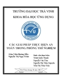
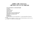
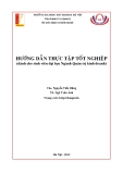
TGF-b1, IL-10, and IL-12. TAMs were popularly stained with HLA-DR, IL-10, sporadically stained with TGF-b1, negatively stained with IL-12, indicating that TAMs were activated without classic M1 or M2 phenotype (Fig. 2).
Relationship between CD68TFHotspot and clinicopathologic characteristics We used the c2 test to assess the relationship between the TAMs and clinicopathologic characteristics. The results showed that the CD68TFHotspot was inversely correlated with TNM stage, the presence of hepatic metastasis, and pathologic classification (Table 1). When hepatic metastasis status was cut into the following three patterns, the CD68TFHotspot was also highly corre- lated with the status of hepatic metastasis: no hepatic metastasis (stage IIIB colon cancer without liver metas- tasis within 5 years of follow-up), metachronous hepatic metastasis (stage IIIB colon cancer with liver metastasis within 5 years of follow-up), and synchronous liver metastasis (stage IV colon cancer with liver metastasis before palliative surgery).
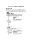
containing small areas among which the infiltration of CD68-positive cells was considerably above the average level of CD68-positive cells was defined as CD68 hot- spots (CD68TFHotspot) [36]. All sections were evaluated far from necrosis areas and H.E. staining was reviewed in case of uncertainty. The CD68TFHotspot of the two highest view fields measured at ×200 magnification was semi-quantitatively graded as no/weak (grade 1), moder- ate (grade 2), strong/robust (grade 3), and massive infil- tration (grade 4). Tumors classified as 1 included completely negative specimens, as well as specimens containing some scattered CD68-positive cells along the tumor margin. Tumors were classified as 2 when CD68 staining was continuous along the tumor margin, but was not extended from the tumor front more than one cell layer on average. CD68 staining that, on average, extended 2-3 cell layers from the tumor margin over the whole section was classified as 3, whereas to be classi- fied as 4, CD68 staining extended several cell layers from the tumor margin in all fields. Each section was scored independently by two independent observers. Interobserver agreements for the CD68TFHotspot were 81%. Disagreements were re-evaluated until a consensus decision was made.
sites of primary
the
Survival analyses By the end of the 5-year follow-up, 68 of patients with stage IIIB colon carcinoma were alive, thus the 5-year survival rate was 69.4%. Based on univariate analysis, including all stage IIIB patients applicable to survival analyses (n = 98), age, gender, tumor invasive depth, pathologic grade, and growth pattern showed no prog- nostic significance for OS and LMFS (Table 2). In con- trast, tumors, pathologic classification, and hepatic metastasis were predictors for OS. The CD68TFHotspot was highly correlated to OS (P = 0.001; log rank test; data not shown), but not LMFS (P = 0.221; log rank test; data not shown).
Statistical analysis The relationship between the various clinicopathologic characteristics and the CD68TFHotspot parameters were compared and analyzed using c2 tests, likelihood ratio, and linear-by-linear association, as appropriate. The cumulative survival time was computed using the Kaplan-Meier method and compared by the log-rank test. Univariate and multivariate analyses were based on the Cox proportional hazards regression model. A two- tailed P < 0.05 was considered to be statistically signifi- cant. All statistical analyses were performed using SPSS 13.0 software for Windows (SPSS Inc., Chicago, IL, USA).
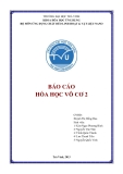
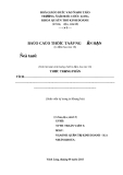
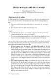
For further analysis, the grade data of the CD68TFHot- spot were divided into 2 groups (grade 1 and 2 versus 3 and 4) according to Forssell’s protocol [36]. Therefore, cases were regrouped into CD68TFHotspot high (3 and 4) versus CD68TFHotspot low (1 and 2) macrophage infiltra- tion. Kaplan-Meier survival curves were then plotted to further investigate the association with OS. The log- rank statistic was used to compare survival rates. There was a positive association between the CD68TFHotspot group and both OS (P < 0.001) and LMFS (P = 0.037; Fig. 3).
Page 3 of 9 Zhou et al. Journal of Translational Medicine 2010, 8:13 http://www.translational-medicine.com/content/8/1/13
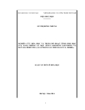
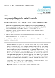
Results CD68 expression TAMs were stained brown in the cytoplasm. The major- ity of CD68-positive cells were located in the stroma, and in particular, along the invasive front. CD68-positive cells were mostly in apparent direct contact with or immediately adjacent to tumor cells lining the invasive front. Although most areas along the invasive front dis- played a fairly homogeneous CD68+ infiltration pattern, there were also tumors containing small areas that showed CD68 infiltration considerably above the average grade (CD68TFHotspot). The CD68TFHotspot was semi- quantitatively graded from 1-4 (Fig. 1).
Multivariate Cox proportional hazards analysis Whether or not the CD68TFHotspot group could serve as an independent predictor of OS and LMFS was ana- lyzed. A multivariate Cox proportional hazards analysis was performed, including gender, age, sites of primary tumors, invasive depth, grade, pathologic classifications,
To identify the phenotype of TAMs, a group of conse- cutive sections was used to stain with CD68, HLA-DR,
Page 4 of 9 Zhou et al. Journal of Translational Medicine 2010, 8:13 http://www.translational-medicine.com/content/8/1/13
Figure 1 Representative pictures of CD68TFHotspot in colon cancer patients (200× magnification). Different grades of macrophage infiltration along the tumor front were examined with immunohistochemical assay: A, no/low, B, moderate, C, high, and D, massive. Arrows point at tumor front.
Figure 2 Representative images of macrophage phenotypes in colon cancer on consecutive sections. Arrows point at tumor front.
Table 1 Correlation between CD68TFHotspot and clinicopathologic characteristics.
Page 5 of 9 Zhou et al. Journal of Translational Medicine 2010, 8:13 http://www.translational-medicine.com/content/8/1/13
Variable P value CD68TFHotspot -/+ ++ +++ + 1 3 4 2 Gender Male 21 37 23 0.939 13 Female 13 27 15 11 Age (years)
Discussion By analyzing the relationship between the density of TAMs and the potential of hepatic metastasis and survi- val, this study showed that a higher density of macro- phages in the invasive front of colon cancer was associated with a higher 5-year survival rate. Most importantly, the CD68TFHotspot was associated with both the incidence of hepatic metastasis and the interval between colon resection and the occurrence of hepatic metastasis.
In contrast to other solid tumors, such as breast can- cer, most studies have shown that TAMs, especially IL- 12-positive TAMs, inhibit the progression of colon can- cers [36-39,41-44]. For example, in Forssell’s study [36] the higher macrophage infiltration along the tumor front correlated with improved survival in colon cancer compared to rectal cancer. In the current study, the Cox model indicated that the CD68TFHotspot was indepen- dently prognostic. A higher 5-year survival rate after radical resection occurred in patients with a higher macrophage infiltration in the invasive front (81.0%) than in those with a lower macrophage infiltration (48.6%), which is in agreement with the previous studies [36-39].
*: p < 0.05. a: Likelihood ratio. b: Exact linear-by-linear association test.
it
The mechanisms behind the antitumor effects of TAMs have not been fully elucidated and could poten- tially be ascribed to the M1 phenotype, which is in part controlled by the CD4+T cells and the death of cancer cells [45-47]. TAMs with the M1 phenotype are charac- terized by a high capacity to present antigen, high IL-12 and IL-23 production, and high production of toxic intermediates, such as nitric oxide and reactive oxygen intermediates. Thus, TAMs with the M1 phenotype are generally considered potent effector cells which kill tumor cells [48-51]. In fact, TAMs showed a spectrum from M1 to M2 phenotypes in murine colon adenocar- cinoma tumors [52]. This study showed that TAMs expressed with HLA-DR and IL-10 rather than TGF-b1 and IL-12, consistent with the previous observation [52]. Although an abundance of evidence relevant to the molecular mechanisms underlying the anti-tumor effect is still of macrophages has been documented, unknown how TAMs exert a protective effect, except that one recent study indicated that TAMs reduce the development of peritoneal colorectal carcinoma metas- tases [36-39,41-44,53]. The current study analyzed the relationship between the infiltration of TAMs and hepa- tic metastasis. The results showed that a higher density of TAMs in the invasive front was associated with lower synchronous and metachronous hepatic metas- tases. Since hepatic metastasis of colon cancer is a key prognostic factor, this study might partly explain the
liver metastasis, growth patterns, and CD68TFHotspot groups. the high In stage IIIB colon cancers, CD68TFHotspot group had a significantly lower risk for OS (hazard ratio [HR], 0.433; 95% confidence interval [CI], 0.194-0.966) and LMFS (HR, 0.265; 95% CI, 0.078- 0.900) than did the low CD68TFHotspot group. Liver metastasis (HR, 8.144; 95% CI, 3.276-20.250) was an independent prognostic factor for OS. Additionally, patients with left colon cancer were prone to have a longer OS, whereas pathologic classification was not associated with OS (Table 3).
17 26 22 0.195 15 < 60 ≥ 60 17 38 16 9 Sites of primary tumors 0.107 Left Right 14 20 40 24 25 13 16 8 TNM stages IIIB 18 46 17 0.025* 17 IV 16 18 21 7 Invasive depth T3 30 53 31 0.422a 17 T4 4 11 7 7 Hepatic metastasis(1) No 14 42 13 0.004* 16 Yes 20 22 25 8 Hepatic metastasis(2) No 14 42 13 0.001*b 16 Metachronous 4 4 4 1 Synchronous 16 18 21 7 Grade 0.124b G1 G2 1 21 1 48 1 23 0 21 G3 11 14 14 2 G4 1 1 0 1 Pathologic classification Papillary + tubular 25 57 28 0.022*a 23 Mucoid + signet ring 9 7 10 1 Growth pattern 0.071 Pushing Infiltrating 8 26 18 46 19 19 8 16
Table 2 Univariate analyses of factors associated with OS and LMFS.
Page 6 of 9 Zhou et al. Journal of Translational Medicine 2010, 8:13 http://www.translational-medicine.com/content/8/1/13
Univariate analysis, Cox proportional hazards regression model. Abbreviations: HR, hazard ratio; CI, confidence interval; NA, not assessment. *: p < 0.05; **: p < 0.001.
reason that macrophage infiltration improves the prog- nosis of patients with colon cancer.
metastasis, showing that the immune microenvironment of the primary tumor modifies the metastatic potential of colon cancer, and the function of TAMs is change- able in different tumor microenvironment [61].
Most immune cells, such as CD45RO+T cells, CD3 +T cells, NK cells, TAMs, and even Treg cells, have shown a protective effect when infiltrated into colon cancer tissue [62-67]. Additionally, an autoimmune response is associated with the efficacy of biochem- otherapy (GOLFIG regimen) for colon cancer [68,69]. The current study has given additional evidence that macrophage infiltration is involved in the inhibition of hepatic metastasis. These data indicate that colon can- cer is an immunogenic tumor. Therefore, more
The molecular mechanisms underlying hepatic metas- tasis of colon cancers is poorly understood. Traditional clinicopathologic indices for hepatic metastasis of color- ectal cancer, which include the depth of invasion, the presence of venous invasion, and lymph node metastasis, have only limited prognostic value [54]. Although multi- ple markers, such as CD10, CD44, VEGF, TGF-a, have been shown to be correlated with hepatic metastasis, the predictive efficacy of these markers is still unclear [55-60]. In the current study, a higher density of TAMs in the invasive front was associated with lower synchro- nous hepatic metastasis and lower metachronous hepatic
Variable OS (n = 98) LMFS (n = 98) HR, (95% CI) P value HR, (95% CI) P value 1.157 (0.562-2.381) 0.693 0.416 (0.114-1.510) 0.182 Gender (female vs. male) Age (< 60 y vs. ≥ 60 y) 0.732 (0.352-1.519) 0.402 0.704 (0.230-2.153) 0.538 Invasive depth (T4 vs. T3) 1.023 (0.392-2.674) 0.962 0.902 (0.200-4.068) 0.893 Sites of primary tumors (right vs. left) 2.271 (1.093-4.717) 0.028* 0.815 (0.267-2.491) 0.720 Grade (G3 vs. G2 vs. G1) 1.519 (0.715-3.224) 0.277 1.036 (0.324-3.311) 0.953 0.023* 1.148 (0.316-4.171) 0.834 Pathologic classification (mucoid + signet ring vs. papillary + tubular) 2.415 (1.129-5.168) Growth pattern (infiltrating vs. pushing) 0.817 (0.389-1.718) 0.595 2.709 (0.600-12.223) 0.195 0.568 (0.393-0.822) 0.003* 0.594 (0.344-1.025) 0.061 CD68TFHotspot (4 vs. 3 vs. 2 vs.1) 0.288 (0.139-0.600) 0.001* 0.324 (0.106-0.991) 0.048* CD68TFHotspot group (high vs. low) Hepatic metastasis (yes vs. no) 5.852 (2.737-12.511) 0.000** NA NA
Figure 3 Kaplan–Meier analysis of overall survival (A) and liver metastasis-free survival (B) for CD68TFHotspot group. The patients with a higher CD68TFHotspot group (solid lines) were associated with longer 5-year overall survival and liver metastasis-free survival than those with a lower CD68TFHotspot group (dashed lines).
Table 3 Multivariate analyses of factors associated with OS and LMFS
Page 7 of 9 Zhou et al. Journal of Translational Medicine 2010, 8:13 http://www.translational-medicine.com/content/8/1/13
Multivariate analysis, Cox proportional hazard regression model. Abbreviations: HR, hazard ratio; CI, confidence interval; NA, not assessment. *: p < 0.05; **: p < 0.001.
analyzed the results. ZXS and ZYX conceived the study, participated in the design, and coordinated and helped draft the manuscript. All authors read and approved the final manuscript.
attention should be paid to exploiting the immune response in an effort to improve conventional therapy for colon cancer [70].
Competing interests The authors declare that they have no competing interests.
Received: 3 November 2009 Accepted: 8 February 2010 Published: 8 February 2010
2.
3.
Additionally, our study main aim is to find if there any relationship between macrophages and liver metastasis in colon cancer which was cut into the following three patterns: no hepatic metastasis, metachronous and syn- chronous liver metastasis. We decided to choose single stage IIIB colon cancer which is the biggest group in our center colon resource database to avoid the influ- ence of different stages factor on relationship between macrophages and liver metastasis. Although this consti- tution minimized confounding factors, it cannot com- pletely represent ordinary setup, so our results, and as such, should be viewed with some caution.
4.
5.
6.
Variable OS (n = 98) LMFS (n = 98) HR, (95% CI) P value HR, (95% CI) P value 1.954 (0.841-4.538) 0.119 0.333 (0.083-1.335) 0.121 Gender (female vs. male) Age (< 60 y vs. ≥ 60 y) 0.504 (0.227-1.116) 0.091 0.881 (0.267-2.906) 0.835 Invasive depth (T4 vs. T3) 1.941 (0.693-5.436) 0.207 0.846 (0.171-4.190) 0.838 Site of primary tumors (right vs. left) 2.184 (0.981-4.859) 0.056 1.009 (0.298-3.414) 0.989 Grade (G3 vs. G2 vs. G1) 1.224 (0.457-3.281) 0.688 1.616 (0.345-7.575) 0.543 0.125 0.537 (0.071-4.061) 0.547 Pathologic Classification (mucoid + signet ring vs. papillary + tubular) 2.364 (0.787-7.100) 0.419 0.224 Growth patterns (infiltrating vs. pushing) 0.700 (0.295-1.662) 2.650 (0.551-12.746) 0.433 (0.194-0.966) 0.041* 0.265 (0.078-0.900) 0.033* CD68TFHotspot group (high vs. low) NA Liver metastasis (yes vs. no) 8.144 (3.276-20.250) 0.000** NA
Conclusion This study demonstrated that TAMs infiltrated in the invasive front are associated with improvement in both hepatic metastasis and OS in colon cancer, implying that TAMs have protective potential in colon cancers and might serve as a novel therapeutic target.
7.
8.
Acknowledgements This study was supported by research grants from the National Nature Science Foundation (30972882) and the Nature Science Foundation of Guangdong Province, China (9151008901000149).
9.
References 1. Mayo SC, Pawlik TM: Current management of colorectal hepatic metastasis. Expert Rev Gastroenterol Hepatol 2009, 3(2):131-144. Duffy MJ, van Dalen A, Haglund C, Hansson L, Holinski-Feder E, Klapdor R, Lamerz R, Peltomaki P, Sturgeon C, Topolcan O: Tumour markers in colorectal cancer: European Group on Tumour Markers (EGTM) guidelines for clinical use. Eur J Cancer 2007, 43(9):1348-1360. Shah A, Alberts S, Adam R: Accomplishments in 2007 in the management of curable metastatic colorectal cancer. Gastrointest Cancer Res 2008, 2(3 Suppl):S13-18. Shrivastav A, Varma S, Saxena A, DeCoteau J, Sharma RK: N- myristoyltransferase: a potential novel diagnostic marker for colon cancer. J Transl Med 2007, 5:58. Lind GE, Ahlquist T, Kolberg M, Berg M, Eknaes M, Alonso MA, Kallioniemi A, Meling GI, Skotheim RI, Rognum TO, Thiis-Evensen E, Lothe RA: Hypermethylated MAL gene - a silent marker of early colon tumorigenesis. J Transl Med 2008, 6:13. Eccles SA, Welch DR: Metastasis: recent discoveries and novel treatment strategies. Lancet 2007, 369(9574):1742-1757. Li CY, Li BX, Liang Y, Peng RQ, Ding Y, Xu DZ, Zhang X, Pan ZZ, Wan DS, Zeng YX, Zhu XF, Zhang XS: Higher percentage of CD133+ cells is associated with poor prognosis in colon carcinoma patients with stage IIIB. J Transl Med 2009, 7:56. Gao YF, Peng RQ, Li J, Ding Y, Zhang X, Wu XJ, Pan ZZ, Wan DS, Zeng YX, Zhang XS: The paradoxical patterns of expression of indoleamine 2,3- dioxygenase in colon cancer. J Transl Med 2009, 7:71. Kopfstein L, Christofori G: Metastasis: cell-autonomous mechanisms versus contributions by the tumor microenvironment. Cell Mol Life Sci 2006, 63(4):449-468.
10. Hallam S, Escorcio-Correia M, Soper R, Schultheiss A, Hagemann T:
Activated macrophages in the tumour microenvironment-dancing to the tune of TLR and NF-kappaB. J Pathol 2009, 219(2):143-152.
Author details 1State Key Laboratory of Oncology in South China, Cancer Center, Sun Yat- Sen University, 651 Dongfeng R E, 510060, Guangzhou, China. 2Biotherapy Center, Cancer Center, Sun Yat-Sen University, 651 Dongfeng R E, 510060, Guangzhou, China. 3Department of Colorectal Oncology, Cancer Center, Sun Yat-Sen University, 651 Dongfeng R E, 510060, Guangzhou, China. 4Department of Pathology, Cancer Center, Sun Yat-Sen University, 651 Dongfeng R E, 510060, Guangzhou, China.
11. Hagemann T, Lawrence T, McNeish I, Charles KA, Kulbe H, Thompson RG, Robinson SC, Balkwill FR: “Re-educating” tumor-associated macrophages by targeting NF-kappaB. J Exp Med 2008, 205(6):1261-1268.
12. Hagemann T, Biswas SK, Lawrence T, Sica A, Lewis CE: Regulation of
macrophage function in tumors: the multifaceted role of NF-kappaB. Blood 2009, 113(14):3139-3146.
Authors’ contributions WXJ, DY, ZQM, PZZ, and WDS carried out the case collection; ZQ, XQ, and HJH carried out the immunohistochemical staining; and PRQ and ZX
32. Welsh TJ, Green RH, Richardson D, Waller DA, O’Byrne KJ, Bradding P:
13. Condeelis J, Pollard JW: Macrophages: obligate partners for tumor cell
14.
33.
migration, invasion, and metastasis. Cell 2006, 124(2):263-266. Lin EY, Nguyen AV, Russell RG, Pollard JW: Colony-stimulating factor 1 promotes progression of mammary tumors to malignancy. J Exp Med 2001, 193(6):727-740.
Macrophage and mast-cell invasion of tumor cell islets confers a marked survival advantage in non-small-cell lung cancer. J Clin Oncol 2005, 23(35):8959-8967. Kataki A, Scheid P, Piet M, Marie B, Martinet N, Martinet Y, Vignaud JM: Tumor infiltrating lymphocytes and macrophages have a potential dual role in lung cancer by supporting both host-defense and tumor progression. J Lab Clin Med 2002, 140(5):320-328.
34. Richardsen E, Uglehus RD, Due J, Busch C, Busund LT: The prognostic
35.
36.
16.
15. Dave SS, Wright G, Tan B, Rosenwald A, Gascoyne RD, Chan WC, Fisher RI, Braziel RM, Rimsza LM, Grogan TM, Miller TP, LeBlanc M, Greiner TC, Weisenburger DD, Lynch JC, Vose J, Armitage JO, Smeland EB, Kvaloy S, Holte H, Delabie J, Connors JM, Lansdorp PM, Ouyang Q, Lister TA, Davies AJ, Norton AJ, Muller-Hermelink HK, Ott G, Campo E, Montserrat E, Wilson WH, Jaffe ES, Simon R, Yang L, Powell J, Zhao H, Goldschmidt N, Chiorazzi M, Staudt LM: Prediction of survival in follicular lymphoma based on molecular features of tumor-infiltrating immune cells. N Engl J Med 2004, 351(21):2159-2169. Siveen KS, Kuttan G: Role of macrophages in tumour progression. Immunol Lett 2009, 123(2):97-102.
impact of M-CSF, CSF-1 receptor, CD68 and CD3 in prostatic carcinoma. Histopathology 2008, 53(1):30-38. Lissbrant IF, Stattin P, Wikstrom P, Damber JE, Egevad L, Bergh A: Tumor associated macrophages in human prostate cancer: relation to clinicopathological variables and survival. Int J Oncol 2000, 17(3):445-451. Forssell J, Oberg A, Henriksson ML, Stenling R, Jung A, Palmqvist R: High macrophage infiltration along the tumor front correlates with improved survival in colon cancer. Clin Cancer Res 2007, 13(5):1472-1479. 37. Nagorsen D, Voigt S, Berg E, Stein H, Thiel E, Loddenkemper C: Tumor-
18.
38.
17. Nishie A, Ono M, Shono T, Fukushi J, Otsubo M, Onoue H, Ito Y, Inamura T, Ikezaki K, Fukui M, Iwaki T, Kuwano M: Macrophage infiltration and heme oxygenase-1 expression correlate with angiogenesis in human gliomas. Clin Cancer Res 1999, 5(5):1107-1113. Leek RD, Lewis CE, Whitehouse R, Greenall M, Clarke J, Harris AL: Association of macrophage infiltration with angiogenesis and prognosis in invasive breast carcinoma. Cancer Res 1996, 56(20):4625-4629.
19. Pollard JW: Macrophages define the invasive microenvironment in breast
39.
20.
cancer. J Leukoc Biol 2008, 84(3):623-630. Shabo I, Stål O, Olsson H, Doré S, Svanvik J: Breast cancer expression of CD163, a macrophage scavenger receptor, is related to early distant recurrence and reduced patient survival. Int J Cancer 2008, 123(4):780-786.
infiltrating macrophages and dendritic cells in human colorectal cancer: relation to local regulatory T cells, systemic T-cell response against tumor-associated antigens and survival. J Transl Med 2007, 5:62. Funada Y, Noguchi T, Kikuchi R, Takeno S, Uchida Y, Gabbert HE: Prognostic significance of CD8+ T cell and macrophage peritumoral infiltration in colorectal cancer. Oncol Rep 2003, 10(2):309-313. Sugita J, Ohtani H, Mizoi T, Saito K, Shiiba K, Sasaki I, Matsuno S, Yagita H, Miyazawa M, Nagura H: Close association between Fas ligand (FasL; CD95L)- positive tumor-associated macrophages and apoptotic cancer cells along invasive margin of colorectal carcinoma: a proposal on tumor-host interactions. Jpn J Cancer Res 2002, 93(3):320-328. 40. National Comprehensive Cancer Network. http://www.nccn.org/
21. Hanada T, Nakagawa M, Emoto A, Nomura T, Nasu N, Nomura Y:
41.
22.
professionals/physician_gls/PDF/colon.pdf. Kuniyasu H, Sasaki T, Sasahira T, Ohmori H, Takahashi T: Depletion of tumor-infiltrating macrophages is associated with amphoterin expression in colon cancer. Pathobiology 2004, 71(3):129-136.
23.
24.
43.
25.
44.
42. Bacman D, Merkel S, Croner R, Papadopoulos T, Brueckl W, Dimmler A: TGF- beta receptor 2 downregulation in tumour-associated stroma worsens prognosis and high-grade tumours show more tumour-associated macrophages and lower TGF-beta1 expression in colon carcinoma: a retrospective study. BMC Cancer 2007, 7:156. Inoue Y, Nakayama Y, Minagawa N, Katsuki T, Nagashima N, Matsumoto K, Shibao K, Tsurudome Y, Hirata K, Nagata N, Itoh H: Relationship between interleukin-12-expressing cells and antigen-presenting cells in patients with colorectal cancer. Anticancer Res 2005, 25(5):3541-3546. Tan SY, Fan Y, Luo HS, Shen ZX, Guo Y, Zhao LJ: Prognostic significance of cell infiltrations of immunosurveillance in colorectal cancer. World J Gastroenterol 2005, 11(8):1210-1214.
Prognostic value of tumor-associated macrophage count in human bladder cancer. Int J Urol 2000, 7(7):263-269. Shabo I, Olsson H, Sun XF, Svanvik J: Expression of the macrophage antigen CD163 in rectal cancer cells is associated with early local recurrence and reduced survival time. Int J Cancer 2009, 125(8):1826-1831. Salvesen HB, Akslen LA: Significance of tumour-associated macrophages, vascular endothelial growth factor and thrombospondin-1 expression for tumour angiogenesis and prognosis in endometrial carcinomas. Int J Cancer 1999, 84(5):538-543. Lee CH, Espinosa I, Vrijaldenhoven S, Subramanian S, Montgomery KD, Zhu S, Marinelli RJ, Peterse JL, Poulin N, Nielsen TO, West RB, Gilks CB, Rijn van de M: Prognostic significance of macrophage infiltration in leiomyosarcomas. Clin Cancer Res 2008, 14(5):1423-1430. Jensen TO, Schmidt H, Møller HJ, Høyer M, Maniecki MB, Sjoegren P, Christensen IJ, Steiniche T: Macrophage markers in serum and tumor have prognostic impact in American Joint Committee on Cancer stage I/ II melanoma. J Clin Oncol 2009, 27(20):3330-3337.
45. Umemura N, Saio M, Suwa T, Kitoh Y, Bai J, Nonaka K, Ouyang GF,
26. Ohno S, Inagawa H, Dhar DK, Fujii T, Ueda S, Tachibana M, Suzuki N,
Okada M, Balazs M, Adany R, Shibata T, Takami T: Tumor-infiltrating myeloid-derived suppressor cells are pleiotropic-inflamed monocytes/ macrophages that bear M1- and M2-type characteristics. J Leukoc Biol 2008, 83(5):1136-1144.
46. Weigert A, Tzieply N, von Knethen A, Johann AM, Schmidt H, Geisslinger G,
27.
Inoue M, Soma G, Nagasue N: The degree of macrophage infiltration into the cancer cell nest is a significant predictor of survival in gastric cancer patients. Anticancer Res 2003, 23(6D):5015-5022. Tanaka Y, Kobayashi H, Suzuki M, Kanayama N, Suzuki M, Terao T: Upregulation of bikunin in tumor-infiltrating macrophages as a factor of favorable prognosis in ovarian cancer. Gynecol Oncol 2004, 94(3):725-734.
Brüne B: Tumor cell apoptosis polarizes macrophages role of sphingosine- 1-phosphate. Mol Biol Cell 2007, 18(10):3810-3819. 47. DeNardo DG, Barreto JB, Andreu P, Vasquez L, Tawfik D, Kolhatkar N,
28. Peng J, Ding T, Zheng LM, Shao JY: Influence of tumor-associated macrophages on progression and prognosis of nasopharyngeal carcinoma. Ai Zheng 2006, 25(11):1340-1345, [Article in Chinese].
Coussens LM: CD4(+) T cells regulate pulmonary metastasis of mammary carcinomas by enhancing protumor properties of macrophages. Cancer Cell 2009, 16(2):91-102.
29. Ding T, Xu J, Wang F, Shi M, Zhang Y, Li SP, Zheng L: High tumor-
48. Mantovani A, Sica A, Allavena P, Garlanda C, Locati M: Tumor-associated
30.
49.
macrophages and the related myeloid-derived suppressor cells as a paradigm of the diversity of macrophage activation. Hum Immunol 2009, 70(5):325-330. Sica A, Schioppa T, Mantovani A, Allavena P: Tumour-associated macrophages are a distinct M2 polarised population promoting tumour progression: potential targets of anti-cancer therapy. Eur J Cancer 2006, 42(6):717-727.
31.
50. Gordon S, Taylor PR: Monocyte and macrophage heterogeneity. Nat Rev
Immunol 2005, 5(12):953-964.
51. Pollard JW: Trophic macrophages in development and disease. Nat Rev
Immunol 2009, 9(4):259-270.
infiltrating macrophage density predicts poor prognosis in patients with primary hepatocellular carcinoma after resection. Hum Pathol 2009, 40(3):381-389. Li YW, Qiu SJ, Fan J, Gao Q, Zhou J, Xiao YS, Xu Y, Wang XY, Sun J, Huang XW: Tumor-infiltrating macrophages can predict favorable prognosis in hepatocellular carcinoma after resection. J Cancer Res Clin Oncol 2009, 135(3):439-449. Kawai O, Ishii G, Kubota K, Murata Y, Naito Y, Mizuno T, Aokage K, Saijo N, Nishiwaki Y, Gemma A, Kudoh S, Ochiai A: Predominant infiltration of macrophages and CD8(+) T Cells in cancer nests is a significant predictor of survival in stage IV nonsmall cell lung cancer. Cancer 2008, 113(6):1387-1395.
Page 8 of 9 Zhou et al. Journal of Translational Medicine 2010, 8:13 http://www.translational-medicine.com/content/8/1/13
52. Umemura N, Saio M, Suwa T, Kitoh Y, Bai J, Nonaka K, Ouyang GF,
Okada M, Balazs M, Adany R, Shibata T, Takami T: Tumor-infiltrating myeloid- derived suppressor cells are pleiotropic-inflamed monocytes/ macrophages that bear M1- and M2-type characteristics. J Leukoc Biol 2008, 83(5):1136-1144.
70.
Cusi MG, Tassone P, Francini G: Immunity feedback and clinical outcome in colon cancer patients undergoing chemoimmunotherapy with gemcitabine + FOLFOX followed by subcutaneous granulocyte macrophage colony- stimulating factor and aldesleukin (GOLFIG-1 Trial). Clin Cancer Res 2008, 14(13):4192-499. Stout RD, Watkins SK, Suttles J: Functional plasticity of macrophages: in situ reprogramming of tumor-associated macrophages. J Leukoc Biol 2009, 86(5):1105-1109.
53. Bij van der GJ, Bögels M, Oosterling SJ, Kroon J, Schuckmann DT, de Vries HE, Meijer S, Beelen RH, van Egmond M: Tumor infiltrating macrophages reduce development of peritoneal colorectal carcinoma metastases. Cancer Lett 2008.
54. Bird NC, Mangnall D, Majeed AW: Biology of colorectal liver metastases: A
55.
doi:10.1186/1479-5876-8-13 Cite this article as: Zhou et al.: The density of macrophages in the invasive front is inversely correlated to liver metastasis in colon cancer. Journal of Translational Medicine 2010 8:13.
review. J Surg Oncol 2006, 94(1):68-80. Fang YJ, Lu ZH, Wang GQ, Pan ZZ, Zhou ZW, Yun JP, Zhang MF, Wan DS: Elevated expressions of MMP7, TROP2, and survivin are associated with survival, disease recurrence, and liver metastasis of colon cancer. Int J Colorectal Dis 2009, 24(8):875-884.
57.
56. Barozzi C, Ravaioli M, D’Errico A, Grazi GL, Poggioli G, Cavrini G, Mazziotti A, Grigioni WF: Relevance of biologic markers in colorectal carcinoma: a comparative study of a broad panel. Cancer 2002, 94(3):647-657. Kato H, Semba S, Miskad UA, Seo Y, Kasuga M, Yokozaki H: High expression of PRL-3 promotes cancer cell motility and liver metastasis in human colorectal cancer: a predictive molecular marker of metachronous liver and lung metastases. Clin Cancer Res 2004, 10(21):7318-7128. 58. Ohji Y, Yao T, Eguchi T, Yamada T, Hirahashi M, Iida M, Tsuneyoshi M:
59.
Evaluation of risk of liver metastasis in colorectal adenocarcinoma based on the combination of risk factors including CD10 expression: multivariate analysis of clinicopathological and immunohistochemical factors. Oncol Rep 2007, 17(3):525-530. Tanami H, Tsuda H, Okabe S, Iwai T, Sugihara K, Imoto I, Inazawa J: Involvement of cyclin D3 in liver metastasis of colorectal cancer, revealed by genome-wide copy-number analysis. Lab Invest 2005, 85(9):1118-1129.
60. Hu H, Sun L, Guo C, Liu Q, Zhou Z, Peng L, Pan J, Yu L, Lou J, Yang Z,
Zhao P, Ran Y: Tumor cell-microenvironment interaction models coupled with clinical validation reveal CCL2 and SNCG as two predictors of colorectal cancer hepatic metastasis. Clin Cancer Res 2009, 15(17):5485-5493.
61. Nonaka K, Saio M, Suwa T, Frey AB, Umemura N, Imai H, Ouyang GF,
62.
Osada S, Balazs M, Adany R, Kawaguchi Y, Yoshida K, Takami T: Skewing the Th cell phenotype toward Th1 alters the maturation of tumor- infiltrating mononuclear phagocytes. J Leukoc Biol 2008, 84(3):679-688. Laghi L, Bianchi P, Miranda E, Balladore E, Pacetti V, Grizzi F, Allavena P, Torri V, Repici A, Santoro A, Mantovani A, Roncalli M, Malesci A: CD3+ cells at the invasive margin of deeply invading (pT3-T4) colorectal cancer and risk of post-surgical metastasis: a longitudinal study. Lancet Oncol 2009, 10(9):877-884.
63. Pagès F, Berger A, Camus M, Sanchez-Cabo F, Costes A, Molidor R,
64.
65.
Mlecnik B, Kirilovsky A, Nilsson M, Damotte D, Meatchi T, Bruneval P, Cugnenc PH, Trajanoski Z, Fridman WH, Galon J: Effector memory T cells, early metastasis, and survival in colorectal cancer. N Engl J Med 2005, 353(25):2654-2666. Salama P, Phillips M, Grieu F, Morris M, Zeps N, Joseph D, Platell C, Iacopetta B: Tumor-infiltrating FOXP3+ T regulatory cells show strong prognostic significance in colorectal cancer. J Clin Oncol 2009, 27(2):186-192. Sandel MH, Speetjens FM, Menon AG, Albertsson PA, Basse PH, Hokland M, Nagelkerke JF, Tollenaar RA, Velde van de CJ, Kuppen PJ: Natural killer cells infiltrating colorectal cancer and MHC class I expression. Mol Immunol 2005, 42(4):54154-6.
Page 9 of 9 Zhou et al. Journal of Translational Medicine 2010, 8:13 http://www.translational-medicine.com/content/8/1/13
Submit your next manuscript to BioMed Central and take full advantage of:
66. Ohtani H: Focus on TILs: prognostic significance of tumor infiltrating lymphocytes in human colorectal cancer. Cancer Immun 2007, 7:4. 67. Gout S, Huot J: Role of cancer microenvironment in metastasis: focus on
colon cancer. Cancer Microenviron 2008, 1(1):69-83.
• Convenient online submission
68. Correale P, Cusi MG, Tsang KY, Del Vecchio MT, Marsili S, Placa ML,
• Thorough peer review
• No space constraints or color figure charges
• Immediate publication on acceptance
• Inclusion in PubMed, CAS, Scopus and Google Scholar
Intrivici C, Aquino A, Micheli L, Nencini C, Ferrari F, Giorgi G, Bonmassar E, Francini G: Chemo-immunotherapy of metastatic colorectal carcinoma with gemcitabine plus FOLFOX 4 followed by subcutaneous granulocyte macrophage colony- stimulating factor and interleukin-2 induces strong immunologic and antitumor activity in metastatic colon cancer patients. J Clin Oncol 2005, 23(35):8950-8958.
• Research which is freely available for redistribution
69. Correale P, Tagliaferri P, Fioravanti A, Del Vecchio MT, Remondo C, Montagnani F, Rotundo MS, Ginanneschi C, Martellucci I, Francini E,
Submit your manuscript at www.biomedcentral.com/submit