
BioMed Central
Page 1 of 9
(page number not for citation purposes)
Virology Journal
Open Access
Research
Murine leukemia virus (MLV) replication monitored with
fluorescent proteins
Katja Sliva1, Otto Erlwein1, Alexandra Bittner1 and Barbara S Schnierle*1,2
Address: 1Institute for Biomedical Research, Georg-Speyer-Haus, Paul-Ehrlich-Str. 42-44, 60596 Frankfurt/Main, Germany and 2Paul-Ehrlich-
Institute, Paul-Ehrlich-Str. 51-59, 63225 Langen, Germany
Email: Katja Sliva - slika@pei.de; Otto Erlwein - erlw1@aol.com; Alexandra Bittner - alexandrabittner@web.de;
Barbara S Schnierle* - schba@pei.de
* Corresponding author
Abstract
Background: Cancer gene therapy will benefit from vectors that are able to replicate in tumor
tissue and cause a bystander effect. Replication-competent murine leukemia virus (MLV) has been
described to have potential as cancer therapeutics, however, MLV infection does not cause a
cytopathic effect in the infected cell and viral replication can only be studied by immunostaining or
measurement of reverse transcriptase activity.
Results: We inserted the coding sequences for green fluorescent protein (GFP) into the proline-
rich region (PRR) of the ecotropic envelope protein (Env) and were able to fluorescently label MLV.
This allowed us to directly monitor viral replication and attachment to target cells by flow
cytometry. We used this method to study viral replication of recombinant MLVs and split viral
genomes, which were generated by replacement of the MLV env gene with the red fluorescent
protein (RFP) and separately cloning GFP-Env into a retroviral vector. Co-transfection of both
plasmids into target cells resulted in the generation of semi-replicative vectors, and the two color
labeling allowed to determine the distribution of the individual genomes in the target cells and was
indicative for the occurrence of recombination events.
Conclusions: Fluorescently labeled MLVs are excellent tools for the study of factors that influence
viral replication and can be used to optimize MLV-based replication-competent viruses or vectors
for gene therapy.
Background
Efficient and long-lasting gene delivery is the major chal-
lenge in the development of vectors for gene therapy. Rep-
lication-competent retroviruses (RCRs) encoding suicide
genes linked via an internal ribosome entry site (IRES)
offer a significant advantage over replication-deficient
vectors in cancer gene therapy, since they are able to
spread efficiently in vivo [1-4]. Uncontrolled virus spread
is, however, associated with serious risk of adverse events
due to viral-integration mutagenesis. Therefore, for a ther-
apeutic application, RCRs have to be equipped with addi-
tional safety features, e.g. transcription controllable by
exogenous agents or viral entry restricted to the diseased
cells. The selective delivery of a therapeutic gene by target-
ing retroviral entry would immensely reduce unfavorable
side effects and ease the clinical application of gene ther-
apy. The ecotropic MLV envelope protein does not recog-
nizes receptors on human cells. An obvious challenge has
Published: 20 December 2004
Virology Journal 2004, 1:14 doi:10.1186/1743-422X-1-14
Received: 26 November 2004
Accepted: 20 December 2004
This article is available from: http://www.virologyj.com/content/1/1/14
© 2004 Sliva et al; licensee BioMed Central Ltd.
This is an Open Access article distributed under the terms of the Creative Commons Attribution License (http://creativecommons.org/licenses/by/2.0),
which permits unrestricted use, distribution, and reproduction in any medium, provided the original work is properly cited.

Virology Journal 2004, 1:14 http://www.virologyj.com/content/1/1/14
Page 2 of 9
(page number not for citation purposes)
been to extend the host range of vectors carrying the eco-
tropic envelope glycoprotein to a predetermined human
cell type. This change in host range requires the inclusion
of a novel attachment site and the induction of fusion via
a novel receptor interaction. It has been shown before that
it is possible to modify ecotropic Env and change its bind-
ing specificity, however, the efficient triggering of the
membrane fusion or the escape from endosomes of viral
particles targeted to e.g. epidermal growth factor (EGF)-
receptor is still missing [5,6]. The further development of
such targeted vectors requires the understanding of the
mechanisms that are involved in adsorption and internal-
ization of retroviruses.
Investigating murine leukemia virus (MLV) replication is
technically inconvenient because MLV infection does not
cause a cytopathic effect in the infected cell. Viral replica-
tion can only be studied by immunostaining, measure-
ment of reverse transcriptase activity or syncytia
formation. We have developed a tool to simplify these
analyses. We generated an MLV tagged with a fluorescent
envelope protein, which allows viral replication and Env
attachment to target cells to be followed by flow cytome-
try. This method will be useful for optimizing RCRs or ret-
roviral vectors for gene therapy.
Results
Construction of GFP-tagged MLVs and their replication
We previously constructed a modified ecotropic murine
leukemia virus (Mo-MLV) bearing the green fluorescent
protein (GFP) from Aequoria victoria in its envelope. A rep-
lication competent ecotropic MLV variant was generated
(GFP-EMO) that had the 53 aas of the epidermal growth
factor (EGF) fused to the N-terminus of Env and the GFP
sequences inserted into the proline-rich region (PRR) [7].
We deleted the EGF sequences by replacing a Pfl MI frag-
ment of GFP-EMO with wt sequences. This resulted in a
replication-competent virus expressing the chimeric GFP-
Env protein (GFP-MOV) (Fig. 1A). NIH3T3 cells were
transfected with 10 µg plasmid DNA encoding GFP-MOV
or GFP-EMO using the calcium-phosphate procedure and
were cultured for 13 days. Viral replication was monitored
as GFP-positive cells by flow cytometry. As indicated in
Figure 1B, both viruses replicate with similar kinetics.
Untransfected NIH3T3 cells did not show green
fluorescence.
Sequestering of EGF-Env-containing viral particles has
been described before [8,9]. Viral particles containing
EGF-Env were rapidly trafficked to endosomes and
became degraded. This effect was dominant over the nor-
mal entry pathway, because mouse cells expressing the
ecotropic receptor and the EGF-receptor showed a severely
decreased infectivity of EGF-Env containing vectors [8].
We were interested, if replication competent GFP-EMO
might be useful to select viral variants able to escape the
degradation in the endosomes. Transfection of GFP-EMO
into cells expressing only the EGF-receptor (A431, COS-7)
did not result in viral replication (data not shown). There-
fore, GFP-EMO and GFP-MOV were transfected into FLY-
Jet cells [10], which express the human EGF-receptor and
the receptor for ecotropic MLV. Viral replication of GFP-
EMO could be observed in FLY-Jet cells, although strongly
delayed, after 10 days only 7.4 % of the cells were GFP-
positive. After 38 days, all cells were GFP-positive and the
N-terminus of the Env gene was analyzed by PCR amplifi-
cation of genomic DNA isolated from infected cells. Pre-
dominantly a band migrating faster than the GFP-EMO
fragment was amplified (Figure 1C), which was verified
by sequence analysis to contain wt Env sequences. The less
abundant, slower migrating fragments still contained the
EGF sequences in Env. This confirms the sequestering of
EGF-Env containing retroviral particles via the EGF-recep-
tor. The selection of viruses able to escape the endosomal
degradation was not possible and shows that degradation
of viral particles in the endosomes favors the selection of
wt Env-containing MLV, which escapes the sequestering
by EGF-receptor.
Cell binding of GFP-tagged MLV
Viral entry is initiated by the binding of the envelope pro-
tein (Env) to the retrovirus receptor at the target cell sur-
face. To test whether labeling of Env with GFP allows viral
attachment to be monitored, we incubated supernatants
of NIH3T3 cells producing GFP-EMO or GFP-MOV with
cells that either express mCAT, the receptor for ecotropic
MLV [11] (NIH3T3), do not express it (293T, A431) or do
express the human EGF receptor (A431). As illustrated in
Figure 2A, NIH3T3 cells incubated with cell culture super-
natants showed a shift to green fluorescence, indicating
specific binding of GFP-tagged Env to mCAT. The shift to
green fluorescence could not be increased by larger
amounts of viral supernatants or longer incubation times
(data not shown), which shows that already after 5 min.
all receptors are occupied by Env. For GFP-MOV superna-
tants a shift in fluorescence was only observed with
mCAT-expressing cells, while GFP-EMO supernatants also
produced a shift with A431 cells. This indicates additional
specific binding to the EGF receptor. The shift was more
pronounced on A431 cells than COS-7 cells, correlating
with the amount of EGF receptor expressed by the target
cells (data not shown).
The specificity of cell staining by supernatants containing
GFP-MOV was further examined using chronically Mo-
MLV-infected NIH3T3 cells (NIH3T3i-MLV). These cells
have only negligible numbers of mCAT molecules on the
cell surface, because Env expression leads to their reten-
tion within the cell (receptor interference). As expected,
NIH3T3i-MLV cells produced no shift when incubated
Virology Journal 2004, 1:14 http://www.virologyj.com/content/1/1/14
Page 3 of 9
(page number not for citation purposes)
with GFP-MOV supernatants (Fig. 2B). Furthermore,
binding of GFP-MOV supernatants could be inhibited by
preincubation of NIH3T3 target cells with a soluble Env
fragment containing the receptor binding domain (sRBD)
derived from the ecotropic Env [12], but not with the
equivalent sRBD derived from the amphotropic Env [12],
which binds to a different receptor (Fig. 2B). This shows
that GFP-tagging can be used to investigate Env-binding
properties by flow cytometry.
Replication of semi-replicative retroviral vectors
The size of a retroviral genome is limited to roughly 11 kb.
The capacity for the insertion of a therapeutic gene for
gene therapy is, however, increased by the use of semi-rep-
licative retroviral vectors (SRRVs), where the gag/pol and
env genes are split between two viral genomes. We con-
structed split viral genomes and used fluorescent proteins
to monitor the replication of the resulting SRRVs.
A packagable MLV Gag/Pol expression vector, GAG/POL-
RFP, was generated by deleting of the env gene and replac-
ing it with the red fluorescent protein (RFP) (Fig. 3). RFP
is encoded by the spliced mRNA and its expression can be
monitored by red fluorescence (Fig. 4C). The GFP-Env
protein was cloned into the retroviral vector pczCFG5
IEGZ (Lindemann, unpublished) (Fig. 3). This vector has
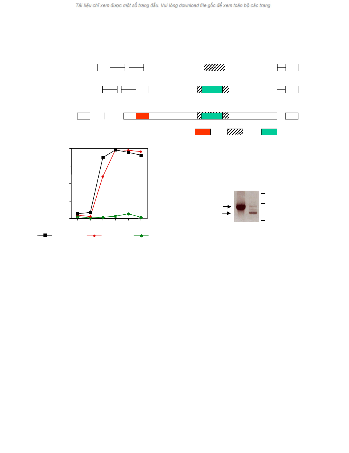
Generation and replication of the GFP-Env-tagged virusesFigure 1
Generation and replication of the GFP-Env-tagged viruses. (A) Schematic representation of the GFP-Env-tagged viruses. EGF,
epidermal growth factor; PRR, proline rich region; GFP, green fluorescent protein; L, signal peptide.(B) Viral replication kinetic
in transfected NIH3T3 cells monitored by the percentage of GFP-positive cells.(C) PCR analysis of genomic DNA from FLY-Jet
cells transfected with GFP-EMO. The N-terminal sequences of the EGF-Env gene were analyzed by PCR using the primers
MLV-5'-Env and BS-5. GFP-EMO plasmid DNA was used as a positive control and gave rise to a 900 bp fragment. Predomi-
nantly faster migrating fragments were amplified from genomic DNA (gDNA) of GFP-EMO transfected FLY-Jet cells 32 days
after transfection.
PRR
PRRPRR
PRR GFP
GFPGFP
GFPEGF
EGFEGF
EGF
LTR
LTRLTR
LTR LTR
LTRLTR
LTRL
LL
L
MLV
MLVMLV
MLV
GFP
GFPGFP
GFP-
--
-EMO
EMOEMO
EMO
LTR
LTRLTR
LTR LTR
LTRLTR
LTRL
LL
L
gag
gaggag
gag-
--
-pol
polpol
pol
aa
aaaa
aa 1
11
1
env
envenv
env
LTR
LTRLTR
LTR LTR
LTRLTR
LTRL
LL
L
GFP
GFPGFP
GFP-
--
-MOV
MOVMOV
MOV
0
00
0
25
2525
25
50
5050
50
75
7575
75
100
100100
100
2
22
23
33
34
44
46
66
69
99
913
1313
13 days
daysdays
days
mock
mockmock
mock
GFP
GFPGFP
GFP-
--
-MOV
MOVMOV
MOV
GFP
GFPGFP
GFP-
--
-EMO
EMOEMO
EMO
% GFP positive cells
% GFP positive cells
% GFP positive cells
% GFP positive cells
A
AA
A
B
BB
B
1.5 kb
1.5 kb1.5 kb
1.5 kb
1 kb
1 kb1 kb
1 kb
0.5 kb
0.5 kb0.5 kb
0.5 kb
EGF
EGFEGF
EGF-
--
-Env
EnvEnv
Env
wt
wtwt
wt-
--
-Env
EnvEnv
Env
GFP
GFP
GFP
GFP-
-
-
-EMO
EMO
EMO
EMO
g DNA
g DNA
g DNA
g DNA
C
CC
C
Virology Journal 2004, 1:14 http://www.virologyj.com/content/1/1/14
Page 4 of 9
(page number not for citation purposes)
additional GFP sequences linked via an IRES element, but
GFP expression derived from IRES-GFP in transduced cells
is barely detectable. GFP expressing cells always showed
staining of the endoplasmatic reticulum (ER)/Golgi and
plasma membrane but not of the nucleus. This is the
expected pattern for Env, indicating that the green fluores-
cence detected derived from GFP-Env (Fig. 4B). Co-trans-
fection of equal amounts of both plasmids into NIH3T3
cells resulted in the spread of both genomes, which was
detecteable by the appearance of green and red fluores-
cence (Fig. 4A, green, red and double positive). Separation
of the viral genomes strongly delayed viral growth and we
did not observe 100% double-positive cells in any of the
transfections. Since the expression of Env in the target cell
leads to receptor down-regulation (receptor interference),
Env-expressing cells should no longer be transducible.
This could explain the selected appearance of GFP-posi-
tive cells, but their rapid increase starting day 12 also
points towards the generation of full-length MLV
genomes containing GFP-Env. We therefore, analyzed the
integrity of the viral genomes by PCR. Both split genomes
were co-transfected in different ratios into NIH3T3 cells
and genomic DNA was isolated at the time points indi-
cated in Figure 5. Primers derived from the pol and the env
regions (p1, p2; Fig. 3) were used to study the generation
of full-length MLV from the split genomes. As indicated in
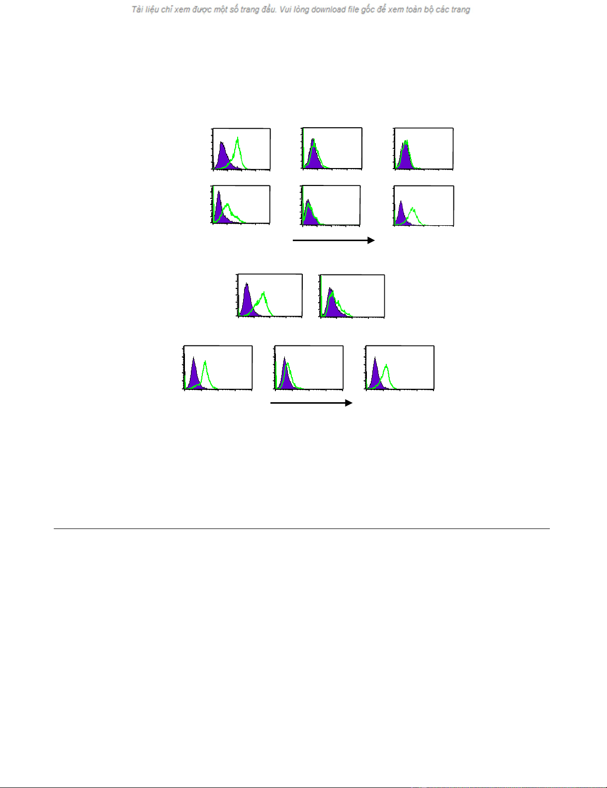
Binding of GFP-Env to cellsFigure 2
Binding of GFP-Env to cells. (A) Supernatants of GFP-EMO- or GFP-MOV-infected NIH3T3 cells were incubated with the indi-
cated target cells and analyzed by flow cytometry. Binding of GFP-Env was detected by a shift to green fluorescence (FL-1).(B)
Supernatants from GFP-MOV-infected NIH3T3 cells were incubated with the indicated target cells and analyzed by flow
cytometry. Soluble receptor binding domains of the ecotropic or the amphotropic MLV Env (E-sRBD, A-sRBD) were added
prior to the virus, as supernatants from 293T cells transfected with the expression constructs. After 5 mins., supernatants of
GFP-MOV-infected NIH3T3 cells were added for an additional 5 mins. Binding of GFP-Env was detected by a shift to green flu-
orescence (FL-1). NIH3T3i-MLV: chronically MLV-infected NIH3T3 cells.
GFP
GFPGFP
GFP-
--
-MOV
MOVMOV
MOV
(wt)
(wt)(wt)
(wt)
GFP
GFPGFP
GFP-
--
-EMO
EMOEMO
EMO
(EGF)
(EGF)(EGF)
(EGF)
GFP
GFPGFP
GFP
10010110 210 310 4
FL1-H
100101102103104
FL1-H
10010 110210 310 4
FL1-H
100101102103104
FL1-H 100101102103104
FL1-H
10
010110210
310
4
FL1-H
150
150
150
150
counts
counts
counts
counts
0
0
0
0
120
120
120
120
counts
counts
counts
counts
0
0
0
0
120
120
120
120
counts
counts
counts
counts
0
0
0
0120
120
120
120
counts
counts
counts
counts
0
0
0
0
120
120
120
120
counts
counts
counts
counts
0
0
0
0
120
120
120
120
counts
counts
counts
counts
0
0
0
0
NIH 3T3
NIH 3T3NIH 3T3
NIH 3T3
(
((
(mCat
mCatmCat
mCat+/EGFR
+/EGFR+/EGFR
+/EGFR-
--
-)
))
)
293T
293T293T
293T
(
((
(mCat
mCatmCat
mCat-
--
-/EGFR
/EGFR/EGFR
/EGFR-
--
-)
))
)
A431
A431A431
A431
(
((
(mCat
mCatmCat
mCat-
--
-/EGFR+)
/EGFR+)/EGFR+)
/EGFR+)
GFP
GFPGFP
GFP-
--
-MOV
MOVMOV
MOV
100101102103104
FL1-H
NIH3T3
NIH3T3NIH3T3
NIH3T3
+ A
+ A+ A
+ A-
--
-sRBD
sRBDsRBD
sRBD
150
150
150
150
counts
counts
counts
counts
0
0
0
0
100101102103104
FL1-H
+ E
+ E+ E
+ E-
--
-sRBD
sRBDsRBD
sRBD
NIH3T3
NIH3T3NIH3T3
NIH3T3
150
150
150
150
counts
counts
counts
counts
0
0
0
0
100101102103104
FL1-H
NIH3T3
NIH3T3NIH3T3
NIH3T3
150
150
150
150
counts
counts
counts
counts
0
0
0
0
100101102103104
FL1-H
NIH3T3
NIH3T3NIH3T3
NIH3T3
120
120
120
120
counts
counts
counts
counts
0
0
0
0
100101102103104
FL1-H
NIH3T3
NIH3T3NIH3T3
NIH3T3
i
iii
-
--
-MLV
MLVMLV
MLV
120
120
120
120
counts
counts
counts
counts
0
0
0
0
GFP
GFPGFP
GFP-
--
-MOV
MOVMOV
MOV
A
AA
A
B
BB
B
GFP
GFPGFP
GFP
Virology Journal 2004, 1:14 http://www.virologyj.com/content/1/1/14
Page 5 of 9
(page number not for citation purposes)
Figure 5A, lane 3, a 600 bp fragment can be amplified
from full-length MLV DNA using these primers. The split
genomes do not give rise to a DNA fragment, because the
primer binding sites are on separate genomes (Fig. 5A,
lane 2). After 13 days of culture, the appearance of a full-
length MLV recombinant could be observed when the vec-
tor genomes were co-transfected in a ratio of 1:1 (gag/
pol:env) (Fig. 5A, lane 5) and after 32 days, wt MLV could
be detected in all samples (Fig. 5A, lanes 9, 10 and 11).
This illustrates that full-length MLV was generated from
the split viral genomes after prolonged passage.
In addition, we examined the stability of the GFP-tagged
Env in the split genome approach. As shown in Figure 5B,
PCR analysis with primers flanking the GFP sequences in
Env (p3, p4; Fig. 3) clearly demonstrated that GFP-Env is
stable and the GFP sequences were not deleted from the
viral genome after 32 days of culture (Fig. 5B, lanes 5, 6
and 7).
Discussion
Our data demonstrate that labeling the MLV Env with a
fluorescent protein is an easy method of monitoring MLV
replication and the attachment of Env to target cells. This
is especially useful for the development of novel cancer
gene therapies that use replication-competent MLV
encoding a cytotoxic gene [3]. Labeling Env with GFP in
the PRR leaves the 3' untranslated region at the Env
boundary available for the insertion of IRES-linked thera-
peutic genes [1]. These recombinant viruses could be
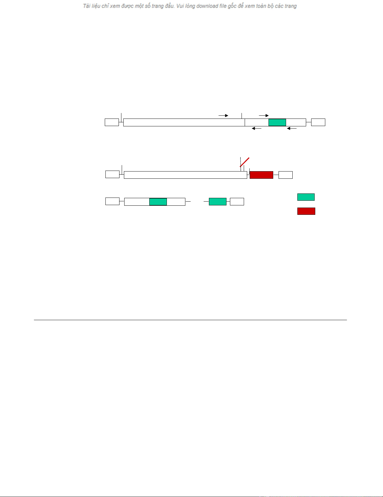
Schematic representation of fluorescently labeled semi-replicative retroviral vectorsFigure 3
Schematic representation of fluorescently labeled semi-replicative retroviral vectors. The env open reading frame was replaced
with the gene for red fluorescent protein (RFP) in the gag/pol-expressing construct, GAG/POL-RFP, and GFP-tagged Env was
expressed from a packagable vector (GFP-Env). Positions of primers used to analyze the appearance of replication-competent
viruses and the stability of the inserted GFP sequences by polymerase chain reactions (PCR) are indicated as p1 to p4. SA:
splice acceptor site; SD: splice donor site.
MLV
MLVMLV
MLV
GFP
GFPGFP
GFP-
--
-Env
EnvEnv
Env
GAG/POL
GAG/POLGAG/POL
GAG/POL-
--
-RFP
RFPRFP
RFP
GFP
GFPGFP
GFP
LTR
LTRLTR
LTR LTR
LTRLTR
LTR
env
envenv
env
RFP
RFPRFP
RFP
RFP
RFPRFP
RFP
LTR
LTRLTR
LTR LTR
LTRLTR
LTR
gag
gaggag
gag-
--
-pol
polpol
pol env
envenv
env
SD
SDSD
SD SA
SASA
SA
LTR
LTRLTR
LTR LTR
LTRLTR
LTR
gag
gaggag
gag-
--
-pol
polpol
pol RFP
RFPRFP
RFP
ATG
ATGATG
ATG ATG
ATGATG
ATG
SA
SASA
SA
SD
SDSD
SD
p1
p1p1
p1
p2
p2p2
p2
p3
p3p3
p3
p4
p4p4
p4
IRES
IRESIRES
IRES GFP
GFPGFP
GFP


![PET/CT trong ung thư phổi: Báo cáo [Năm]](https://cdn.tailieu.vn/images/document/thumbnail/2024/20240705/sanhobien01/135x160/8121720150427.jpg)


%20--%3e%3cdefs%3e%3cstyle%3e%20.st0%20{%20fill:%20%23fff;%20}%20.st1%20{%20fill:%20%237800fa;%20}%20%3c/style%3e%3c/defs%3e%3cpath%20class='st1'%20d='M117.78,12.18H43.11c2.9,3.47,4.65,7.94,4.65,12.82,0,5.6-2.3,10.66-6.01,14.29h76.02l7.22-13.56-7.22-13.56Z'/%3e%3cg%3e%3cpath%20class='st0'%20d='M53.58,26.17h-.59v-1.46h.59v-4.96h2.83c1.78,0,2.67.94,2.67,2.82v5.76c0,1.87-.89,2.81-2.67,2.81h-2.83v-4.96ZM55.36,21.37v3.34h1.1v1.46h-1.1v3.34h1.01c.61,0,.91-.37.91-1.1v-5.93c0-.74-.3-1.1-.91-1.1h-1.01Z'/%3e%3cpath%20class='st0'%20d='M65.99,31.14h-1.8l-.31-2.07h-2.19l-.31,2.07h-1.64l1.82-11.39h2.62l1.82,11.39ZM65.28,18.04c-.25.46-.51.77-.75.94-.21.15-.47.22-.79.22-.26,0-.57-.07-.92-.22l-.38-.15c-.14-.05-.26-.07-.37-.07-.3,0-.53.18-.71.54l-.91-.68c.25-.46.51-.77.75-.94.21-.14.48-.21.79-.21.26,0,.57.07.92.21l.38.15c.14.05.26.07.37.07.3,0,.53-.18.71-.54l.91.68ZM61.91,27.52h1.73l-.87-5.76-.87,5.76Z'/%3e%3cpath%20class='st0'%20d='M74.53,26.89v1.52c0,1.91-.89,2.86-2.67,2.86s-2.67-.95-2.67-2.86v-5.93c0-1.91.89-2.86,2.67-2.86s2.67.95,2.67,2.86v1.11h-1.69v-1.22c0-.75-.31-1.12-.93-1.12s-.93.37-.93,1.12v6.15c0,.74.31,1.11.93,1.11s.93-.37.93-1.11v-1.63h1.69Z'/%3e%3cpath%20class='st0'%20d='M81.4,31.14h-1.8l-.31-2.07h-2.19l-.31,2.07h-1.64l1.82-11.39h2.62l1.82,11.39ZM75.9,19.2l1.52-1.91h1.71l1.51,1.91h-1.61l-.76-.95-.75.95h-1.61ZM77.32,27.52h1.73l-.87-5.76-.87,5.76ZM83.1,15.99l-1.76,1.91h-1.26l1.17-1.91h1.86Z'/%3e%3cpath%20class='st0'%20d='M84.86,19.75c1.78,0,2.67.94,2.67,2.82v1.48c0,1.87-.89,2.81-2.67,2.81h-.85v4.28h-1.79v-11.39h2.64ZM84.01,21.37v3.86h.85c.58,0,.87-.36.87-1.08v-1.71c0-.71-.29-1.07-.87-1.07h-.85Z'/%3e%3cpath%20class='st0'%20d='M93.51,19.75c1.78,0,2.67.94,2.67,2.82v1.48c0,1.87-.89,2.81-2.67,2.81h-.85v4.28h-1.79v-11.39h2.64ZM92.66,21.37v3.86h.85c.58,0,.87-.36.87-1.08v-1.71c0-.71-.29-1.07-.87-1.07h-.85Z'/%3e%3cpath%20class='st0'%20d='M98.8,31.14h-1.79v-11.39h1.79v4.88h2.03v-4.88h1.83v11.39h-1.83v-4.88h-2.03v4.88Z'/%3e%3cpath%20class='st0'%20d='M105.36,24.55h2.46v1.62h-2.46v3.34h3.09v1.63h-4.88v-11.39h4.88v1.63h-3.09v3.18ZM108.17,17.29l-1.76,1.91h-1.26l1.17-1.91h1.86Z'/%3e%3cpath%20class='st0'%20d='M112.2,19.75c1.78,0,2.67.94,2.67,2.82v1.48c0,1.87-.89,2.81-2.67,2.81h-.85v4.28h-1.79v-11.39h2.64ZM111.35,21.37v3.86h.85c.58,0,.87-.36.87-1.08v-1.71c0-.71-.29-1.07-.87-1.07h-.85Z'/%3e%3c/g%3e%3ccircle%20class='st1'%20cx='25'%20cy='25'%20r='20'/%3e%3cpath%20class='st0'%20d='M32.78,19.27c2.92,0,4.43,2.55,5.28,5.33l.71,2.17c.14.38-.33.75-.71.75h-5.61c.19-.33.24-.71.09-1.08l-.75-2.45c-.43-1.32-.99-2.64-1.79-3.77.75-.57,1.65-.94,2.78-.94h0ZM25,18.38c3.25,0,4.9,2.78,5.89,5.89l.76,2.45c.14.42-.33.8-.8.8h-11.69c-.42,0-.94-.38-.8-.8l.75-2.45c.99-3.11,2.64-5.89,5.89-5.89h0ZM25,11.35c1.74,0,3.11,1.37,3.11,3.11s-1.37,3.11-3.11,3.11-3.11-1.41-3.11-3.11,1.41-3.11,3.11-3.11h0ZM17.27,19.27c1.08,0,1.98.38,2.73.94-.8,1.13-1.37,2.45-1.74,3.77l-.8,2.45c-.14.38-.05.75.09,1.08h-5.56c-.42,0-.9-.38-.75-.75l.71-2.17c.9-2.78,2.41-5.33,5.33-5.33h0ZM17.27,12.91c1.51,0,2.78,1.27,2.78,2.83s-1.27,2.83-2.78,2.83-2.83-1.27-2.83-2.83,1.27-2.83,2.83-2.83h0ZM32.78,12.91c1.56,0,2.78,1.27,2.78,2.83s-1.23,2.83-2.78,2.83-2.83-1.27-2.83-2.83,1.27-2.83,2.83-2.83h0ZM27.07,28.56v.09c0,.57-.24,1.08-.61,1.46h0v.05c-.38.33-.9.57-1.46.57s-1.08-.24-1.46-.61h0c-.38-.38-.61-.9-.61-1.46v-.09h1.41v.09c0,.19.05.38.19.47v.05c.09.09.28.19.47.19s.38-.09.47-.19v-.05c.14-.09.24-.28.24-.47t-.05-.09h1.41ZM30.99,28.56v.09c0,1.65-.66,3.16-1.74,4.24-1.08,1.08-2.59,1.79-4.24,1.79s-3.16-.71-4.24-1.79l-.05-.05c-1.04-1.08-1.7-2.55-1.7-4.2v-.09h1.41v.09c0,1.27.47,2.4,1.27,3.25h.05c.85.85,1.98,1.37,3.25,1.37s2.4-.52,3.25-1.37c.85-.8,1.37-1.98,1.37-3.25v-.09h1.37ZM34.99,28.56v.09c0,2.78-1.13,5.28-2.92,7.07-1.79,1.79-4.29,2.92-7.07,2.92s-5.23-1.13-7.07-2.92c-1.79-1.79-2.92-4.29-2.92-7.07v-.09h1.41v.09c0,2.4.94,4.53,2.5,6.08,1.56,1.56,3.72,2.5,6.08,2.5s4.52-.94,6.08-2.5c1.56-1.56,2.5-3.68,2.5-6.08v-.09h1.41Z'/%3e%3c/svg%3e)