
Int.J.Curr.Microbiol.App.Sci (2020) 9(4): 218-224
218
Original Research Article https://doi.org/10.20546/ijcmas.2020.904.026
In-vitro Evaluation of Some Plant Leaf Extract against Coconut Leaf Spot
Caused by Pestalotia palmarum (Cooke) in Bastar Plateau of Chhattisgarh
Vandana Chadar*, R. R. Bhanwar, Rajendra Kashyap,
Shraddha Karcho and Luchika Rana
Department of Plant Pathology, S. G. College of Agriculture and Research Station,
Jagdalpur, IGKV, Chhattisgarh, 494001, India
*Corresponding author
A B S T R A C T
Introduction
Coconut is one of the major plantation crop in
india. Coconut tree (Cocos nucifera) is a
member of the palm tree family (Arecaceae).
In India, coconut farming is inseparably
embedded in the socio-historical culture as
well as the ethnic identity. Considering the
versatile nature of the crop and the multi-uses
of its products, the coconut palm is eulogized
as Kalpavriksha (Tree of Heaven). In India
with a total cultivated area of 1975.81
thousand hectares with a production of 21,665
million nuts which makes India stand 3rd in
the world.
India occupies the premier position in the
world with an annual production of 13 billion
nuts, overtaking Indonesia and the Philippines,
the other two prominent coconut-growing
International Journal of Current Microbiology and Applied Sciences
ISSN: 2319-7706 Volume 9 Number 4 (2020)
Journal homepage: http://www.ijcmas.com
In the present investigation 30 Plant leaf extract were evaluated in in vitro
condition against P. palmarum adopting poisoned food technique. The per
cent inhibition of pathogen was 100 per cent by Dhatura, Anjwain and
Tobacco at 10 per cent concentration followed by the turmeric (95.84),
safed musli (90.62), garlic (87.5), hathjodh (86.5), kalmegh (86.46),
jetropha (83.34), neem bark (79.18), satavar (78.12), lemongrass (69.81),
laung (67.71), ashoka (65.62), aadusa (63.68), karanj bark (63.06),
bhringraj (62.5), karanj (62.5), dalchini (61.15), beshram (60.43), brijdanti
(57.46), arandi (56.25), neem (54.18), nilgiri (54.18), pattharchatta (52.09),
tulsi (46.87), marigold (40.62) and aloevera (40.62) whereas the lowest
inhibition was recorded in amari (Gangura) with 36.46 per cent.
Keywords
Coconut leaf spot,
plant leaf extract,
In vitro, Pestalotia
palmarum,
Poisoned food
technique
Accepted:
04 March 2020
Available Online:
10 April 2020
Article Info

Int.J.Curr.Microbiol.App.Sci (2020) 9(4): 218-224
219
countries (Raghavi et al., 2019). Yield of the
coconut also reduces day by day due to the
causes of various diseases. Such as, sooty
mould, stem bleeding, leaf spot, white thread
blight, root rot, brown root rot and bud rot
disease which are caused by different fungus.
Among the diseases every year grey leaf spot
disease caused by Pestalotia palmarum
(Cooke.) attacks the gardens and decreases the
growth and development of the tree as well as
the yield of the fruit. The symptom is only
developed in the mature leaves in the form of
grayish white spots surrounded by brown
margin. Several of the spots coalesce together
and form irregular grey necrotic patches and
show burnt or blighted appearances. The
upper surface of the affected leaves reveals
dark grey eruptions like pin heads. This
disease is a serious problem all over the
coconut growing regions of Bangladesh
(Rahman et al., 2013).
Materials and Methods
Collection of sample
Diseased leaves of Coconut with typical leaf
spot symptoms were collected from AICRP on
Palms Research field of SGCARS, Jagdalpur
(C.G.) (Fig. 2).
Preparation of potato dextrose agar (PDA)
medium
The basic medium, PDA was prepared
following the standard procedure (Anon.
1968). At first 200 g peeled potato is cut into
slice and then boiled in 1000 ml water. After
that it was sieved and 15 gm agar were mixed
with it in a water bath, after few minutes 20 g
dextrose were mixed with it and stirred
properly so that it cannot be coagulated. The
pH was adjusted to 6.5 of the media by using
pH meter with the help of 1N HCL and
sterilized in autoclave at 121°C temperature
for 20 minutes.
Isolation of the fungus
The fungus was isolated from the infected leaf
of coconut following tissue planting technique
(Tuite, 1969). The infected diseased samples
along with healthy tissues were cut into small
pieces and surface sterilized by dipping in
0.1% sodium hypochloride (NaOCl) solution
for two minutes. NaOCl on the surface of the
leaf pieces was decanted by soaking with
sterilized blotting paper. The cut pieces were
then placed onto sterilized potato dextrose
agar (PDA) in glass petridishes (20 ml/
petridish) and incubated in an incubator at 27
± 1°C until mycelium formation. The hyphal
tips were transferred onto PDA plate after
growing the mycelium.
Identification of fungus
The fungus was then identified on the basis
the morphological of characteristics with the
help of identifying key book (Barnett and
Hunter, 1972).
Purification
To obtain pure culture of the pathogen, the
hyphal tips were transferred aseptically onto
PDA plate by using the flame sterilized tip of
an inoculation needle. The plate was incubated
at room temperature for seven days.
Multiplication of P. palmarum
PDA was poured in sterilized petridishes, 25
ml in each. After solidification, the plates
were inoculated by placing 5 mm discs of
three days old PDA culture of P. palmarum.
The discs were cut with flame sterilized cork
borer (5 mm diameter). The inoculated
petridishes were kept in the growth chamber at
a temperature of 28 ± 1ºC for few days. All
the works were undertaken under the laminar
air flow cabinet.
Int.J.Curr.Microbiol.App.Sci (2020) 9(4): 218-224
220
Evaluation of different plant extract used in
this experiment
These plant extracts were tested initially under
in-vitro condition by using poison food
technique (Schmitz, 1930). The fresh leaves
were grounded in a blender with distilled
water. The extract was filtered through double
layered muslin cloth. The extracts were tried
at concentration of 10 per cent for seed
treatment, prepared by diluting the extract in
distilled water (Table 1).
Different plant extract were evaluated in in
vitro condition against P. palmarum following
poison food technique (Dhingra and Sinclair,
1985). All the plant extract were tested at
recommended by adopting poisoned food
technique. The test pathogen was grown on
PDA medium in Petri plates for seven days
prior to setting up of experiment. The required
plant extract was added to the melted PDA
medium to obtain the desired concentration.
20 ml of poisoned medium was poured in each
Petri plate. Suitable checks were maintained
without addition of fungicides. A mycelial
disc of five mm diameter was taken from the
periphery of 7 days old colony and placed in
the centre and incubated at 28 ± 2℃ for full
growth of the fungus.
Three replications were maintained for each
treatment. The radial growth of the colony was
measured in two directions and average was
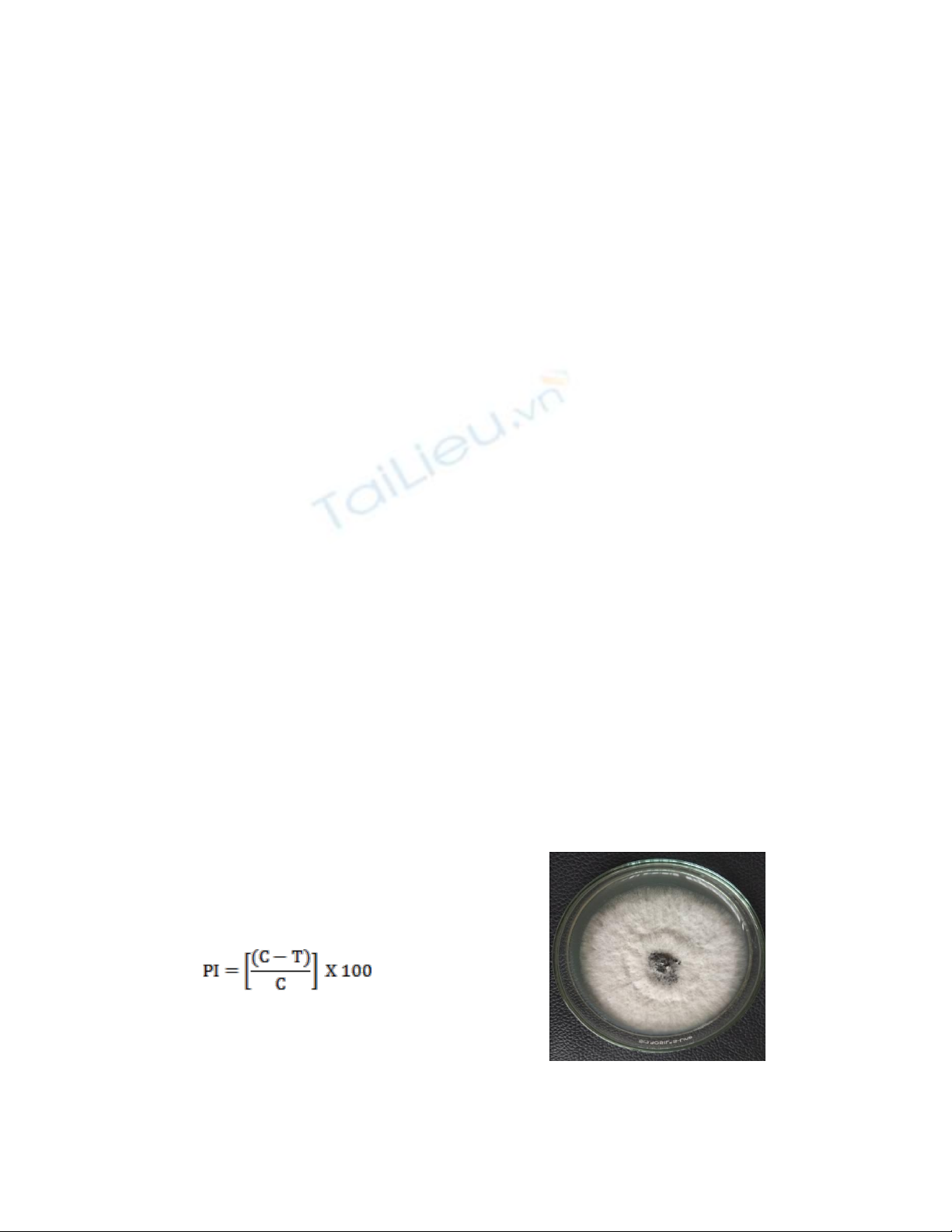
recorded. Per cent inhibition was recorded by
using the formula given by Vincent (1947) as
under:
Where,
PI = Per cent inhibition, C = Growth in control
and T = Growth in treatment.
Results and Discussion
Among the 30 plant leaf extract were
evaluated against coconut leaf spot (P.
palmerum) adopting poisoned food technique.
Observations of the radial growth of the
pathogen were recorded after 7 day after
inoculation. The percent inhibition of the
pathogen over control was calculated and
presented in Table 2, Fig. 3 and Chart 1. The
superiority in controlling the inhibition of
pathogen was managed by Dhatura, Anjwain
and Tobacco inhibited the growth of P.
palmarum was 100 per cent.
No growth was found at given concentration.
the turmeric (95.84), safed musli (90.62),
garlic (87.5), hathjodh (86.5), kalmegh(86.46),
jetropha (83.34), neem bark (79.18), satavar
(78.12), lemongrass (69.81), laung (67.71),
ashoka (65.62), aadusa (63.68), karanj bark
(63.06), bhringraj (62.5), karanj (62.5),
dalchini (61.15), beshram (60.43), brijdanti
(57.46), arandi (56.25), neem (54.18), nilgiri
(54.18), pattharchatta (52.09), tulsi (46.87),
marigold (40.62) and aloevera (40.62)
whereas the lowest inhibition was recorded in
amari (Gangura) with 36.46 per cent.
Islam et al., (2004) revealed that the two doses
(4 and 5 %) of garlic extract were found most
effective in inhibiting the redial growth of the
fungus i. e. 88.76 per cent which favors the
present study.
Fig.1 Pure culture of P. palmarum

Int.J.Curr.Microbiol.App.Sci (2020) 9(4): 218-224
221
Table.1 List of botanicals used in the experiment
S.N.
Treatment
Name of Botanicals
Botanical name
1.
T1
Tulsi
Ocimum tenuiflorum
2.
T2
Turmeric
Curcuma longa
3.
T3
Marigold
Tagetes spp.
4.
T4
Garlic
Allium sativam
5.
T5
Jetropha
Jatropha curcas
6.
T6
Dhatura
Datura stramonium
7.
T7
Caster
Ricinus communis
8.
T8
Pattharchatta
Bryophyllum pinnatum
9.
T9
Vringraj
Eclipta prostrate
10.
T10
Neem
Azadirachta indica
11.
T11
Karanj
Millettia pinnata
12.
T12
Neem bark
Azadirachta indica
13.
T13
Kalmegh
Andrographis paniculata
14.
T14
Satavar
Asparagus racemosus
15.
T15
Ashoka
Saraca asoca
16.
T16
Nilgiri
Eucalyptus spp.
17.
T17
Laung
Syzygium aromaticum
18.
T18
Anjwain
Trachyspermum ammi
19.
T19
Tobacco
Nicotiana tabacum
20.
T20
Lemongrass
Cymbopogan spp.
21.
T21
Beshram
Ipomoea carnea
22.
T22
Brijdanti
Banteria prionitis
23.
T23
Karanj bark
Millettia pinnata
24.
T24
Aloevera
Aloe barbadensis
25.
T25
Amari (Gangura)
Hibiscus sabdariffa
26.
T26
Aadusa
Justicia adhatoda
27.
T27
Dalchini
Cinnamamum verum
28.
T28
Safed musli
Chlorophytum borivilianum
29.
T29
Hathjodh
Cissus quadrangularis
30.
T30
Control
Without phyto extract

Int.J.Curr.Microbiol.App.Sci (2020) 9(4): 218-224
222
Table.2 Percent inhibition of the radial growth of the pathogen of coconut leaf spot in in-vitro
Treatment
Mean growth (mm) of
pathogens
Percent inhibition of
pathogens (%)
T1
17.000
46.87
T2
01.333
95.84
T3
19.000
40.62
T4
04.000
87.5
T5
05.333
83.34
T6
00.000
100
T7
17.333
56.25
T8
15.333
52.09
T9
12.000
62.50
T10
14.667
54.18
T11
12.000
62.50
T12
06.667
79.18
T13
06.667
86.46
T14
07.000
78.12
T15
11.000
65.62
T16
14.667
54.18
T17
10.333
67.71
T18
00.000
100
T19
00.000
100
T20
09.667
69.81
T21
12.667
60.43
T22
13.667
57.46
T23
11.667
63.06
T24
19.000
40.62
T25
20.333
36.46
T26
11.667
63.68
T27
12.333
61.15
T28
03.000
90.62
T29
04.333
86.50
T30
32.000
-
C.D at 5 %
4.339
SE(m) ±
1.530


![Tiềm năng sản xuất hè của giống đậu xanh [Vigna radiata (L) wilczek]: Các biện pháp quản lý dinh dưỡng khác nhau tại điều kiện Nam Gujarat](https://cdn.tailieu.vn/images/document/thumbnail/2020/20200722/angicungduoc6/135x160/6761595379748.jpg)


%20--%3e%3cdefs%3e%3cstyle%3e%20.st0%20{%20fill:%20%23fff;%20}%20.st1%20{%20fill:%20%237800fa;%20}%20%3c/style%3e%3c/defs%3e%3cpath%20class='st1'%20d='M117.78,12.18H43.11c2.9,3.47,4.65,7.94,4.65,12.82,0,5.6-2.3,10.66-6.01,14.29h76.02l7.22-13.56-7.22-13.56Z'/%3e%3cg%3e%3cpath%20class='st0'%20d='M53.58,26.17h-.59v-1.46h.59v-4.96h2.83c1.78,0,2.67.94,2.67,2.82v5.76c0,1.87-.89,2.81-2.67,2.81h-2.83v-4.96ZM55.36,21.37v3.34h1.1v1.46h-1.1v3.34h1.01c.61,0,.91-.37.91-1.1v-5.93c0-.74-.3-1.1-.91-1.1h-1.01Z'/%3e%3cpath%20class='st0'%20d='M65.99,31.14h-1.8l-.31-2.07h-2.19l-.31,2.07h-1.64l1.82-11.39h2.62l1.82,11.39ZM65.28,18.04c-.25.46-.51.77-.75.94-.21.15-.47.22-.79.22-.26,0-.57-.07-.92-.22l-.38-.15c-.14-.05-.26-.07-.37-.07-.3,0-.53.18-.71.54l-.91-.68c.25-.46.51-.77.75-.94.21-.14.48-.21.79-.21.26,0,.57.07.92.21l.38.15c.14.05.26.07.37.07.3,0,.53-.18.71-.54l.91.68ZM61.91,27.52h1.73l-.87-5.76-.87,5.76Z'/%3e%3cpath%20class='st0'%20d='M74.53,26.89v1.52c0,1.91-.89,2.86-2.67,2.86s-2.67-.95-2.67-2.86v-5.93c0-1.91.89-2.86,2.67-2.86s2.67.95,2.67,2.86v1.11h-1.69v-1.22c0-.75-.31-1.12-.93-1.12s-.93.37-.93,1.12v6.15c0,.74.31,1.11.93,1.11s.93-.37.93-1.11v-1.63h1.69Z'/%3e%3cpath%20class='st0'%20d='M81.4,31.14h-1.8l-.31-2.07h-2.19l-.31,2.07h-1.64l1.82-11.39h2.62l1.82,11.39ZM75.9,19.2l1.52-1.91h1.71l1.51,1.91h-1.61l-.76-.95-.75.95h-1.61ZM77.32,27.52h1.73l-.87-5.76-.87,5.76ZM83.1,15.99l-1.76,1.91h-1.26l1.17-1.91h1.86Z'/%3e%3cpath%20class='st0'%20d='M84.86,19.75c1.78,0,2.67.94,2.67,2.82v1.48c0,1.87-.89,2.81-2.67,2.81h-.85v4.28h-1.79v-11.39h2.64ZM84.01,21.37v3.86h.85c.58,0,.87-.36.87-1.08v-1.71c0-.71-.29-1.07-.87-1.07h-.85Z'/%3e%3cpath%20class='st0'%20d='M93.51,19.75c1.78,0,2.67.94,2.67,2.82v1.48c0,1.87-.89,2.81-2.67,2.81h-.85v4.28h-1.79v-11.39h2.64ZM92.66,21.37v3.86h.85c.58,0,.87-.36.87-1.08v-1.71c0-.71-.29-1.07-.87-1.07h-.85Z'/%3e%3cpath%20class='st0'%20d='M98.8,31.14h-1.79v-11.39h1.79v4.88h2.03v-4.88h1.83v11.39h-1.83v-4.88h-2.03v4.88Z'/%3e%3cpath%20class='st0'%20d='M105.36,24.55h2.46v1.62h-2.46v3.34h3.09v1.63h-4.88v-11.39h4.88v1.63h-3.09v3.18ZM108.17,17.29l-1.76,1.91h-1.26l1.17-1.91h1.86Z'/%3e%3cpath%20class='st0'%20d='M112.2,19.75c1.78,0,2.67.94,2.67,2.82v1.48c0,1.87-.89,2.81-2.67,2.81h-.85v4.28h-1.79v-11.39h2.64ZM111.35,21.37v3.86h.85c.58,0,.87-.36.87-1.08v-1.71c0-.71-.29-1.07-.87-1.07h-.85Z'/%3e%3c/g%3e%3ccircle%20class='st1'%20cx='25'%20cy='25'%20r='20'/%3e%3cpath%20class='st0'%20d='M32.78,19.27c2.92,0,4.43,2.55,5.28,5.33l.71,2.17c.14.38-.33.75-.71.75h-5.61c.19-.33.24-.71.09-1.08l-.75-2.45c-.43-1.32-.99-2.64-1.79-3.77.75-.57,1.65-.94,2.78-.94h0ZM25,18.38c3.25,0,4.9,2.78,5.89,5.89l.76,2.45c.14.42-.33.8-.8.8h-11.69c-.42,0-.94-.38-.8-.8l.75-2.45c.99-3.11,2.64-5.89,5.89-5.89h0ZM25,11.35c1.74,0,3.11,1.37,3.11,3.11s-1.37,3.11-3.11,3.11-3.11-1.41-3.11-3.11,1.41-3.11,3.11-3.11h0ZM17.27,19.27c1.08,0,1.98.38,2.73.94-.8,1.13-1.37,2.45-1.74,3.77l-.8,2.45c-.14.38-.05.75.09,1.08h-5.56c-.42,0-.9-.38-.75-.75l.71-2.17c.9-2.78,2.41-5.33,5.33-5.33h0ZM17.27,12.91c1.51,0,2.78,1.27,2.78,2.83s-1.27,2.83-2.78,2.83-2.83-1.27-2.83-2.83,1.27-2.83,2.83-2.83h0ZM32.78,12.91c1.56,0,2.78,1.27,2.78,2.83s-1.23,2.83-2.78,2.83-2.83-1.27-2.83-2.83,1.27-2.83,2.83-2.83h0ZM27.07,28.56v.09c0,.57-.24,1.08-.61,1.46h0v.05c-.38.33-.9.57-1.46.57s-1.08-.24-1.46-.61h0c-.38-.38-.61-.9-.61-1.46v-.09h1.41v.09c0,.19.05.38.19.47v.05c.09.09.28.19.47.19s.38-.09.47-.19v-.05c.14-.09.24-.28.24-.47t-.05-.09h1.41ZM30.99,28.56v.09c0,1.65-.66,3.16-1.74,4.24-1.08,1.08-2.59,1.79-4.24,1.79s-3.16-.71-4.24-1.79l-.05-.05c-1.04-1.08-1.7-2.55-1.7-4.2v-.09h1.41v.09c0,1.27.47,2.4,1.27,3.25h.05c.85.85,1.98,1.37,3.25,1.37s2.4-.52,3.25-1.37c.85-.8,1.37-1.98,1.37-3.25v-.09h1.37ZM34.99,28.56v.09c0,2.78-1.13,5.28-2.92,7.07-1.79,1.79-4.29,2.92-7.07,2.92s-5.23-1.13-7.07-2.92c-1.79-1.79-2.92-4.29-2.92-7.07v-.09h1.41v.09c0,2.4.94,4.53,2.5,6.08,1.56,1.56,3.72,2.5,6.08,2.5s4.52-.94,6.08-2.5c1.56-1.56,2.5-3.68,2.5-6.08v-.09h1.41Z'/%3e%3c/svg%3e)