
BioMed Central
Page 1 of 22
(page number not for citation purposes)
BMC Plant Biology
Open Access
Research article
The PTI1-like kinase ZmPti1a from maize (Zea mays L.) co-localizes
with callose at the plasma membrane of pollen and facilitates a
competitive advantage to the male gametophyte
Markus M Herrmann1, Sheena Pinto1,2, Jantjeline Kluth1, Udo Wienand1 and
René Lorbiecke*1
Address: 1Biozentrum Klein-Flottbek und Botanischer Garten, Universität Hamburg, Ohnhorststrasse 18, 22609 Hamburg, Germany and
2Deutsches Krebsforschungszentrum, Im Neuenheimer Feld 580, 69120 Heidelberg, Germany
Email: Markus M Herrmann - mmh@antikoerper-welten.de; Sheena Pinto - s.pinto@dkfz.de; Jantjeline Kluth - jantje.Kluth@web.de;
Udo Wienand - udo.wienand@uni-hamburg.de; René Lorbiecke* - lorbiecke@botanik.uni-hamburg.de
* Corresponding author
Abstract
Background: The tomato kinase Pto confers resistance to bacterial speck disease caused by Pseudomonas
syringae pv. tomato in a gene for gene manner. Upon recognition of specific avirulence factors the Pto kinase
activates multiple signal transduction pathways culminating in induction of pathogen defense. The soluble
cytoplasmic serine/threonine kinase Pti1 is one target of Pto phosphorylation and is involved in the
hypersensitive response (HR) reaction. However, a clear role of Pti1 in plant pathogen resistance is
uncertain. So far, no Pti1 homologues from monocotyledonous species have been studied.
Results: Here we report the identification and molecular analysis of four Pti1-like kinases from maize
(ZmPti1a, -b, -c, -d). These kinase genes showed tissue-specific expression and their corresponding
proteins were targeted to different cellular compartments. Sequence similarity, expression pattern and
cellular localization of ZmPti1b suggested that this gene is a putative orthologue of Pti1 from tomato. In
contrast, ZmPti1a was specifically expressed in pollen and sequestered to the plasma membrane, evidently
owing to N-terminal modification by myristoylation and/or S-acylation. The ZmPti1a:GFP fusion protein
was not evenly distributed at the pollen plasma membrane but accumulated as an annulus-like structure
which co-localized with callose (1,3-β-glucan) deposition. In addition, co-localization of ZmPti1a and
callose was observed during stages of pollen mitosis I and pollen tube germination. Maize plants in which
ZmPti1a expression was silenced by RNA interference (RNAi) produced pollen with decreased
competitive ability. Hence, our data provide evidence that ZmPti1a plays an important part in a signalling
pathway that accelerates pollen performance and male fitness.
Conclusion: ZmPti1a from maize is involved in pollen-specific processes during the progamic phase of
reproduction, probably in crucial signalling processes associated with regions of callose deposition. Pollen-
sporophyte interactions and pathogen induced HR show certain similarities. For example, HR has been
shown to be associated with cell wall reinforcement through callose deposition. Hence, it is hypothesized
that Pti1 kinases from maize act as general components in evolutionary conserved signalling processes
associated with callose, however during different developmental programs and in different tissue types.
Published: 06 October 2006
BMC Plant Biology 2006, 6:22 doi:10.1186/1471-2229-6-22
Received: 07 June 2006
Accepted: 06 October 2006
This article is available from: http://www.biomedcentral.com/1471-2229/6/22
© 2006 Herrmann et al; licensee BioMed Central Ltd.
This is an Open Access article distributed under the terms of the Creative Commons Attribution License (http://creativecommons.org/licenses/by/2.0),
which permits unrestricted use, distribution, and reproduction in any medium, provided the original work is properly cited.

BMC Plant Biology 2006, 6:22 http://www.biomedcentral.com/1471-2229/6/22
Page 2 of 22
(page number not for citation purposes)
Background
Protein kinases in plants have been found to be involved
in basic features of plant defense and plant fertilization.
Increasing knowledge about the underlying molecular
mechanisms suggests several parallels between both proc-
esses [1-3]. Plant-pathogen recognition has been studied
extensively in tomato in which gene for gene resistance
against certain Pseudomonas syringae pv. tomato strains is
conferred by the serine/threonine kinase Pto. Upon recog-
nition of bacterial avirulence factors, Pto acts in concert
with the Prf protein resulting in the activation of multiple
signal transduction pathways culminating in the induc-
tion of defense responses including HR [4]. Several Pto-
interacting (Pti) proteins were identified to act in Pto-
mediated signal transduction including the protein kinase
Pti1 and three transcription factors (Pti4/5/6), respec-
tively [5,6]. Pti1 (here referred to as SlPti1 for clarity rea-
sons) is a cytoplasmic protein kinase capable of
autophosphorylation in vitro [5] and moreover also be
phosphorylated by Pto. Tobacco plants over-expressing
S1Pti show enhanced HR in leaves in response to aviru-
lence factor treatment indicating a functional role of
SlPti1 in Pto-mediated disease response [5]. However, a
precise role of SlPti1 in plant pathogen resistance has
remained unclear, owing to functional redundancy of dif-
ferent/additional Pti1 kinases. Three SlPti1 homologous
kinases have been cloned from soybean [7,8], sPti1a,
sPti1b and GmPti1. The former two do not display in vitro
autophosphorylation activity [7], whereas the latter,
GmPti1, possesses autophosphorylation activity. GmPti1
gene expression was found to accelerate in response to
wounding and salicylic acid treatment in seedling leaves
[8]. These findings suggest different Pti1-like kinases to
possess different properties and biological functions in
plants.
Cell-cell recognition and signal response reactions during
plant-pathogen interaction are thought to be molecularly
related to certain steps of plant reproduction, e.g. pollen-
pistil recognition, compatibility reactions, and pollen
tube growth. In studies of the genetic and molecular basis
of pollen development and function more than 150 pol-
len-expressed genes from more than 28 species have been
identified [9-11]. Classification of pollen expressed genes
identified a high number of genes which are involved in
signal transduction. Many of these genes encode putative
protein kinases [10,12,13]. Accordingly, leucine-rich
repeat (LRR) Ser/Thr-type plant receptor kinases (PRK)
LePRK1 to 3 from tomato and several interacting proteins
like KPP, LAT52 and LeSHY have already been attributed
to signaling processes during pollen tube growth [14-17].
Mutations of a number of such gametophytically impor-
tant genes often result in altered Mendelian segregation
ratios due to an abolished or reduced transmission of a
linked marker through pollen. Such genes include SEC8,
ROP2, LIMPET POLLEN and TTD genes [17-21]. Most of
these mutations cause obvious defects in the pollen grain
and affect early stages of pollen development. In contrast,
only few mutations are known that are transmitted
through the male at low frequencies but cause no obvious
defects in pollen morphology. These genes appear to
affect more pollen competitiveness rather than develop-
ment, e.g. TTD41 and ROP2 [18,21].
In this study we report the identification and molecular
analyses of four Pti1 kinases from maize (ZmPti1a, -b, -c,
-d). The genes were expressed in different tissues and
showed different subcellular localizations. Phylogenetic
analysis revealed the existence of three conserved Pti1
kinase subgroups in higher plants. Based on its sequence
similarity, expression profile and subcellular localization
ZmPti1b was suggested to be a putative SlPti1 ortholog. In
contrast, the functional kinase ZmPti1a was specific to
pollen and targeted to the plasma membrane, evidently
owing to N-terminal acylation. ZmPti1a co-localizes with
regions of callose deposition at stages of pollen matura-
tion and germination. Silencing of the ZmPti1a gene
resulted in a significant decrease in the competitive ability
of pollen. These findings provide evidences of ZmPti1a to
play an important role in influencing pollen fitness.
Our data further suggest that Pti1 kinases from maize act
in various tissues and in different but mechanistically con-
served plant response pathways which likely involve sim-
ilar signals and/or signal transduction molecules.
Results
Pti1-like kinases of maize
A 217 bp partial cDNA of ZmPti1a was cloned in a molec-
ular approach with the aim to identify genes that are spe-
cifically expressed in maize pollen. Using this clone as a
hybridization probe, two nearly identical 1.6 kb full-
length cDNAs [GenBank:AY554281, Gen-
Bank:AY554282] were isolated from a λ-cDNA library of
in vitro germinated pollen from white pollen (whp) plants
[22] expressing the c2 gene. Both cDNAs probably repre-
sent different alleles of the same gene. The cDNA clone
AY554281 was further analyzed in this study. AY554281
contains an open reading frame (ORF) of 1122 bp, a 207
bp 5' untranslated region and a 304 bp 3' untranslated
region including a poly(A)+ tail. The putative protein of
AY554281 is 374 amino acids (aa) in length with a molec-
ular mass of 40.8 kDa (Fig. 1A). Database search revealed
69% identity and 75% similarity to the Pto-interactor 1
(Pti1) protein kinase of Solanum lycopersicum [5]. There-
fore the cloned gene was named Zea mays Pti1a (ZmPti1a).
The putative catalytic kinase domain of ZmPti1a starts
approximately 75 aa after the first methionine and con-
tains 11 canonical subdomains that are typical of serine/
BMC Plant Biology 2006, 6:22 http://www.biomedcentral.com/1471-2229/6/22
Page 3 of 22
(page number not for citation purposes)
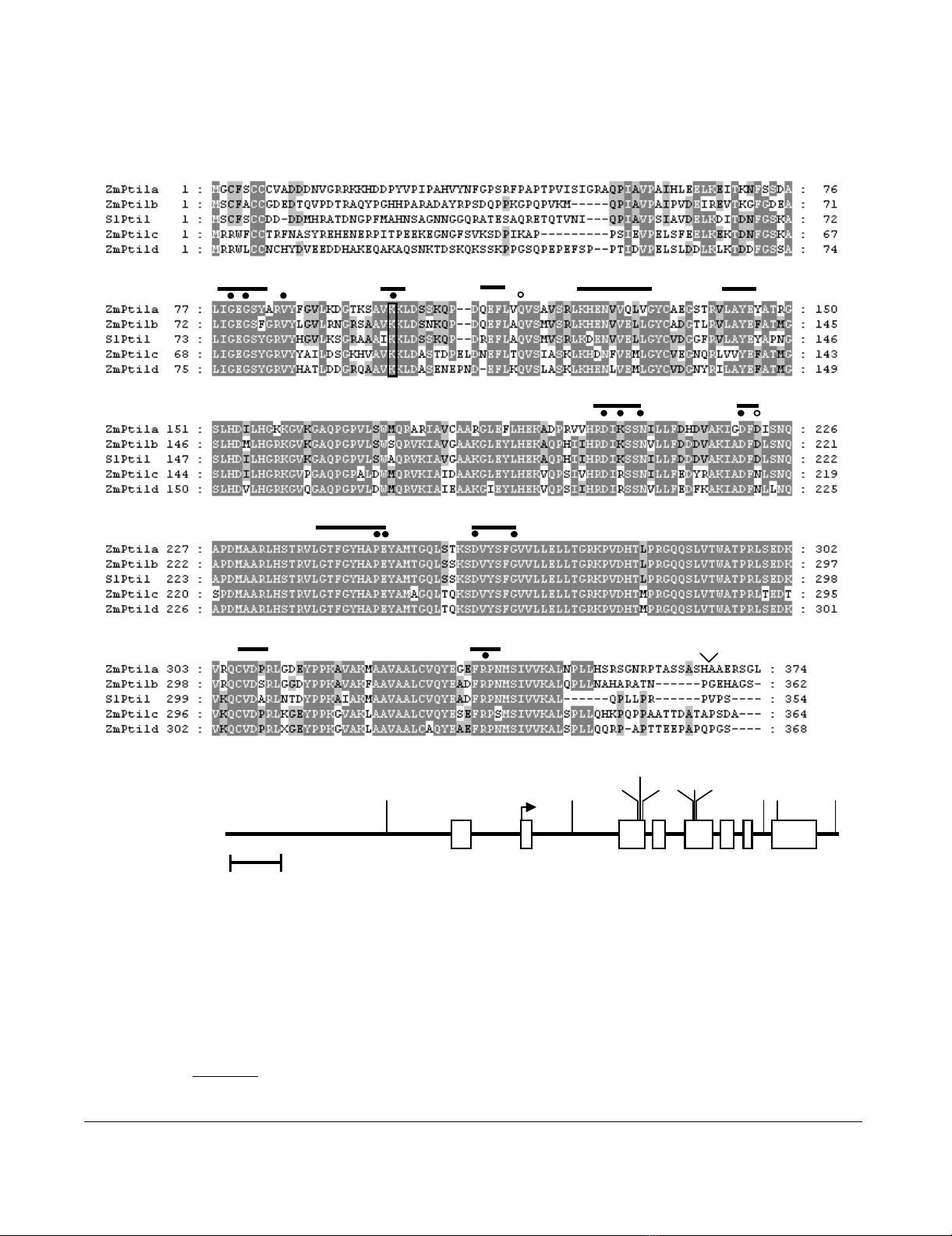
Similarity and predicted genomic structure of ZmPti1aFigure 1
Similarity and predicted genomic structure of ZmPti1a. (A) Alignment of Pti1 kinases from maize with SlPti1 from
tomato. Amino acids identical in at least three of the sequences are highlighted in grey. The 11 canonical subdomains con-
served in serine/threonine kinases are indicated with Roman numerals. Invariant residues common to the majority of protein
kinases are marked with black dots. Invariant residues that are conserved in other protein kinases but not in Pti1 kinases are
marked with open circles. The highly conserved lysine residue in subdomain II which is required for activity in SlPti1 and most
protein kinases is boxed. Threonine 233 has been identified as the major site of SlPti1 phosphorylation by SlPto and is marked
with an asterisk. Amino acids which differ between ZmPti1a and the deduced protein sequence of the second cloned ZmPti1a
cDNA [GenBank:AY554282] are indicated above the sequences. (B) Genomic locus and restriction map of the ZmPti1a gene.
Exons are indicated as boxes with Roman numerals. Start and stop of the open reading frame are marked with an arrow and
asterisk, respectively. E, EcoRI; H, HindIII; P, PstI, X, XhoI.
500 bp
III III IV V VIVII VIII
*
ZmPti1a
EE
E
EPP
H
XX X X
B
III III IV V
VI VII
VIII IX
XXI
*
AAA
L
T
V
A

BMC Plant Biology 2006, 6:22 http://www.biomedcentral.com/1471-2229/6/22
Page 4 of 22
(page number not for citation purposes)
threonine kinases (Fig. 1A; [23]). Out of the 15 invariant
amino acid residues common to the majority of protein
kinases, 13 were found to be conserved in ZmPti1a (Fig.
1A). A glutamine in subdomain III is substituted with a
glutamate at position 115 and a conserved glycine in sub-
domain VII is substituted with an aspartate at position
222. Identical substitutions are present in the kinase
SlPti1 [5] suggesting that ZmPti1a is also a functional
kinase. A corresponding full-length genomic clone [Gen-
bank:AY554283] spanning the entire transcribed region
as well as 2.2 kb of the promoter of ZmPti1a was isolated
from a λ-phage library of the maize inbred LC by plaque
screening and inverse PCR. The gene consists of 8 exons
and 7 introns (Fig. 1B). The nucleotide sequence of the
deduced transcribed region was found to be nearly identi-
cal to the previously cloned cDNAs with the exception of
line specific single nucleotide polymorphisms that
changed three aa in less conserved regions of the deduced
protein. An insertion of 9 bp resulted in the addition of
three alanine residues in the c-terminus (Fig. 1A). The pro-
posed translation start is located in exon 2. Hybridizing
bands in genomic Southern analyses with probes specific
for the promoter, 5'-UTR, ORF, and 3'-UTR of ZmPti1a
correlated well with the predicted restriction patterns of
the cloned gene and suggested that ZmPti1a is a single
copy gene (data not shown).
Database search led to the identification of additional
ESTs coding for ZmPti1a homologues from maize. These
sequences were found to be well conserved at the nucle-
otide level (41 to 52%), and even more conserved at the
the protein level (71 to 78%). Corresponding ORF and 3'
UTRs were amplified by RT-PCR from lines A188 and LC,
respectively. All cloned sequences were identical to their
corresponding EST with the exception of few line specific
SNPs. Accordingly, these sequences were named ZmPti1b
[Genbank:DQ647388], ZmPti1c [Genbank: DQ647389],
and ZmPti1d [Genbank: DQ647390], respectively. An EST
clone [Genbank:AY708048] which resembles ZmPti1c
was annotated previously as a salt-inducible putative ser-
ine/threonine/tyrosine kinase (Zou et al., unpublished
data). Data mining of genomic BAC and MAGI sequences
containing ZmPti1b and -d indicated that the correspond-
ing genes possess nearly identical exon/intron structures
as compared to ZmPti1a (data not shown). This indicates
that the maize Pti1 gene family most likely originates from
a single ancestor gene. Out of the four putative ZmPti1
kinases, ZmPti1b showed highest protein similarity to
Pti1 from tomato (77% identity, 85% similarity). All
ZmPti1 proteins possess conserved kinase catalytic
domains. However, their N – and C-terminal regions are
highly variable and only some Pti1 kinases, including
ZmPti1a, were predicted to contain a putative myristoyla-
tion signal at their N-termini. Such protein modifications
in which the saturated fatty acid myristate is covalently
but reversibly attached to an N-terminal Gly after co-
translational cleavage of the first Met residue can fulfill
several functions, e.g. mediating membrane association.
Phylogenetic relationship of ZmPti kinases
Phylogenetic comparison of ZmPti1 proteins from maize
and putative Pti1 kinases from other plants indicated
three major Pti1 subgroups in angiosperms (I, II & III)
with the known maize proteins belonging to subgroups II
and III, respectively (Fig 2). Each subfamily possesses a
conserved N-terminal domain with a specific consensus
sequence and consists of proteins from mono – as well as
dicotyledonous species. The N-terminal domains are rich
in polar or aromatic residues and contain at least two con-
served cysteines. Some of the kinases, e.g. ZmPti1a, sPti1a,
sPti1b and At3g17410 are predicted to contain a putative
N-terminal myristoylation signal. Gene organization of
most of the Pti1 kinases from Arabidopsis thaliana were
found to be similar to that of ZmPti1a, i.e. 8 exons and a
predicted translation start in exon 2 (data not shown).
Based on these findings, Pti1 genes appear to represent an
ancient kinase family in higher plants. Amino acid
sequences of the different N-terminal regions are con-
served in a broad spectrum of monocotyledonous and
dicotyledonous species (Fig. 2). Thus, it is feasible to spec-
ulate that the conserved N-terminal motifs of the different
Pti1 subfamilies were retained during evolution because
of specific relevant biological functions.
ZmPti1 proteins localize to different subcellular
compartments
To investigate the subcellular localization of ZmPti1 pro-
teins in situ, we transiently expressed in-frame coding
sequences of ZmPti1 kinases fused to green fluorescent
protein (GFP) in onion epidermal cells and in in vitro ger-
minating pollen, respectively. When expressed under con-
trol of the ubiquitin promoter, ZmPti1a:GFP was targeted
to the cell periphery suggesting ZmPti1a to localize to the
plasma membrane (Fig 3A). This pattern was clearly dif-
ferent from that observed when GFP was expressed alone
(Fig 3E). Association of ZmPti1a:GFP with the plasma
membrane was also proven by confocal laser scanning
microscopy (data not shown). Twenty-four amino acids
of the ZmPti1a N-terminus were found to be sufficient to
target GFP entirely to the cell periphery (Myr:GFP, Fig.
3B). Truncation of twenty amino acids at the N-terminus
of ZmPti1a abolished cell periphery targeting coinciding
with cytoplasmic and nuclear localization of the fusion
protein (ΔZmPti1a, Fig 3C). Identical results were
observed for these three ZmPti1a fusion constructs when
expressed ectopically in stably transformed maize plants
(Fig. 6 and data not shown). These findings are in agree-
ment with the assumption that ZmPti1a is targeted to the

BMC Plant Biology 2006, 6:22 http://www.biomedcentral.com/1471-2229/6/22
Page 5 of 22
(page number not for citation purposes)
plasma membrane by N-terminal acylation, likely myris-
toylation.
To study the structural basis of ZmPti1a being targeted to
the plasma membrane in detail, potential myristoylation
and/or palmitoylation sites, i.e. Gly2/Cys3 and Cys6/
Cys7 were subjected to site-directed mutagenesis (Table
Fig. 3). Conjugation of myristate to proteins is absolutely
dependent on a glycine residue at position 2.
Exchange of Gly2 or Cys3 with Ala prevented targeting of
the ZmPt1a:GFP fusion to the cell periphery. Instead, GFP
fluorescence appeared in the nucleus and as small cyto-
plasmic granules (Fig. 3F and data not shown). The same
GFP pattern was seen when both, Cys3 and Cys6, were
replaced by Ala (data not shown).
Combined replacement of the adjacent amino acids Gly2
and Cys3 with Ala residues also caused nuclear localiza-
tion. However, GFP fluorescence was evenly distributed in
the cytoplasm and no granules were observed (Fig. 3G). A
similar distribution of GFP fluorescence was observed
when Cys6 and Cys7 in the second motif were replaced
with alanine residues (data not shown).
These results indicate that combined mutation of single
residues in each of the two motifs (Gly2/Cys3 or Cys6/
Cys7) resulted in GFP fluorescence associated with cyto-
plasmic granules. This localization pattern might reflect
an imperfect targeting or mistargeting of mutated
ZmPti1a to membranes. Combined replacement of both
adjacent residues in either one of the two motifs seems to
strengthen mistargeting and completely prevents ZmPti1a
membrane association.
ZmPti1b, c and d from maize and SlPti1 from tomato nat-
urally lack a Gly2 residue that would serve as a potential
target site of myristoylation (Fig. 1A). Accordingly, Zhou
et al. [5] predicted the tomato SlPti1 to be a cytoplasmic
kinase. Expression of a SlPti1:GFP fusion protein con-
Phylogenetic analysis of ZmPti1 kinasesFigure 2
Phylogenetic analysis of ZmPti1 kinases. Similarity and phylogenetic relationship of Pti1 proteins from maize, rice,
tobacco, soybean and tomato were calculated using ClustalX and visualized using Treeview. SlPto [gi 626010/pir:A49332] was
used as the outgroup. Consensus sequences of the N-termini are given for each subgroup. Highly conserved residues are indi-
cated in bold. Ambiguities are given in brackets with residues of high appearance in bold and of less appearance in subscribed
letters.
0.1
gi|50725347 Oryza sativa
ZmPti1c
gi|56784334 Oryza sativa
gi|34907668 Oryza sativa
ZmPti1d
gi|50920049 Oryza sativa
gi|29838544 GmPti1 Glycine max
At2g43230 Arabidopsis thaliana
At3g59350 Arabidopsis thaliana
At2g30740
Arabidopsis
thaliana
At1g06700
Arabidopsis
thaliana
gi|626010 SlPto Lycopersicon esculentum
gi|50909605 Oryza sativa
gi|38488407 Nicotiana tabacum
gi|38488409 Nicotiana tabacum
gi|50540700 Oryza sativa
gi|34902310 Oryza sativa
ZmPti1a
gi|34894710 Oryza sativa
ZmPti1b
gi|51038251 Oryza sativa
At2g47060 Arabidopsis thaliana
At3g62220
Arabidopsis thaliana At3g17410
Arabidopsis
thaliana
At1g48210
Arabidopsis
thaliana
At1g48220 Arabidopsis thaliana
SlPti1 gi|3668069 Solanum lycopersicum
gi|1586940 Solanum lycopersicum
gi|9651969 sPti1a Glycine max
gi|9651971 sPti1b Glycine max
At2g41970 Arabidopsis thaliana
M-[GS]-C-F-[AGS]-[C
FW
]-C
M-[S
FIW
]-C-C-[G
S
]-G
M-[R
LV
]-[R
QK
]-[W
R
]-[WRFLI]-[C
FR
]-C
I
II
III