VNU Journal of Science: Mathematics – Physics, Vol. 41, No. 1 (2025) 9-16
9
Original Article
Phase Tailoring of Dense Tantalum Films
for Improving Hardness of Titanium Implants Using Negative
Substrate Bias During DC Sputtering at Low Temperature
Dang Minh Duc1, Dao Hong Bach1, Nguyen Duc Trung Kien2,
Truong Quoc Phong1, Mai Xuan Dung3, Truong Nguyen Tuan Minh4,
Pham Thi Mai Phuong1, Le Thai Hung1, Pham Hung Vuong1,5,*
1Hanoi University of Science and Technology (HUST), 1 Dai Co Viet, Hanoi, Vietnam
2Phenikaa University, Yen Nghia, Ha Dong, Hanoi, Vietnam
3Hanoi Pedagogical University 2, Vinh Phuc, Vietnam
4Tampere University, Hervanta Campus, Korkeakoulunkatu 7 Kampusareena, 33720 Tampere, Finland
5Laboratory of Biomedical Materials, Hanoi University of Science and Technology (HUST),
1 Dai Co Viet, Hanoi, Vietnam
Received 27th July 2024
Revised 05th September 2024; Accepted 15th February 2025
Abstract: This work reports the deposition of a dense α-Ta and β-Ta films to enhance the surface
hardness and biocompatibility of Ti by applying a different negative substrate bias during direct
current (DC) sputtering. When a Ta film was deposited with a negative substrate bias voltage of 70
V, the microstructure of the film exhibited a single β-Ta phase. Whereas, a negative substrate bias
of 150 V resulted in the formation of a single α-Ta phase. The deposition of the dense Ta film onto
Ti significantly improved the hardness of the Ti film. Specifically, β-Ta films possessed a hardness
as large as 21.0 ± 1.5 GPa, substantially higher than that of bare Ti (3.6 ± 0.3 GPa) and α-Ta films
(8.3 ± 0.3 GPa). Additionally, the obtained results also showed that bydepositing the dense Ta film,
one can get the biocompatible surface.
Keywords: Biocompatibility, Ta coating, hardness, substrate bias, hardness.
________
* Corresponding author.
E-mail address: vuong.phamhung@hust.edu.vn
https//doi.org/10.25073/2588-1124/vnumap.4958

D. M. Duc et al. / VNU Journal of Science: Mathematics – Physics, Vol. 41, No. 1 (2025) 9-16
10
1. Introduction
While titanium (Ti) is generally acknowledged for its biocompatibility [1, 2], its utilization in
implants can occasionally lead to complications. This is attributed to the naturally occurring thin layer
of titanium oxide on its surface, which tends to possess high defect density. Consequently, this can result
in the release of metal ions into the surrounding tissue [3, 4], potentially compromising the intended
functionality and longevity of the implant. The presence of significant quantities of Ti has been observed
to potentially impede the recovery process by exacerbating local inflammation [5]. Despite this concern,
there is a considerable industrial interest in developing coatings aimed at enhancing the surface
characteristics of Ti implants. Such thin films technology is aimed to enhance the augment properties of
the coatings such as hardness and biocompatibility, thereby potentially mitigating adverse effects and
improving overall performance in clinical applications.
Tantalum (Ta) emerges as a highly sought-after refractory metal due to its impressive attributes,
including a notably high melting point, low electrical resistivity, exceptional chemical inertness,
magnetic resonance, and biocompatibility. Its versatile nature renders it appealing across various
domains, ranging from serving as a protective coating for corrosion and wear resistance to finding
applications in electrical and medical fields [6, 7].
Huang et al., [8] demonstrated the formation and development of α-Ta and β-Ta phases in thin films
using hybrid high-power pulsed and DC magnetron co-sputtering, along with their impact on hardness,
elastic modulus, and wear resistance. The results revealed significant differences between the properties
of the two phases, providing valuable information for industrial and biomedical applications. Motemani
et al., [9] investigated Ti–Ta films engineered to exhibit a characteristic nanostructure and analyzed the
impact of Ta composition on the structure and properties of the films. The study showed that the films
exhibited high hardness, uniform nanostructure, and significant potential for applications in fields
requiring superior mechanical and biological properties. Wang et al., [10] demonstrated that variations
in sputtering power significantly affect the crystalline structure, grain size, and Ta doping concentration
in the films. The results indicate that adjusting sputtering power can optimize the hardness and wear
resistance of the films, thereby enhancing the performance of Ta-doped TiN films for applications
demanding high durability.
Recent in vitro investigations have highlighted the enhanced bioactivity of Ta coatings compared to
both Ti substrates and Ti-deposited glass [11, 12]. Ta exhibits dual phases: an α-phase, characterized by
a body-centered cubic structure, known for its softness, ductility, and chemical resistance, and a β-phase,
possessing a tetragonal structure, distinguished by its hardness and brittleness [13, 14]. Depending on
the specific application requirements, the precise control over either α-Ta or β-Ta phase becomes
particularly significant. Previous research has documented the formation of β-Ta film on Cu substrates
[15], while α-Ta film was observed on Al substrates [16]. Furthermore, certain investigations have
underscored the formation of α-Ta films under high-temperature conditions [17]. These findings
collectively contribute to a deeper understanding of the nuanced behavior and applications of tantalum
in diverse contexts.
This research introduces an innovative approach aimed at manipulating the phase of Ta to enhance
the hardness of Ti by applying a coating of either dense α-Ta or β-Ta film onto Ti surface. This
manipulation is achieved through the application of a negative substrate bias during direct current (DC)
sputtering [18]. The microstructure and crystalline structure of the Ta-deposited Ti substrate were
meticulously examined using field emission scanning electron microscopy (FE-SEM) and X-ray
diffraction (XRD) techniques, respectively. Additionally, the hardness and elastic modulus of the coated
surfaces were quantified through ultra-low load microhardness tests. Moreover, to evaluate the
biocompatibility of the dense α-Ta or β-Ta film on Ti implants, cell attachment studies utilizing MC3T3-
E1 cells were conducted by confocal laser scanning microscopy (CLSM) technique. By delving into

D. M. Duc et al. / VNU Journal of Science: Mathematics – Physics, Vol. 41, No. 1 (2025) 9-16
11
these comprehensive analyses, this work endeavors to shed light on the potential of tailored Ta coatings
to not only enhance the mechanical properties of Ti but also ensure its compatibility with biological
systems, thereby advancing the realm of biomaterials and implant technologies.
2. Experimental Procedure
Tantalum films were deposited onto Ti substrates using direct current (DC) sputtering with a
variation of a negative substrate bias, to control the phase of Ta films. Ti substrates with dimensions of
10 mm x 10 mm x 1 mm (Kahee Metal, Korea) that were polished with a diamond suspension down to
1 µm, were subjected to ion bombardment in argon flow discharge with a negative bias voltage of 600
V for 30 min using a DC magnetron sputter (Ultech, Daegu, Korea) to remove any residual surface
contamination. Ta films were deposited using a Ta target (diameter 75 mm, thickness 5 mm, purity
99.99%, Kojundo, Japan) at a deposited power of 60 W for 90 minutes in high purity argon (99.998%
pure). The deposition of Ta films was carried out by varying the application of a negative substrate bias
(Vb) up to 150 V to the Ti substrates. For all the films studied herein, the working pressure of 0.6 Pa
was employed during reactive sputtering, while the substrate temperature of 100 oC was maintained
using a halogen heater with a programmable temperature controller.
The crystalline structures of the Ta films were characterized by an X-ray diffractometer (M18XHF-
SRA, Mac Science Co, Yokohama, Japan). The microstructure and thickness of the deposited films were
determined by FE-SEM (SUPRA 55 VP, CARL ZEISS, Germany). Microhardness tests were performed
to evaluate the mechanical properties, such as hardness and elastic modulus, of the Ta-deposited Ti
substrates and compared to those of the bare Ti substrate. Ultra-low load microindentation (Fischerscope
H100, Germany) was performed under a Vicker’s diamond indenter at a load of 10 mN. The load-
indentation depth profiles were recorded automatically during the indentations. The microhardness (H)
and elastic modulus (E) were evaluated from the load/upload curves using a computer-controlled
microhardness tester. Ten indentations were tested to obtain an average and standard deviation.
In this work, we used the contact angle measurement method to assess the surface properties of the
samples after modification. This method determines the level of hydrophilicity or hydrophobicity,
clarifying structural changes after treatment and supporting optimization for specific applications.
Before cell attachment on the tested samples, bare Ti, α-Ta, or β-Ta films were sterilized by
autoclaving at 121 °C for 60 minutes. Preosteoblast cells, MC3T3-E1, were cultured in DMEM at 37
°C in a humidified atmosphere with 5% CO2. Cell suspensions with a density of 1104 cells/mL were
used for attachment onto the bare Ti, α-Ta, or β-Ta films. After culturing for 48 hours, the cells on the
samples were fixed in 4% paraformaldehyde for 10 minutes, washed with PBS, permeabilized with 0.1%
Triton X-100 for 7 minutes, washed again with PBS, and stained with phalloidin 555 for 60 minutes.
The nuclei of the cells were labeled with DAPI for 10 minutes. The stained cells on the samples were
mounted on glass slides and observed using a confocal laser scanning microscope (Olympus;
FV3000RS, Japan).
3. Results and Discussion
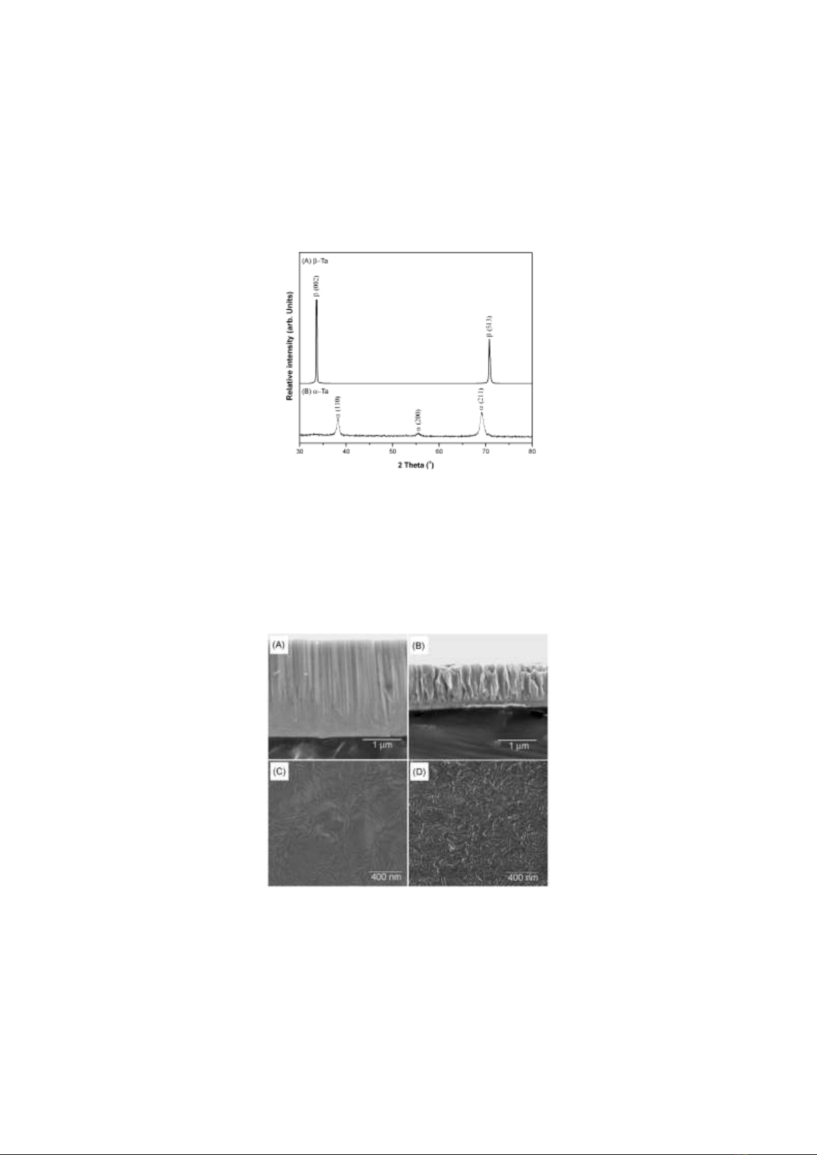
Fig. 1 (A) and (B) display typical XRD patterns of the Ta films deposited on Ti substrates with Vb
of 70 V and 150 V during reactive DC sputtering, respectively. On the one hand, the Ta film deposited
with Vb of 70 V exhibited a relatively strong peak at 2θ ~33.6o, corresponding to the (002) plane of the
crystalline β-Ta structure (JCPDS 25-1280), along with an additional peak at 2θ ~ 70.7o, corresponding
to (513) plane (pattern "A" in Fig. 1 ). On the other hand, when a negative substrate bias voltage of 150
D. M. Duc et al. / VNU Journal of Science: Mathematics – Physics, Vol. 41, No. 1 (2025) 9-16
12
V was applied, the Ta film showed a prominent peak at 2θ ~ 69.5o, corresponding to (211) plane of the
crystalline α-Ta structure (JCPDS 04-0788), as well as peaks at 2θ ~38.2o and 55.5o, associated with
(110) and (200) planes, respectively (pattern "B" in Fig. 1). These results demonstrated that the Ta films
deposited with Vb of 70 V and 150 V predominantly exhibited the single β-Ta and the α-Ta crystalline
phases, respectively.
Figure 1. XRD patterns of the β-Ta film deposited at Vb of 70 V (A) and α-Ta film deposited at Vb of 150 V (B).
The microstructures of the Ta films deposited with Vb of 70 V and 150 V are illustrated in Figs. 2
(A)-(D). With a negative substrate bias of 70 V, the Ta film showed a dense and smooth columnar
microstructure (Fig. 2A) with a smooth surface of β-Ta (Fig. 2C), whereas a negative substrate bias of
150 V resulted in a relatively rough columnar microstructure (Fig. 2B) with a rough surface of α-Ta.
The overall thicknesses of the Ta films deposited with Vb of 70 V and 150 V were approximately
2.5 µm and 1.2 µm, respectively.
Figure 2. Microstructures of the β-Ta films deposited with Vb of 70V ((A),(C)) and α-Ta films deposited with Vb
of 150 V ((B),(D)), ((A)-(B)) : cross-sectional view, (C)-(D) : surface morphology)
The surface hardness and elastic modulus of the Ta-deposited Ti substrates were assessed using an
ultra-low load microhardness test, which is an appropriate method to evaluate the mechanical property
D. M. Duc et al. / VNU Journal of Science: Mathematics – Physics, Vol. 41, No. 1 (2025) 9-16
13
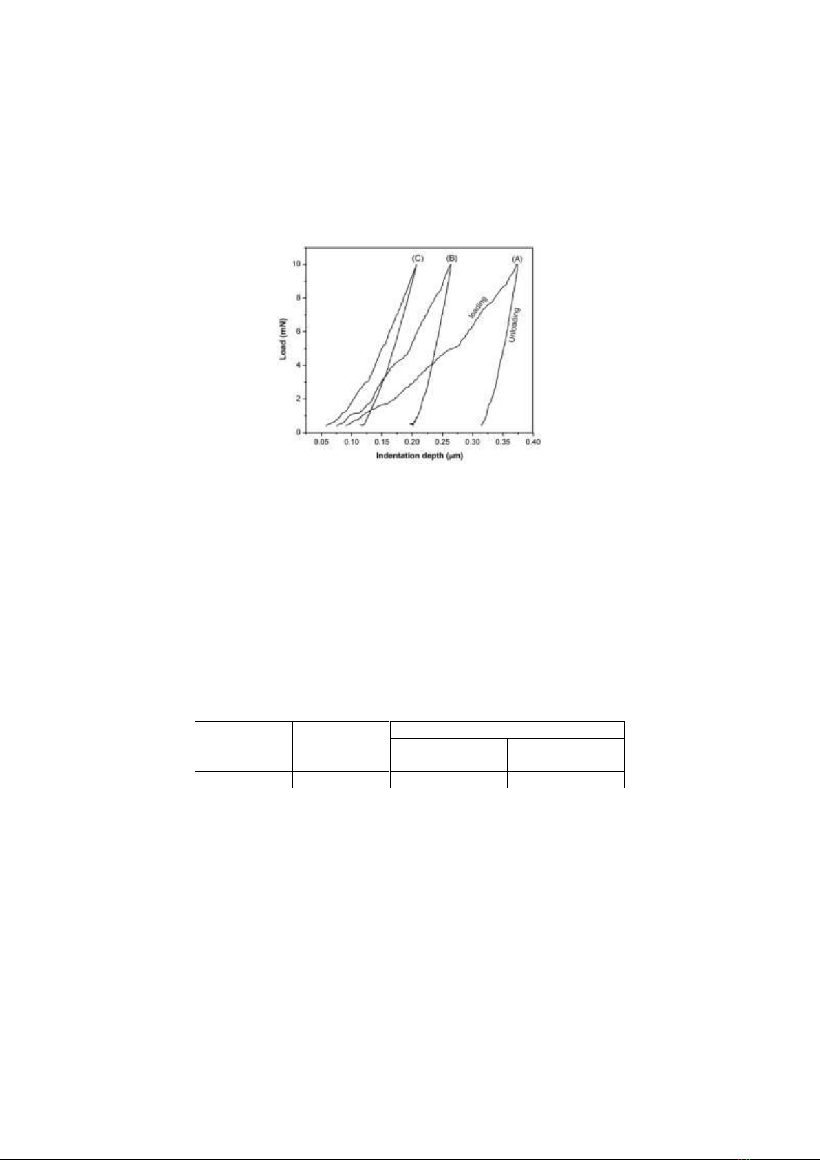
of thin film [19, 20]. The typical loading-indentation depth curves of the substrates (bare Ti and Ta-
deposited substrates with Vb of 70 V and 150 V) are presented in Fig. 3. Compared to the bare Ti, the
Ta-deposited substrates exhibited smaller indentation depths under the same load, indicating effective
surface hardening due to the deposition of the Ta films via DC sputtering.
Figure 3. Load-indentation depth curves of (A) the bare Ti, (B) the α-Ta-deposited Ti substrate
with Vb of 150 V, and (C) the β-Ta-deposited Ti substrate with a Vb of 70 V.
The hardness and elastic modulus derived from the loading-indentation depth curves are summarized
in Table 1. The deposition of the Ta films on the Ti substrate resulted in a significant increase in both
hardness and elastic modulus. Notably, the hardness of the β-Ta film deposited with a negative substrate
bias of 70 V was much higher than that of the α-Ta film deposited with a negative substrate bias of 150
V by approximately a factor of 2.5. While the hardness of α-Ta film in this study is lower than that of
α-Ta film reported in [18], the hardness of β-Ta film is higher than that of β-Ta film reported in [21].
The higher hardness of β-Ta was primarily attributed to its complicated four-layer stacking arrangement
of atoms, impeding dislocation motion [22].
Table 1. Hardness (H) and elastic modulus (E) of the bare Ti, Ta-deposited Ti substrates with Vb
of 70 V (β-Ta) and with Vb of 150 V (α-Ta)
Bare Ti
Ta-Deposited Ti Substrates
α- Ta
β-Ta
H [GPa]
3.6 ± 0.3
8.3 ± 0.3
21.0 ± 1.5
E [GPa]
117.2 ± 7.1
186.6 ± 8.9
171.0 ± 9.3
Fig. 4 illustrates the wettability images of three samples: bare Ti, α-Ta, and β-Ta. The contact angles
of these samples are 65°, 18°, and 21°, respectively, highlighting significant differences in the surface
properties of these materials. The bare Ti sample has the largest contact angle at 65°, indicating that this
surface is more hydrophobic compared to the samples coated with α-Ta and β-Ta. This suggests that
water is less likely to spread on the bare Ti surface, implying it has lower surface energy compared to
the coated samples.
The α-Ta sample has a contact angle of 18°, significantly lower than that of bare Ti. This reduction
in contact angle indicates that the α-Ta coating makes the surface more hydrophilic, possibly due to
changes in surface structure after coating α-Ta on the Ti substrate. The β-Ta sample, with a contact angle
of 21°, is also more hydrophilic than bare Ti, though slightly less than α-Ta. This indicates that the β-