
VNU Journal of Science: Mathematics – Physics, Vol. 41, No. 1 (2025) 49-60
49
Original Article
Preparation of Ag/Au Bimetallic Nanodecahedra through
Galvanic Displacement Method
Pham Thi Nga1,2, Pham Thi Thu Ha1,*, Nguyen Dac Dien3, Doan Thi Thao Anh1,4,
Tran Thu Trang1, Nguyen Vu Anh Tuyet5, Vuong Truong Xuan1, Vu Xuan Hoa1
1Institute of Science and Technology, TNU-University of Sciences, Tan Thinh, Thai Nguyen, Vietnam
2Hoa Lu University, 2 Xuan Thanh, Ninh Nhat, Hoa Lu, Ninh Binh, Vietnam
3Vietnam Trade Union University, 169 Tay Son, Quang Trung, Dong Da, Hanoi, Vietnam
4Bac Duyen Ha High School, 3 Hung Ha, Hung Ha, Thai Binh, Vietnam
6Hung Yen High School for the Gifted, 1 Chu Van An, An Ta, Hung Yen, Vietnam
Received 21st October 2024
Revised 26th November 2024; Accepted 15th February 2025
Abstract: In this work, Ag/Au bimetallic nanodecahedra was successfully synthesized using the
reduction process and photochemical method. Polyvinylpyrrolidone was used as a shape-directing
agent, AgNO3, HAuCl4 as precursor raw materials, sodium borohydride as a reducing agent for the
growth of Ag metal. Ag+ ions were reduced to Ag nanoparticles (AgNPs) as seeds using sodium
borohydride (NaBH4), and then Ag nanodecahedra (AgND) were prepared by a photochemical
seeding growth method under the illumination of blue light emitting diode (BLED) with a
wavelength of 480 nm. Next, an Au nanofilm was formed by chemical reduction on the surface of
the Ag nanodecahedra from precursors of HAuCl4 and ascorbic acid (C6H8O6). At the same time,
Ag was replaced by Au through a galvanic replacement reaction. As-prepared products have been
characterized by X-ray diffraction, scanning electron microscope (SEM), transmission electron
microscope (TEM), high-resolution transmission electron microscope (HRTEM), energy dispersive X-
ray spectroscopy (EDS), EDX mapping, and ultraviolet-visible (UV-Vis) absorption spectroscopy. The
intensity and position of UV-Vis peaks of AgND and Ag/Au changed as BLED illumination time and
HAuCl4 amount changed.
Keywords: Au/Ag bimetallic, galvanic replacement reaction, nanodecahedra, photochemical reduction.
1. Introduction*
An intrinsic property of metallic nanoparticles is localized surface plasmon resonance (LSPR). It
has been widely applied in photothermal therapy [1], medicine [2], biomedicine [3], surface-enhanced
________
* Corresponding author.
E-mail address: haptt@tnus.edu.vn
https//doi.org/10.25073/2588-1124/vnumap.4972

P. T. Nga et al. / VNU Journal of Science: Mathematics – Physics, Vol. 41, No. 1 (2025) 49-60
50
Raman scattering (SERS) substrates [4], antibacterial [5], etc. The plasmon optical properties of metallic
nanoparticles can be modulated by adjusting their shape, size, and composition. Single-element noble
metal materials such as gold (Au) and silver (Ag) have some limitations in terms of chemical stability
and reproducibility. Although Ag nanostructures were applied in surface-enhanced Raman scattering
(SERS) substrates owing to their reusability, biocompatibility, and high SERS performance, their
surface is highly sensitive to oxidation, so Ag-based SERS substrates have poor stability. To improve
the stability of Ag, an Au thin nanolayer that does not influence the sensitivity of Ag-based SERS
substrates has been coated on Ag core. Therefore, coating an Au nanolayer on Ag nanostructure is highly
desirable to overcome the drawback of pristine Ag. The combination of Au and Ag to form Au@Ag or
Ag@Au core-shell or Ag/Au bimetallic alloy has been reported in numerous works. Core-shell
nanostructures contain a metal core surrounded by other metal shell [6]. The position of the localized
surface plasmon resonance (LSPR) band has been shifted as the radius of the bimetallic spherical or
thickness of the shell increases [7].
For example, Yin et al., prepared Ag@Au core-shell dendrites via a two-step process. Ag dendrites
were grown on Si wafer using the hydrothermal corrosion method, and Au nanofilm was then fabricated
on the surface of Ag dendritic nanostructure by chemical reduction [8]. Huang et al., synthesized
Au@Ag dendritic nanoforests on silicon by fluoride-assisted galvanic replacement reaction method [9].
Yang et al., prepared gold-coated silver (Ag-Au) and silver-coated gold (Au-Ag) composite
nanoparticles by a seeding growth method [10]. Pham et al. developed an Au-Ag alloy on SiO2
nanoparticles, in which AgNO3 was reduced by ascorbic acid and using polyvinylpyrrolidone (PVP) as
the structure-directing agent, then Ag shell was grown on the surface of the Au seeds [11]. Fu et al.
synthesized Au-Ag alloy nano-garlands with multiple petal-like branches using HAuCl4 and AgNO3 as
precursors, ascorbic acid as a reducing agent, and 4-NTP as a shape-directing agent [12]. Liu et al.,
manufactured Ag core-embedded Au film by thermally depositing Ag/Au alloy target, and the
monodisperse Ag nanoparticles were embedded within the Au film [13]. Attila et al., used spark
discharge generation in the gas phase to fabricate Au/Ag nanoparticles onto glass microfiber filters [14].
Khan et al. fabricated Au-Ag alloy nanoparticles on filter paper by soaking filter paper in a mixture of
HAuCl4 and AgNO3 solution [15]. Lee et al., developed bimetallic Au/Ag nanocavities by galvanic
engineering [16]. Liu also prepared Ag-Au bimetals through galvanic replacement reactions [17], etc.
There is still a huge space for developing this kind of Ag/Au bimetallic nanostructure, especially
through the galvanic displacement route. Ag nanoparticles were commonly used as sacrificial templates
to synthesize alloy nanoparticles. For instance, Wang et al. synthesized Au-Ag nanocages with hollow
interiors and porous walls through galvanic replacement using Ag nanocubes as templates [5]. The
elemental composition and the shell layer-specific surface area can be controlled by varying the amount
of precursors and reaction time [18]. The cell dimension and crystal structure of Au and Ag metals are
almost the same. Ag has a higher plasmonic response than Au, but Au has better chemical stability
comparing to Ag. Combining Ag and Au leads to better SERS performance than pure Au or Ag
nanoparticles.
Herein, we prepared a new bimetallic Ag/Ag nanodecahedra composed of Ag core covered by Au
shell with various Au/Ag ratios. The samples were synthesized in two steps containing the
photochemical reduction and galvanic replacement reaction using silver nanodecahedra as a sacrificial
template to obtain hollow silver-gold alloy nanoshells. The dimension of the hollow cavities can be
controlled by varying the amount of HAuCl4. The prepared Ag/Au decahedra can be applied to the SERS
analysis.

P. T. Nga et al. / VNU Journal of Science: Mathematics – Physics, Vol. 41, No. 1 (2025) 49-60
51
2. Methods
2.1. Materials
Silver nitrate (AgNO3, 99.98%), trisodium citrate dihydrate (TSC, Na3C6H5O7.2H2O, 99%),
polyvinylpyrrolidone (PVP, molar weight MW = 40,000 g/mol), sodium borohydride (NaBH4, 99%),
sodium hydroxide (NaOH, 99%), L-ascorbic acid (L-AA, C6H8O6, 99.96%), chloroauric acid
(HAuCl4.4H2O, 99.9%) were purchased from Merck Co. (Germany). All the chemicals were of
analytical grade and used as received from the supplier without further purification. Double-distilled
water and absolute ethanol were used as solvents for solution preparation and rinsing of prepared
products. In this work, Ag was synthesized before Au.
2.2. Preparation of Ag Nanodecahedra
The Ag nanodecahedra was synthesized via a two-step experimental process. In the first step, Ag
nanoparticles were prepared using a reduction route. Typically, 2 ml AgNO3 solution (0.01 M), 0.4 ml
trisodium citrate (TSC, 0.6 M), and 0.6 ml polyvinylpyrrolidone (PVP, 0.0067 M) were mixed in 150
ml distilled water in an ice bath under continuous stirring for 15 min. The reaction temperature was
controlled at 4 C. Then, 0.5 ml NaBH4 solution (0.1 M) was added dropwise into the above mixture
under continuous stirring for 1 hour, and the color of the reaction solution gradually changed to grey-
yellow. The Ag+ ions from AgNO3 were reduced to produce metallic Ag nanoparticles.
3 4 2 3 3
2AgNO 2NaBH 2Ag H 2BH 2NaNO
(1)
Afterward, 0.4 ml NaOH solution (0.05 M) was added to the mixture and further stirring for 15 min.
In the second step, the growth of Ag nanodecahedra from Ag nanoparticles was performed in a
photochemical reaction apparatus. A blue light emitting diode (BLED, wavelength of 480 nm) was used
as a stimulating visible light source. The light power was maintained at 22.5 mW, and exposure time
was set at 5, 10, 20, 30, 40, 60, and 80 min. The corresponding Ag nanodecahedra were signed as
AgND5, AgND10, AgND20, AgND30, AgND40, AgND60, and AgND80, respectively. Ag
nanoparticles were self-assembled through a diffusion-limited aggregation process. Ag nanoparticles
adhere to form clusters governed by the lowest surface energy, allowing the formation of Ag
nanodecahedra. Finally, the obtained products were filtered and washed several times with deionized
water and absolute ethanol to remove excess sodium citrate, sodium borohydride remnant, and by-
products such as BH3 and NaNO3. After filtration, the samples were resuspended in deionized water and
stirred for 2 h to replace the surfactant (PVP) to achieve Ag nanodecahedra.
2.3. Formation of Au Nanolayer on the Surface of Ag Nanodecahedra
The reaction was carried out at room temperature (20 - 25 C). Firstly, the full amount of the as-
prepared AgND20, 0.4 ml ascorbic acid (L-AA, 0.01 M), and 0.6 ml polyvinylpyrrolidone (PVP, 0.0067
M) were dispersed in 50 ml deionized water using magnetic stirring. Then, 10 µl HAuCl4 with different
amounts of 0.15, 0.25, 0.35, 0.45, 0.55, and 0.65 µmol was slowly added into the above mixture under
magnetic stirring for 1 hour. The reaction solution changed color abundantly by adjusting the addition
of HAuCl4. The obtained Ag/Au bimetallic products were signed as Ag/Au1, Ag/Au2, Ag/Au3, Ag/Au4,
Ag/Au5, and Ag/Au6, respectively. Au nanolayer would be formed via the reduction of HAuCl4 by
ascorbic acid as the following equation:
6 8 6 4 2 2
3C H O 20HAuCl 18H O 20Au 80HCl 18CO
(2)
P. T. Nga et al. / VNU Journal of Science: Mathematics – Physics, Vol. 41, No. 1 (2025) 49-60
52
Spontaneously, Ag was replaced by Au through galvanic displacement. Finally, the obtained product
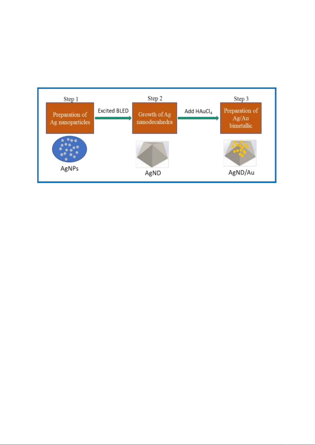
was washed with deionized water several times. Fig. 1 illustrates the fabrication process of Ag/Au
bimetallic nanodecahedra.
Figure 1. Illustration of Ag nanodecahedra and Ag/Au bimetallic fabrication procedure.
2.4. Characterizations
The phase identification of the samples was characterized via powder X-ray diffraction (XRD,
Bruker D8 Advance, Germany) operated at 30 kV with Cu-Kα radiation (wavelength of = 0.154056
nm) in the scanning angle 2 in the range of 30-80. The morphologies and microstructures were
observed via scanning electron microscopy (SEM, Hitachi S4800, Japan) operating at 10 kV,
transmission electron microscopy (TEM, JEOL JEM-1010, Japan) operating at 80 kV, high-resolution
TEM (HRTEM, JEOL JEM-1010, Japan) operating at 200 kV. A particle size analyzer (Micro 100C,
3P Instruments, Germany) was also utilized to measure the size distribution of samples. The chemical
composition was executed by energy-dispersive X-ray spectroscopy (EDS, Nova 450). The elemental
distribution in the nanostructures was examined through energy-dispersive X-ray (EDX) spectroscopy
mapping (Hitachi SU 8020) operating at an accelerating voltage of 200 kV. The optical properties were
monitored by recording the ultraviolet-visible (UV-Vis) absorbance spectra using a UV-Vis
spectrophotometer (JASCO V770, Japan) in the wavelength of 200–800 nm.
3. Results and Discussion
3.1. Samples Morphology and Size
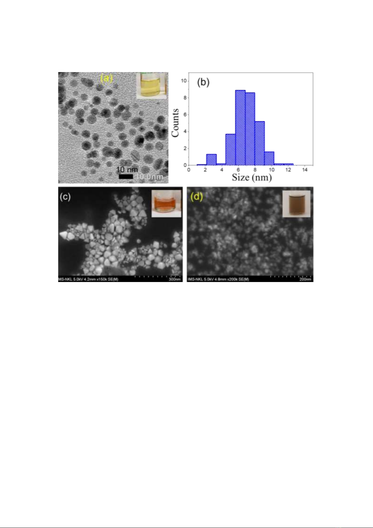
Fig. 2a shows a TEM image of AgNPs with homogeneous shapes and good dispersion. The average
diameter of the AgNPs is almost 7 nm (Fig. 2b). The TEM image of the Ag nanodecahedra (AgND)
before loading Au nanolayer is shown in Fig. 2c. The diameter of AgND is around 80 nm, and the
surface is smooth. Figure 2d is a SEM image of Ag/Au bimetallic nanostructure, showing that only
nanodecahedra can be clearly seen.
P. T. Nga et al. / VNU Journal of Science: Mathematics – Physics, Vol. 41, No. 1 (2025) 49-60
53
Figure 2. (a) TEM image of Ag nanoparticles, (b) size distribution of Ag seed nanoparticles; (c) SEM image of
AgND20, (d) SEM image of Ag/Au bimetallic, insets in (a, c, d) are the corresponding solutions.
After Au3+ ions reduction process, an Au nanolayer with a thickness of approximately 4 nm appears
on the Ag surface. The size of the Ag/Au bimetallic nanostructure is almost the same as that of the
primordial Ag nanodecahedra. To study the effect of HAuCl4 on the formation of bimetallic
nanostructure, the different amounts of HAuCl4 (i.e., 0.15, 0.25, 0.35, 0.45, 0.55, and 0.65 µmol) were
added into mixture of AgND, L-AA, and PVP. Figure 3a shows that when a small amount (0.15 µmol)
HAuCl4 is added, a thin layer is formed, and the arrangement between the nanodecahedra is close and
partially overlapping. The nanodecahedra have no obvious hollow cores. When increasing the HAuCl4
amount to 0.35 µmol, the morphology of the nanodecahedra changes dramatically, and adjacent
nanodecahedra with internal hollow cavities stacked together, the round edges and apices are instead of
sharp ones (Fig. 3c). When the amount of HAuCl4 was increased to 0.55 µmol, the average size of the
nanodecahedra increases from 80 to 90 nm due to the formation of Au nanofilms (Fig. 3e). The
nanodecahedra become larger and coarser, the gaps between neighboring nanodecahedra decrease. As the
Au layer continues to grow, it gradually expands outwards, wrapping around twelve sides to form an
almost rounded shell layer. With the increase of the amount of HAuCl4 upto 0.65 µmol, the difference
in the shapes and sizes of the nanodecahedra decreases, and they become more uniform. The distribution
of nanodecahedra was denser, the overlap between neighboring nanodecahedra increased, the particle
size of the nanodecahedra increased, and their sharpness decreased.


![Thí nghiệm Vật lí (BKEM-012): Tài liệu [Mô tả/Hướng dẫn/Bài tập,...]](https://cdn.tailieu.vn/images/document/thumbnail/2025/20251219/thanhlong020907@gmail.com/135x160/54561766129946.jpg)