
64 Nguyen Thanh Thu, Dinh Binh Duong, Tran Thi Thao, Pham Van Truong
AC-MLP: AXIAL CONVOLUTION-MLP MIXER FOR NUCLEI SEGMENTATION
IN HISTOPATHOLOGICAL IMAGES
Nguyen Thanh Thu, Dinh Binh Duong, Tran Thi Thao*, Pham Van Truong
School of Electrical and Electronic Engineering, Hanoi University of Science and Technology, Vietnam
*Corresponding author: thao.tranthi@hust.edu.vn
(Received: July 11, 2024; Revised: September 01, 2024; Accepted: September 27, 2024)
DOI: 10.31130/ud-jst.2024.332E
Abstract - Recent MLP-Mixer has a good ability to handle long-
range dependencies, however, to have a good performance, one
requires huge data and expensive infrastructures for the pre-training
process. In this study, we proposed a novel model for nuclei image
segmentation namely Axial Convolutional-MLP Mixer, by
replacing the token mixer of MLP-Mixer with a new operator,
Axial Convolutional Token Mix. Specifically, in the Axial
Convolutional Token Mix, we inherited the idea of axial depthwise
convolution to create a flexible receptive field. We also proposed a
Long-range Attention module that uses dilated convolution to
extend the convolutional kernel size, thereby addressing the issue
of long-range dependencies. Experiments demonstrate that our
model can achieve high results on small medical datasets, with Dice
scores of 90.20% on the GlaS dataset, 80.43% on the MoNuSeg
dataset, and without pre-training. The code will be available at
https://github.com/thanhthu152/AC-MLP.
Key words - Depthwise convolution; MLP-Mixer; Nuclei
segmentation; Token mixing
1. Introduction
The distribution and density of nuclei in the tissues are
important and necessary markers in cancer diagnosis.
Therefore, the detection and segmentation of cell nuclei is
getting more and more attention and is an essential task in
biomedical engineering. Nuclei segmentation aids in tissue
structure determination, cell growth analysis, and the study
of cell responses to environmental changes. On this basis,
researchers can derive cell characteristics, diagnose disease
severity, and research drugs.
Studies on cell nuclei identification have been around
for a long time. First of all, there is the appearance of the
microscope, which allows people to see cells with a
microscopic size. Microscopy supports the acquisition of
images of cells, from which traditional methods such as
threshold-based, region-based, and edge-based are widely
utilized to segment cell nuclei. However, the nuclei often
have tiny sizes and high distribution densities, which
causes many difficulties for the traditional segmentation
techniques.
The emergence of deep learning techniques in the field
of Computer Vision brings a novel approach to image
segmentation, particularly in the realm of medical imaging.
The presence of Convolutional Neural Networks (CNNs) is
a leap and opens a series of related studies in image
processing. In 2015, the U-Net model was introduced, which
utilized a U-shaped architecture to effectively segment
medical images. This model extracts multi-scale context
information through its encoder and reconstructs the input
size through its decoder, while skip connections are used to
avoid information loss. Since then, many variants of this
architecture have been proposed to improve performance.
Some typical models can be mentioned as Double Unet [2],
Attention Unet [3], Unet++ [4], ResUnet ++ [5], etc. On the
other hand, concerned with computational cost and real-life
applications, researchers began to focus on lightweight
models. Therefore, depthwise separable convolutions are
now more commonly used as alternatives for conventional
convolutions to reduce the number of parameters while still
achieving high performance. For example, DSCA-Net [6]
combines depthwise separable convolution with an attention
mechanism in a U-shape architecture to create a lightweight
network for accurate medical image segmentation.
MobileNets [7] is another lightweight model which
employed depthwise convolutions and was successfully
embedded in mobile visual applications. Most recently, U-
Lite [8] was introduced as an effective model with less than
1 million parameters, using axial depthwise convolution,
which can give promising results on medical datasets.
Recently, the arrival of Vision Transformer [9] has
attracted a lot of research and overwhelmed CNNs
dominance in computer vision. Vision Transformer (ViT)
applied the Transformer from the Natural Language
Processing (NLP) domain to Computer Vision by dividing
an image into many patches and flattening them to vectors
before taking them as input of the Transformer. With the
self-attention mechanism, ViT has a better ability to learn
long-range dependencies and extract global context
information than CNNs. However, Transformer has a large
amount of computation and may require a lot of resources
during the training process. Swin Transformer [10] was
proposed then to reduce the computational cost by limiting
self-attention computation to non-overlapping local
windows. Besides Transformers, Multi-layer Perceptrons
(MLPs) have also been applied to vision tasks, but are less
common. MLP-Mixer inherits the patch partition of ViT
and passes embedded patches through several layers of the
token mixer and channel mixer. Both these mixing
operators utilize pure MLP, while they can still achieve
comparable results to those of CNN-based models and
transformer-based models on classification tasks. Similar
to ViT, MLP-Mixer is also successful in extracting global
information from input images. However, both require a
large enough dataset to achieve good performance.
In the field of medical image segmentation, obtaining a
large data set, particularly for nuclei, can be challenging.
In the nuclei segmentation task, because the density of cells
is very thick and the size of a cell is small, experts must

ISSN 1859-1531 - THE UNIVERSITY OF DANANG - JOURNAL OF SCIENCE AND TECHNOLOGY, VOL. 22, NO. 12, 2024 65
take great care and spend a significant amount of time
accurately segmenting cell nuclei in order to produce a
high-quality dataset. Moreover, because of the small size
of nuclei, local receptive fields may help aggregate context
information from nuclei better. To address these
challenges, we develop a new model that leverages ideas
from the MLP-Mixer and axial depthwise separable
convolution of the U-Lite model. By using axial depthwise
separable convolution to replace the MLP-Mixer token
mix, our proposed model has achieved the following
contributions:
• Propose Axial Convolution Mixer module based on
the concept of MLP-Mixer.
• Propose AC-MLP model for image segmentation task
with two branches of the encoder and adapt attention to the
decoder.
• Experimentally, AC-MLP achieves the state-of-the-
art (SOTA) performance on small datasets like GlaS and
MoNuSeg using a low number of parameters without pre-
trained.
2. Related work
2.1. Axial Depthwise Convolution
U-Lite [8] takes the usage of axial depth-wise
convolution as the main operator to aggregate spatial
information of the feature maps. This module is established
by simply taking the sum of two separated operators 1× 7
and 7×1 depth-wise convolution, axial depth-wise
convolution does not overly increase the number of
learnable parameters as well as the model's complexity.
However, it can result in a comparable or even slightly
better performance due to the inductive bias in the
receptive field.
2.2. MLP-Mixer
Besides Transformers and CNNs, MLP-like models
have been widely used and are known as a recently
emerging paradigm for Computer Vision. MLP-Mixer is
the first study that utilized pure MLP as token-mixers on
spatial and channel representations of the feature map.
Specifically, for the image classification task, the image is
first passed through a per-patch fully-connected layer for
patch embedding. After that, they are adapted to several
numbers of mixer layers. Each layer comprises two stages
separately, one is Token-mixer for spatial feature
extraction, and the remaining one is responsible for
Channel-mixing, i.e., encoding features along their channel
dimension. Finally, a classification header is designed in
the last layer for the classification tasks. Despite having a
very straightforward architecture, MLP-Mixer can achieve
promisingly comparable results on ImageNet's
classification benchmarks. This new paradigm has inspired
various architectures for performance improvements,
including ViP [12], CycleMLP [13], and AS-MLP [14].
2.3. Progressive Atrous Pyramid Pooling (PASPP)
PASPP [15] utilizes multiple atrous convolutional
layers with different dilation rates and progressive
concatenations to capture multi-scale representations of an
object in feature maps. It has been experimented that this
module can impressively achieve a better performance on
image segmentation tasks compared to the previously
proposed module ASPP [16]. Based on the belief that the
larger the dilation rate is, the more global information the
model can aggregate, PASPP not only provides a larger
receptive field to the model compared to traditional 3×3
convolutions but also retains the number of computational
parameters within a limited resource.
2.4. Convolutional Block Attention Module (CBAM)
Inspired by Squeeze and Excitation [17], CBAM [18]
is regarded as a lightweight efficient attention module that
considerably improves the performance of CNN
architectures. A CBAM contains two main stages: channel
attention and spatial attention. The channel attention
module applies max-pooling and average pooling on every
spatial dimension to aggregate important information per
each channel of the feature maps. They are next passed
through double fully-connected layers, a sigmoid layer,
and then multiplied again with the input. This mechanism
helps to determine which channels are more important than
other ones. The spatial attention module is implemented
similarly, however, 7×7 convolutions are utilized instead
of fully-connections due to the limitation of calculation,
which allows the model to precisely focus on spatial
representations of the feature maps.
3. Methodology
Inspired by the architecture of Axial Attention MLP-
Mixer [19], in this study, we proposed a similar
architecture with some new enhancements, namely Axial
Convolution-MLP Mixer (AC-MLP). The specific
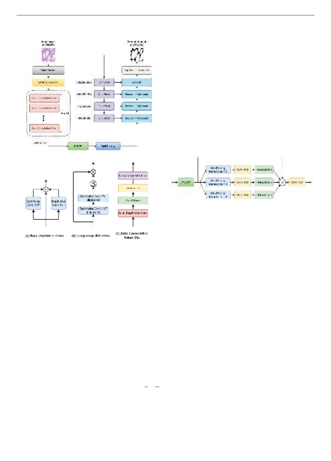
architecture of our proposed model is shown in Figure 1.
Given an input image 𝐼𝑜∈ ℝ3×𝐻×𝑊. The encoder of our
proposed model consists of two branches to extract
features. In the first branch, the image is divided into many
non-overlapping patches of size 𝑝× 𝑝. After that, all
patches are projected to vectors of the same size and
become the input of a network with 12 successive Axial
Convolution Mixer blocks to produce context information.
The output of this network undergoes a bottleneck block
comprising a PASPP module and Multi-Pooling layers,
working together to synthesize beneficial features. Parallel
to the first branch, in the second branch, the input image is
passed through consecutive Conv Block (Figure 4b) and
MaxPooling layers to extract context information on
multiple scales. Finally, after having the features from the
input image, we combine these features in both branches of
the model and pass them through the decoder and upscale
to select information and reconstruct the original size of the
input image before giving the final prediction mask.
3.1. Proposed Axial Convolution Mixer
The effectiveness of Axial Depthwise Convolution
throughout the U-Lite model has motivated us to exploit
this module to develop a novel spatial mixing operator,
serving as a replacement for the token mixing mechanism
of the MLP-Mixer, with the aim of adapting to the task of
nucleus segmentation. While global information is
undoubtedly important in image segmentation, we
hypothesize that, for nucleus segmentation, local
66 Nguyen Thanh Thu, Dinh Binh Duong, Tran Thi Thao, Pham Van Truong
information and dense granularity are the features that
warrant greater focus. Consequently, the parallel use of
horizontal and vertical kernels, as implemented in Axial.
Firgure 1. General structure of the proposed model
Figure 2. Proposed Axial Convolution Token Mixing module
Depthwise Convolution, offers a more flexible
approach to learning local information, in contrast to
utilizing MLP networks to capture global spatial
relationships between pixels, as in the token-mixer
architecture of the MLP-Mixer. Furthermore, compared to
Transformer-based models, Axial Depthwise Convolution
enhances the focus on learning local information along the
horizontal and vertical dimensions of pixels, which aligns
well with the compact and densely structured nature of
nuclei. To address the limitations in learning long-range
dependencies, we propose a long-range attention module
(Figure 2b) subsequent to Axial Depthwise Convolution,
employing dilated convolution to expand the receptive
field. The mathematical formulation of this module is
presented as follows:
𝑦 = GELU(BN(ADC(𝑥))) (1)
𝑧 = 𝑦 × Sigmoid(DW1×7,𝑟=1 (DW7×1,𝑟=2(𝑦))) (2)
where, 𝑥 is the input features with the shape 𝐶 ×𝐻
𝑝×𝑊
𝑝;
𝑧 is the output features; BN, ADC and DW stand for Batch
Normalization, Axial Depthwise Convolution and
Depthwise Convolution, respectively, and 𝑟 is the dilation
rate of the convolution. The new spatial mixer mentioned
above is used to replace token mixer of MLP-Mixer
architecture and we have a new module, namely Axial
Convolution Mixer as show in Figure 2 and Figure 4a.
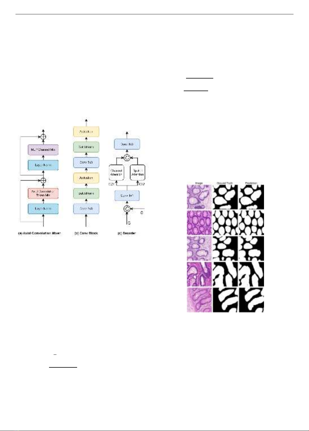
3.2. Bottleneck Block
In the bottleneck of AC-MLP, we utilize the PASPP
[15] and Multi-Pooling to capture multi-scale
representations of high-level feature maps. As depicted in
Figure 3, the Multi-Pooling uses three max-pooling layers
with different sizes of kernel 𝑘 = 2,4 and 8. These max-
pooled features are passed through point-wise
convolutions and then interpolated back to the original
shape. Finally, we concatenate them with the output of
PASPP, and one gain feed the encoded features to 1 × 1
convolution to recover the features' dimension. Different
from PASPP, Multi-Pooling can focus on the most
representative characteristics of an image, surpass non-
essential information, and thus give the model an inductive
bias. The intuition here is that depending on each image
and data, the nuclei may have different shapes and they can
stand alone or gather in groups, then a combination of
convolution and max-pooling operators, where the kernel
size varies flexibly, is an effective way to detect them.
Figure 3. Bottleneck design of AC-MLP model
3.3. Decoder Block
After the initial feature extraction process, wherein
feature maps are obtained from two branches of the
encoder, we propose a novel decoder architecture that
capitalizes on an attention mechanism, as illustrated in
Figure 4c. To begin, we concatenate the feature maps
derived from the two branches. Subsequently, a pointwise
convolution operation is employed to transform and adjust
the channel dimensions, effectively enhancing the
subsequent processing. Inspired by the Channel and Spatial
Attention Module (CBAM) concept, our approach
differentiates itself by reimagining the arrangement of
Channel Attention and Spatial Attention into parallel
blocks, in contrast to the linear configuration seen in
CBAM. This innovative parallel configuration help the
model to extract and emphasize crucial insights from both
the spatial and channel dimensions of the feature maps. The
outputs of these parallel blocks are then concatenated,
facilitating the cohesive integration of the identified salient
features. This unified representation then undergoes
convolution, utilizing a 3 ×3 kernel, as a pivotal step in
the subsequent stages of feature refinement.
4. Experiment
4.1. Implementation Detail
4.1.1. Dataset
To evaluate the effectiveness and efficiency of our
proposed method, we utilize two histopathological nuclei
datasets for the image segmentation task. The Gland
Segmentation (GlaS) dataset [20] introduced in the Colon
Histology Images Challenge Contest, was created to
promote research in segmentation algorithms on images of
ISSN 1859-1531 - THE UNIVERSITY OF DANANG - JOURNAL OF SCIENCE AND TECHNOLOGY, VOL. 22, NO. 12, 2024 67
hematoxylin and Eosin (H&E) stained slides. This dataset
consists of 74 benign images and 91 malignant images in
total, which is divided into 85 images for training and 80
images for testing. Multi-organ Nucleus Segmentation
(MoNuSeg) dataset [21], another nuclei dataset, aims to
look for the best nuclei segmentation techniques on a
diverse set of H&E stained histology images obtained from
multiple organs of patients. This dataset includes 30
images for training and 14 images for testing, with nearly
30,000 nuclear boundary annotations. In our experiment,
all the images are resized to 256×256. Furthermore, the
training images are pre-processed before feeding to the
model through augmentation techniques including image
rotating, horizontal flipping, and vertical flipping to enrich
the training data and avoid over-fitting.
Figure 4. The structure of Axial Convolution Mixer,
Double Convolution and Decoder Block
4.1.2. Training strategy
We conducted the proposed model on the PyTorch
framework running on NVIDIA Tesla T4 GPU with 16GB
of memory. The training process was implemented on 100
epochs with a batch size of 16, where the learning rate was
initialized at 0.001 and decayed by a factor of 2 after every
10 epochs. During the training phase, we adopted the
Adam optimizer [22] with the composite loss function
between Binary Cross-Entropy (BCE) loss and Dice loss.
Define 1 × 𝐻 × 𝑊 as the shape of the predicted mask and
𝑁 = 𝐻 ×𝑊 presents its total number of pixels. The loss
function employed in the experiment is presented as
follows:
𝐿Total(𝑦,𝑦)= 𝛾𝐿BCE(𝑦,𝑦)+(1− 𝛾)𝐿Dice(𝑦,𝑦) (3)
𝐿BCE(𝑦,𝑦)= −1
𝑁∑ [𝑦𝑖log𝑦𝑖+(1−𝑦𝑖)log(1−𝑦𝑖)]
𝑁
𝑖=1 (4)
𝐿Dice = 1− 2∑𝑦𝑖 𝑦
𝑖
𝑁
𝑖=1
∑(𝑦𝑖+𝑦
𝑖)
𝑁
𝑖=1 +𝜀 (5)
Where, 𝑦 = {𝑦1,𝑦2,…,𝑦𝑁} and 𝑦 = { 𝑦1,𝑦2,…,𝑦𝑁} are
respectively sets of pixels on the ground truth and the
predicted mask, where 𝑦𝑖∈{0,1} and 𝑦𝑖∈(0,1) for all
𝑖 = 1,2,…,𝑁. Besides, 𝜀 was added to avoid the zero-
denominator. In our experiment, 𝛾 = 0.5, and 𝜀 = 1.0.
4.1.3. Evaluation metric
To quantitatively evaluate the performance, we utilized
Dice Similarity Coefficient (Dice) and Intersect over
Union (IoU) metrics, which are standard evaluation
indicators typically used for calculating the overlap
between the ground truth and the predicted mask. The
mathematical representations of Dice and IoU are
expressed as follows:
Dice = 2TP
2TP+FP+FN (6)
IoU =TP
TP+FP+FN (7)
where TP, FP, FN respectively stand for True Positives,
False Positives and False Negatives between the ground
truth and the prediction of an image.
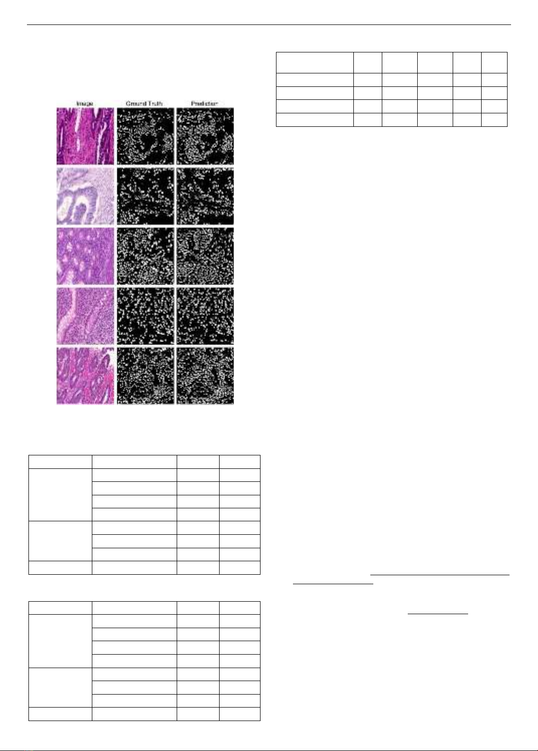
4.2. Representative Results
Figure 5 and Figure 6 show some segmentation results
of AC-MLP model on GlaS and MoNuSeg datasets,
respectively. It is observed that the predictions from our
model match well with the ground truths. Specifically, the
model can properly detect the boundary of each nucleus on
GlaS, and maintain the spatial arrangement of the multi-
organ nuclei on MoNuSeg, even though they are more
numerous and varied.
Figure 5. Some representative results of AC-MLP for
Gland Segmentation (GlaS)
4.3. Comparative Results
Table 1 and Table 2 evaluate the quantitative results on
two nuclei datasets GlaS and MoNuSeg, respectively. We
compared our model with other state-of-the-art
architectures including both the CNN-based and
Transformer-based models. As can be seen from the tables,
our proposed method outperforms the previously proposed
models in terms of Dice and IoU metrics on both datasets.
Specifically, AC-MLP reaches Dice scores of 90.20% on
the GlaS dataset and 80.43% on the MoNuSeg dataset. This
demonstrates the effectiveness of our approach in
accurately segmenting nuclei in histopathology images.
68 Nguyen Thanh Thu, Dinh Binh Duong, Tran Thi Thao, Pham Van Truong
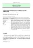
In Table 3, we further demonstrate the efficiency of the
AC-MLP model, where its Dice and IoU results surpass
those of the other MLPs-based variants. One noticeable
realization is that AC-MLP has significantly fewer
parameters than MLP-Mixer, with a total of 8.5M
parameters, while still delivering good performance.
Figure 6. Some representative results of AC-MLP for
Multi-organ Nucleus Segmentation (MoNuSeg)
Table 1. Quantitative evaluation results on GlaS dataset
compared to previously proposed model
Type
Model
Dice
IoU
CNNs baselines
UNet [1]
77.78
65.34
Unet++ [4]
78.03
65.55
ConvUNeXt [23]
78.04
64.42
UneXt [24]
86.49
77.77
Transformers
baselines
Axial Attn Unet [19]
76.30
63.03
MedT [25]
81.02
69.61
Swin Unet [26]
88.25
79.86
Ours
AC-MLP
90.20
82.89
Table 2. Quantitative evaluation results on MoNuSeg dataset
compared to previously proposed models
Type
Model
Dice
IoU
CNNs baselines
UNet [1]
76.45
62.86
Unet++ [4]
77.57
66.20
ConvUNeXt [23]
73.70
60.07
UneXt [24]
78.04
64.42
Transformers
baselines
Axial Attn Unet [19]
76.83
62.49
MedT [25]
79.55
64.42
Swin Unet [26]
78.49
64.72
Ours
AC-MLP
80.43
67.46
Table 3. Quantitative comparisions with variants of
MLPs on GlaS dataset
Methods
Patch
size
Depth
(layer)
Params
(M)
Dice
IoU
MLP-Mixer [11]
16
24
100
82.83
70.81
Permutator [12]
8
36
74
84.21
72.80
AxialAtt-MLP [19]
8
24
29
84.99
73.97
Ours
8
12
8.5
90.20
82.89
5. Conclusion
In this paper, we leverage the primary knowledge about
Axial Depthwise Convolution and MLP-Mixer
architecture to propose a new model AC-MLP for
histopathological nuclei image segmentation. Our novel
module, Axial Convolution Token Mixing, is designed to
capture large-scale information and preserve long-range
dependencies. Experimental results show that AC-MLP
can achieve SOTAs performance meanwhile having an
efficient number of computational parameters. In the
future, we will thoroughly consider the encoder's CNN-
based branch to further improve the model's performance
for the large use case of medical images on the
segmentation task.
Acknowledgement: This research is funded by Vietnam
National Foundation for Science and Technology
Development (NAFOSTED) under grant number
102.05-2021.34.
REFERENCES
[1] O. Ronneberger, P. Fischer, and T. Brox, “U-net: Convolutional
networks for biomedical image segmentation”, Medical Image
Computing and Computer-Assisted Intervention–MICCAI 2015:
18th International Conference, Munich, Germany, October 5-9,
Proceedings, Part III 18. Springer International Publishing, 2015.
[2] D. Jha, M. A. Riegler, D. Johansen, P. Halvorsen, and H.D.
Johansen, “Doubleu-net: A deep convolutional neural network for
medical image segmentation”, 2020 IEEE 33rd International
symposium on computer-based medical systems (CBMS), 2020.
[3] O. Oktay et al., “Attention u-net: Learning where to look for the
pancreas”, arXiv preprint arXiv:1804.03999, 2018.
[4] Z. Zhou, M.M.R. Siddiquee, N. Tajbakhsh, and J. Liang, “Unet++:
A nested u-net architecture for medical image segmentation”, Deep
Learning in Medical Image Analysis and Multimodal Learning for
Clinical Decision Support: 4th International Workshop, DLMIA
2018, and 8th International Workshop, ML-CDS 2018, Held in
Conjunction with MICCAI 2018, Granada, Spain, September 20,
2018, Proceedings 4. Springer International Publishing, 2018.
[5] D. Jha et al., “Resunet++: An advanced architecture for medical
image segmentation”, 2019 IEEE International Symposium on
Multimedia (ISM). IEEE, 2019.
[6] T. Shan, J. Yan, X. Cui, and L. Xie, “DSCA-Net: A depthwise
separable convolutional neural network with attention mechanism
for medical image segmentation”, Math Biosci Eng., vol. 20, pp.
365-382, 2022.
[7] AG. Howard et al., “Mobilenets: Efficient convolutional neural
networks for mobile vision applications”, arXiv preprint
arXiv:1704.04861, 2017.
[8] D.B. Dinh, T.T Nguyen, T.T. Tran, and V.T Pham, “1M parameters are
enough? A lightweight CNN-based model for medical image
segmentation”, 2023 Asia Pacific Signal and Information Processing
Association Annual Summit and Conference (APSIPA ASC). IEEE, 2023.
[9] A. Dosovitskiy et al., “An image is worth 16x16 words:
Transformers for image recognition at scale”, arXiv preprint
arXiv:2010.11929, 2020.
![Máy lạnh trung tâm (Central Air Conditioner Units) và ngành lạnh (Refrigeration): [Thông tin chi tiết/Hướng dẫn/ Tư vấn]](https://cdn.tailieu.vn/images/document/thumbnail/2012/20120202/luly_meo1/135x160/refrigeration_and_air_conditioning_equipment_cooling_split_2_9572.jpg)


![Mạch đo và khống chế nhiệt độ P3: Đề tài [Mới nhất]](https://cdn.tailieu.vn/images/document/thumbnail/2010/20100922/goixanh/135x160/mach_do_va_khong_che_nhiet_do_p3_5214.jpg)
![Mạch đo và khống chế nhiệt độ P2: Đề tài [Mô tả/Định tính]](https://cdn.tailieu.vn/images/document/thumbnail/2010/20100922/goixanh/135x160/mach_do_va_khong_che_nhiet_do_p2_6524.jpg)


![Đề ôn tập cuối kỳ môn Kỹ thuật nhiệt - Nhiệt động học [mới nhất]](https://cdn.tailieu.vn/images/document/thumbnail/2026/20260310/hoaphuong0906/135x160/60681773197823.jpg)

![Bài giảng thang máy và thang cuốn: Tổng hợp kiến thức [chuẩn nhất]](https://cdn.tailieu.vn/images/document/thumbnail/2026/20260310/hoaphuong0906/135x160/41471773283876.jpg)
