RESEARC H Open Access
The effect of acyclovir on the tubular secretion of
creatinine in vitro
Patrina Gunness
1,2
, Katarina Aleksa
1
, Gideon Koren
1,2*
Abstract
Background: While generally well tolerated, severe nephrotoxicity has been observed in some children receiving
acyclovir. A pronounced elevation in plasma creatinine in the absence of other clinical manifestations of overt
nephrotoxicity has been frequently documented. Several drugs have been shown to increase plasma creatinine by
inhibiting its renal tubular secretion rather than by decreasing glomerular filtration rate (GFR). Creatinine and
acyclovir may be transported by similar tubular transport mechanisms, thus, it is plausible that in some cases, the
observed increase in plasma creatinine may be partially due to inhibition of tubular secretion of creatinine, and not
solely due to decreased GFR. Our objective was to determine whether acyclovir inhibits the tubular secretion of
creatinine.
Methods: Porcine (LLC-PK1) and human (HK-2) renal proximal tubular cell monolayers cultured on microporous
membrane filters were exposed to [2-
14
C] creatinine (5 μM) in the absence or presence of quinidine (1E+03 μM),
cimetidine (1E+03 μM) or acyclovir (22 - 89 μM) in incubation medium.
Results: Results illustrated that in evident contrast to quinidine, acyclovir did not inhibit creatinine transport in
LLC-PK1 and HK-2 cell monolayers.
Conclusions: The results suggest that acyclovir does not affect the renal tubular handling of creatinine, and hence,
the pronounced, transient increase in plasma creatinine is due to decreased GFR, and not to a spurious increase in
plasma creatinine.
Background
Acyclovir is an antiviral agent that is commonly used to
treat severe viral infections including herpes simplex and
varicella zoster, in children [1]. Acyclovir is generally well
tolerated [2], however, in some cases, severe nephrotoxi-
city has been reported [2-8]. Acyclovir - induced nephro-
toxicity is typically evidenced by elevated plasma
creatinine and urea levels, the occurrence of abnormal
urine sediments or acute renal failure [2-5,7,8].
Crystalluria leading to obstructive nephropathy is
widely believed to be the mechanism of acyclovir -
induced nephrotoxicity [9]. However, there are several
documented cases of acyclovir - induced nephrotoxicity
in the absence of crystalluria [7,8,10]; suggesting that
acyclovir induces direct insult to tubular cells. Recently,
we provided the first in vitro experimental evidence
which supports existing clinical evidence of direct renal
tubular damage induced by acyclovir [11].
A systematic review of the literature reveals a pro-
nounced, transient elevation (up to 9 fold in some
cases) of plasma creatinine levels in children, often with-
out any other clinical evidence of overt nephrotoxicity
(Table 1). Similar to the cases described in Table 1; a
marked, transient increase in plasma creatinine levels
has been observed in some patients who received the
non-nephrotoxic drugs, cimetidine [12-16], trimetho-
prim [17-19], pyrimethamine [20], dronedarone [21] and
salicylates [22].
Creatinine, a commonly used biomarker that is used to
assess renal function, is eliminated by the kidney via both
glomerular filtration and tubular secretion [23]. The
mechanisms underlying the renal tubular transport of
creatinine has not been fully elucidated. As explained by
Urakami and colleagues [24], both acid and base secret-
ing mechanisms may play a role in the renal tubular
transport of creatinine [13-15,17-22,25-27]. Hence, some
* Correspondence: gkoren@sickkids.ca
1
Division of Clinical Pharmacology and Toxicology, The Hospital for Sick
Children, 555 University Avenue, Toronto, Ontario, M5G 1X8, Canada
Full list of author information is available at the end of the article
Gunness et al.Journal of Translational Medicine 2010, 8:139
http://www.translational-medicine.com/content/8/1/139
© 2010 Gunness et al; licensee BioMed Central Ltd. This is an Open Access article distributed under the terms of the Creative
Commons Attribution License (http://creativecommons.org/licenses/by/2.0), which permits unrestricted use, distribution, and
reproduction in any medium, provided the original work is properly cited.

drugs may share similar renal tubular transport mechan-
isms with creatinine. Drugs that share transport mechan-
isms with creatinine may compete with it for tubular
transport, and subsequently inhibit creatinine secretion
to result in a ungenuine elevation of plasma creatinine
that may not be due to decreased glomerular filtrate rate
(GFR). Cimetidine [12-16], trimethoprim [17-19], pyri-
methamine [20], dronedarone [21] and salicylates [22]
are examples of drugs that share similar renal tubular
transport mechanisms with creatinine and induce spur-
ious increases in plasma creatinine by competing with
and subsequently inhibiting its secretion.
Similar to creatinine, both acid and base secreting
pathways may be involved in the renal tubular transport
of acyclovir [28]. Additionally, it is likely that creatinine
[24-26] and acyclovir [28] may be transported by similar
organic anion transporters (OAT) and organic cation
transporters (OCT). Therefore, it is plausible that acy-
clovir may compete with and successively inhibit renal
secretion of creatinine, resulting in elevations in plasma
creatinine that may be disproportional to the degree of
renal dysfunction.
Employing plasma creatinine levels to estimate GFR,
results from previous studies [29,30] have illustrated
that acyclovir - induced nephrotoxicity induces a signifi-
cant reduction in GFR in children. However, based on:
(1) the cases presented in Table 1, (2) the awareness
that several non-nephrotoxic drugs are known to induce
transient increases in plasma creatinine [12-22] and (3)
the knowledge that acyclovir and creatinine may share
similar renal tubular transport mechanisms; we hypothe-
sized that the pronounced, transient increase in plasma
creatinine levels observed in some patients may be par-
tially due to the inhibition of renal tubular secretion of
creatinine by acyclovir, and not entirely the result of
decreased GFR. To the best of our knowledge, the effect
of acyclovir on the renal tubular secretion of creatinine
in vitro has not been previously evaluated. Thus, the
objective of the study was to determine whether acyclo-
vir inhibits the renal tubular secretion of creatinine. It is
important to determine whether acyclovir inhibits the
tubular transport of creatinine, because if this is the
case, then in addition to creatinine, other biomarkers
should always be employed to assess renal function in
patients receiving acyclovir treatment.
In the present study we were specifically interested in
determining the possible interaction between creatinine
and acyclovir during renal tubular transport by the OCT
pathway. The porcine renal tubular cell line, LLC-PK1,
has been used as an in vitro renal tubular model in a
vast array of transepithelial transport studies. Further-
more, the LLC-PK1 cells are an appropriate in vitro
model for specifically studying renal tubular transport of
organic cations because they are known to possess func-
tional OCTs [31-33]. However, although the LLC-PK1
cells retain similar physiological and biochemical prop-
erties compared to human renal proximal tubular cells
[34], interspecies differences in drug disposition exists
[35-37]. Hence, the use of a human renal proximal tub-
ular cell line, such as the HK-2 cell line, would be a
more suitable in vitro model to study the mechanisms
of renal tubular drug transport in humans. Porcine
LLC-PK1 and human HK-2 cells were employed in our
transepithelial transport studies.
Methods
Cell culture
The LLC-PK1 cells (American Type Culture Collection
(ATCC), USA) were cultured in growth medium which
consisted of Minimum Essential Medium (MEM) alpha
modified (Fisher Scientific, Canada), supplemented with
2 mM L-glutamine, 100 units/mL penicillin, 100 μg
Table 1 Cases of elevated plasma creatinine levels in children who received intravenous acyclovir
Patient Magnitude of increase in plasma
creatinine
(from baseline)
Relevant clinical details References
1 child 5 fold increase within 2 days Creatinine returned to normal in 4 days
Elevated urea
No other pathology reported
[4]
10
children
transient elevation No further impairment reported [2]
3
children
4 fold increase within 4 days Mild reduction in urine output
Creatinine returned to normal 1 week following acyclovir discontinuation
[3]
1 child 2 fold increase within 6 days Creatinine continued to increase following acyclovir discontinuation. Creatinine
returned to normal within 1 week
Elevated urea
Mild proteinuria
[7]
3
children
9 fold increase within 2 to 3 days High urea
Urinary a
1
-microglobulin and albumin
Creatinine returned to normal in 3 - 9 days
[8]
1 child 3 fold increase within 4 days No other information provided [5]
Gunness et al.Journal of Translational Medicine 2010, 8:139
http://www.translational-medicine.com/content/8/1/139
Page 2 of 11

streptomycin and 10% (v/v) fetal bovine serum (Invitro-
gen Canada Inc., Canada). The HK-2 cells (ATCC) were
cultured in growth medium which consisted of Kerati-
nocyte-Serum Free Medium, supplemented with human
recombinant epidermal growth factor 1-53 (5 ng/mL)
and bovine pituitary extract (0.05 mg/mL) (Invitrogen
Canada Inc.) The LLC-PK1 and HK-2 cells were main-
tained at 37°C in a sterile, humidified atmosphere of 5%
CO
2
and 95% O
2
.
Transepithelial transport studies
The transepithelial transport studies were conducted as
outlined by Urakami et al. [33] with modifications. The
LLC-PK1 and HK-2 cells were seeded at densities of
4.5E+05 cells/0.9 cm
2
and 5.0E+05 cells/0.9 cm
2
, respec-
tively, on microporous membrane filter inserts (3 μm
pore size, 0.9 cm
2
growtharea)thatwereplacedinside
cell culture chambers (VWR International, Canada). A
consistent (1 mL) volume of growth or incubation med-
ium (containing no substrates, radiolabeled or non-radi-
olabeled substrates) was placed in the apical and
basolateral compartments of the cell culture chambers
during culturing of the cells or during all transport
experiments. The LLC-PK1 and HK-2 cell monolayers
used for transport studies were cultured in growth med-
ium for 6 and 3 days, respectively, after seeding. All
transepithelial transport studies were conducted on con-
fluent cell monolayers.
At the time of commencement of the transport
experiments, the growth medium from the cell culture
chamber was removed and both sides of the cell mono-
layers were washed twice with incubation medium (145
mM NaCl, 3 mM KCl, 1 mM CaCl
2
,0.5mMMgCl
2
,5
mM D-glucose and 5 mM HEPES (pH 7.4)). Incubation
medium was used for all transport experiments. Cell
monolayers were incubated with medium for 10 min-
utes. Following the 10 minute incubation period, the
medium was removed and the cell monolayers were
incubated with medium as follows: the medium added
to the basolateral compartment of the cell culture cham-
ber contained respective radiolabeled and non-radiola-
beled substrates and the medium added to the apical
compartment of the cell culture chamber contained
neither radiolabeled nor non-radiolabeled substrates.
The radiolabeled and non-radiolabeled substrates used
in the transport studies are outlined below.
The transepithelial transport (basolateral-to-apical) of
radiolabeled substrates across the cell monolayers was
assessed at specific intervals (LLC-PK1: 0, 15, 30, 45 and
60 minutes; HK-2: 0, 7.5, 15, 22.5 and 30 minutes) over
60 and 30 minutes, respectively. Studies were conducted
over different duration of times in LLC-PK1 and HK-2
cells due to differences in the integrity of the cell mono-
layers. The paracellular flux (basolateral-to-apical) of D-
[1-
3
H(N)] mannitol (PerkinElmer, Canada) across the
cell monolayers was used to assess the integrity of cell
monolayers. A priori decision was made to eliminate the
results from any cell monolayers where the paracellular
flux of D-[1-
3
H(N)] mannitol across LLC-PK1 or HK-2
cell monolayers was greater than 5% over the respective
experimental period.
The transport of radiolabeled substrates was assessed
by measuring the radioactivity of 50 μL aliquots of med-
ium that were sampled from the apical and basolateral
compartments of the cell culture chamber, at the afore-
mentioned specified time intervals for the respective cell
line. Radioactivity was measured as disintegrations per
minutes (DPM) using a LS 6500 liquid scintillation
(Beckman Coulter Canada Inc., Canada).
Tetraethylammonium (TEA) transport across cell
monolayers
In order to determine whether the LLC-PK1 and HK-2
cells used in the present studies possessed functional
organic cation transporters; TEA transport across cell
monolayers was assessed. The TEA is a classical organic
cation substrate for OCTs [31,32,38]. The transport of
TEA across LLC-PK1 and HK-2 cell monolayers was
assessed in the presence and absence of the known inhi-
bitor of organic cation transport [24,31-33], quinidine
(Sigma-Aldrich Canada Ltd., Canada). Cell monolayers
were incubated with medium (containing [ethyl-1-
14
C]
TEA (5 μM) (American Radiolabeled Chemicals Inc.,
USA) in the presence or absence of quinidine (1E+03
μM). The transport of TEA was assessed as described
above.
Acyclovir transport across cell monolayers
The transport of acyclovir across LLC-PK1 or HK-2 cell
monolayers was assessed in the presence or absence of
quinidine. Cell monolayers were incubated with medium
(containing [8-
14
C] acyclovir (5E-05 μM) (American
Radiolabeled Chemicals Inc.)) in the presence or
absence of quinidine (1E+03 μM). The transport of acy-
clovir was assessed as described above.
The effect of acyclovir on creatinine transport across cell
monolayers
The transport of creatinine was assessed across LLC-
PK1 or HK-2 cell monolayers in the presence or absence
of acyclovir. Cell monolayers were incubated with med-
ium (containing [2-
14
C] creatinine (5 μM) (American
Radiolabeled Chemicals Inc.)) in the presence or
absence of quinidine (1E+03 μM), cimetidine (1E+03
μM) (Sigma-Aldrich Canada Ltd.) or acyclovir (22 to 89
μM) (Pharmacy at the Hospital for Sick Children,
Canada). The acyclovir concentrations used in the
experiments are representative of concentrations of acy-
clovir that are found in the plasma and hence, are the
concentrations which creatinine may encounter in
plasma.
Gunness et al.Journal of Translational Medicine 2010, 8:139
http://www.translational-medicine.com/content/8/1/139
Page 3 of 11
Statistical analyses
Statistical analyses were performed using ANOVA fol-
lowed by Tukey’s HSD post hoc tests. Statistical analyses
were performed on substrate radioactivity (DPM) data.
Data are presented as the mean ± standard error (SE)
from 3 cell monolayer experiments. Data were consid-
ered statistically significant if p < 0.05.
Results
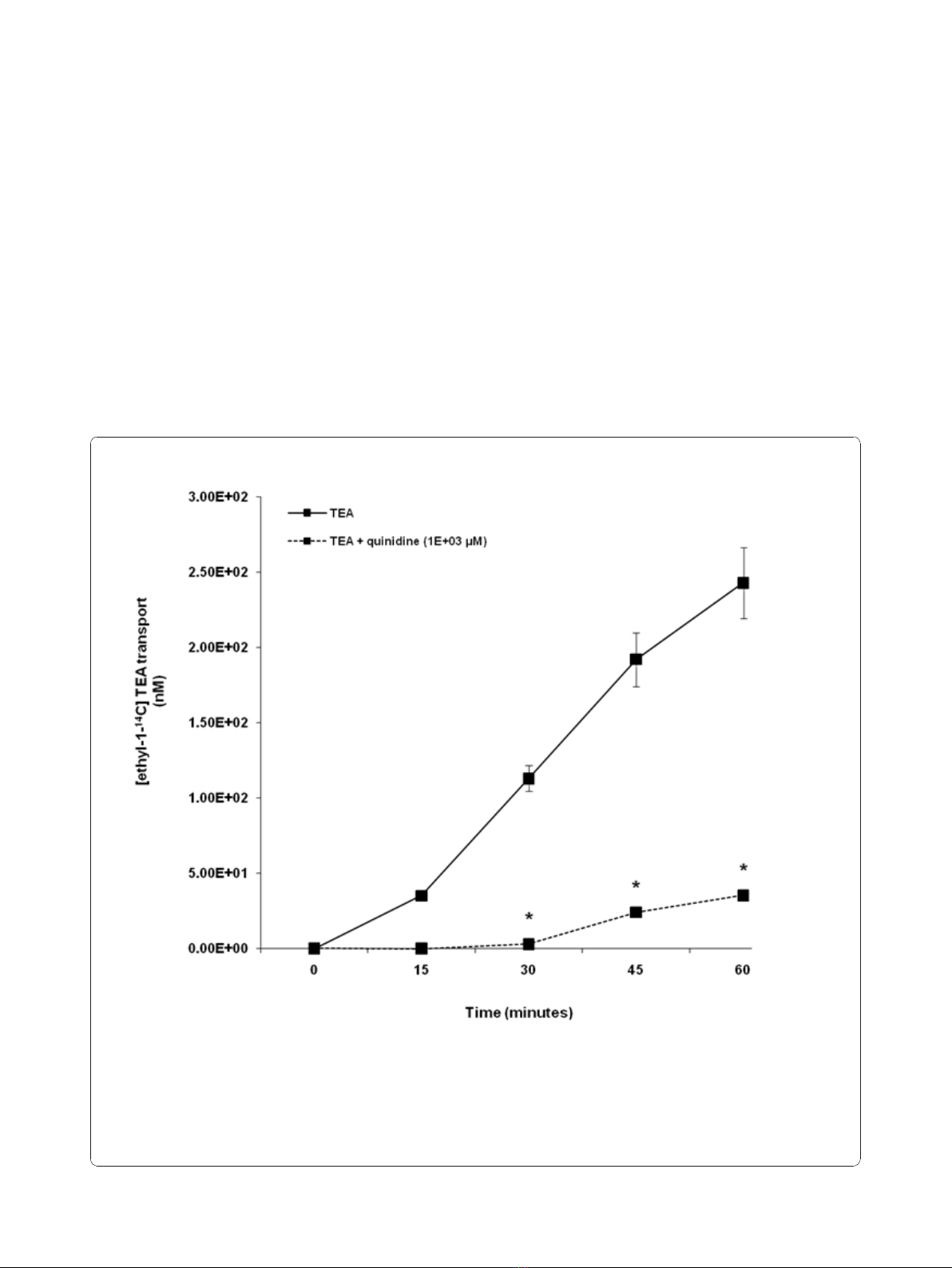
TEA transport across LLC-PK1 and HK-2 cell monolayers
The TEA was transported across LLC-PK1 cell mono-
layers in a time - dependent manner over the experimen-
tal study period (Figure 1). The results illustrate that
there was a significant (p < 0.05) decrease in the
concentration of [ethyl-
14
C] TEA in the apical compart-
mentinthepresenceofquinidineat30,45and60
minutes.
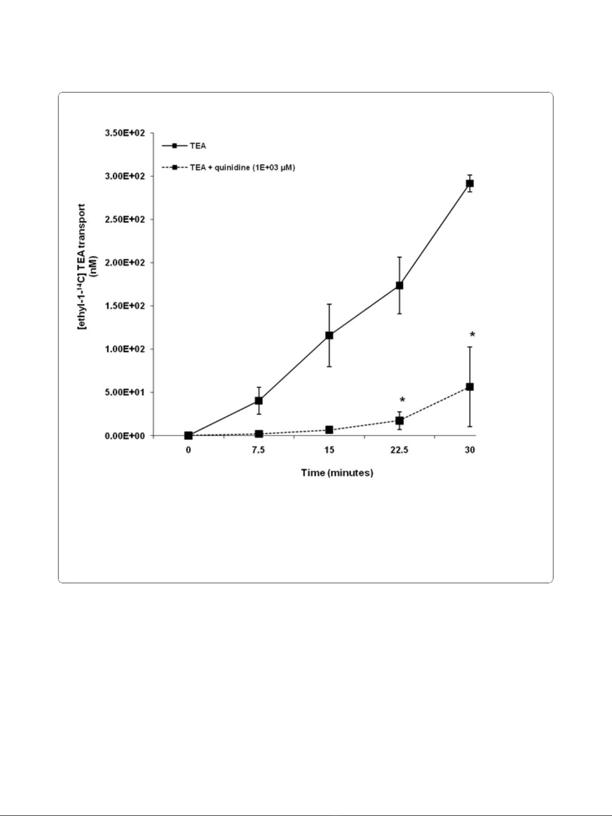
Our results illustrate that TEA was transported across
HK-2 cell monolayers in a time - dependent manner
over the experimental period (Figure 2). The concentra-
tion of [ethyl-
14
C] TEA in the apical compartment was
significantly (p < 0.05) decreased in the presence of qui-
nidine at 22.5 and 30 minutes.
Acyclovir transport across LLC-PK1 and HK-2 cell
monolayers
Acyclovir appeared to be transported across LLC-PK1
cell monolayers in a time - dependent manner from 30
Figure 1 Tetraethylammonium (TEA) transport across porcine renal proximal tubular cell (LLC-PK1) monolayers. The transport
(basolateral-to-apical) of TEA was assessed in LLC-PK1 cells monolayers. Cell monolayers were exposed to [ethyl-1-
14
C] TEA (5 μM) in the
presence or absence of quinidine (1E+03 μM) for 60 minutes. The transport of TEA was assessed by measuring the appearance of [ethyl-1-
14
C]
TEA radioactivity in the apical compartment at specific time intervals (0, 15, 30, 45 and 60 minutes) for 60 minutes. Radioactivity was measured
as disintegrations per minute (DPM). The TEA transport is expressed as the concentration of [ethyl-1-
14
C] TEA in the apical compartment. Results
are presented as the mean (±standard error (SE)) from 3 cell monolayer experiments. * p < 0.05, compared to [ethyl-1-
14
C] TEA radioactivity in
the apical compartment in the absence of quinidine.
Gunness et al.Journal of Translational Medicine 2010, 8:139
http://www.translational-medicine.com/content/8/1/139
Page 4 of 11
to 60 minutes (Figure 3). There was a trend of
decreased concentration of [8-
14
C] acyclovir in the api-
cal compartment in the presence of quinidine over the
experimental study period. Acyclovir transport was not
significantly (p > 0.05) inhibited in the presence of
quinidine.
Acyclovir was transported across HK-2 cell mono-
layers in a time - dependent manner over the experi-
mental study period (Figure 4). Results illustrate that the
concentration of [8-
14
C] acyclovir in the apical compart-
ment was significantly (p < 0.05) decreased in the pre-
sence of quinidine at 15, 22.5 and 30 minutes.
The effect of acyclovir on creatinine transport across LLC-
PK1 and HK-2 cell monolayers
Figure 5 illustrates that in contrast to quinidine and
cimetidine, acyclovir (22 to 89 μM) did not inhibit creati-
nine transport across LLC-PK1 cell monolayers. The
concentration of [2-
14
C] creatinine in the apical compart-
ment over the experimental study period was similar
between cell monolayers exposed to creatinine in the
presence or absence of acyclovir (22 to 89 μM). In con-
trast, there was a decrease in the concentration of [2-
14
C]
creatinine in the apical compartment in the presence of
quinidine or cimetidine, compared to the concentration
Figure 2 Tetraethylammonium (TEA) transport across human renal proximal tubular cell (HK-2) monolayers. The transport (basolateral-
to-apical) of TEA was assessed in HK-2 cells monolayers. Cell monolayers were exposed to [ethyl-1-
14
C] TEA (5 μM) in the presence or absence
of quinidine (1E+03 μM) for 30 minutes. The transport of TEA was assessed by measuring the appearance of [ethyl-1-
14
C] TEA radioactivity in the
apical compartment at specific time intervals (0, 7.5, 15, 22.5 and 30 minutes) for 30 minutes. Radioactivity was measured as disintegrations per
minute (DPM). The TEA transport is expressed as the concentration of [ethyl-1-
14
C] TEA in the apical compartment. Results are presented as the
mean (±standard error (SE)) from 3 cell monolayer experiments. * p < 0.05, compared to [ethyl-1-
14
C] TEA radioactivity in the apical
compartment in the absence of quinidine.
Gunness et al.Journal of Translational Medicine 2010, 8:139
http://www.translational-medicine.com/content/8/1/139
Page 5 of 11


![Báo cáo seminar chuyên ngành Công nghệ hóa học và thực phẩm [Mới nhất]](https://cdn.tailieu.vn/images/document/thumbnail/2025/20250711/hienkelvinzoi@gmail.com/135x160/47051752458701.jpg)


%20--%3e%3cdefs%3e%3cstyle%3e%20.st0%20{%20fill:%20%23fff;%20}%20.st1%20{%20fill:%20%237800fa;%20}%20%3c/style%3e%3c/defs%3e%3cpath%20class='st1'%20d='M117.78,12.18H43.11c2.9,3.47,4.65,7.94,4.65,12.82,0,5.6-2.3,10.66-6.01,14.29h76.02l7.22-13.56-7.22-13.56Z'/%3e%3cg%3e%3cpath%20class='st0'%20d='M53.58,26.17h-.59v-1.46h.59v-4.96h2.83c1.78,0,2.67.94,2.67,2.82v5.76c0,1.87-.89,2.81-2.67,2.81h-2.83v-4.96ZM55.36,21.37v3.34h1.1v1.46h-1.1v3.34h1.01c.61,0,.91-.37.91-1.1v-5.93c0-.74-.3-1.1-.91-1.1h-1.01Z'/%3e%3cpath%20class='st0'%20d='M65.99,31.14h-1.8l-.31-2.07h-2.19l-.31,2.07h-1.64l1.82-11.39h2.62l1.82,11.39ZM65.28,18.04c-.25.46-.51.77-.75.94-.21.15-.47.22-.79.22-.26,0-.57-.07-.92-.22l-.38-.15c-.14-.05-.26-.07-.37-.07-.3,0-.53.18-.71.54l-.91-.68c.25-.46.51-.77.75-.94.21-.14.48-.21.79-.21.26,0,.57.07.92.21l.38.15c.14.05.26.07.37.07.3,0,.53-.18.71-.54l.91.68ZM61.91,27.52h1.73l-.87-5.76-.87,5.76Z'/%3e%3cpath%20class='st0'%20d='M74.53,26.89v1.52c0,1.91-.89,2.86-2.67,2.86s-2.67-.95-2.67-2.86v-5.93c0-1.91.89-2.86,2.67-2.86s2.67.95,2.67,2.86v1.11h-1.69v-1.22c0-.75-.31-1.12-.93-1.12s-.93.37-.93,1.12v6.15c0,.74.31,1.11.93,1.11s.93-.37.93-1.11v-1.63h1.69Z'/%3e%3cpath%20class='st0'%20d='M81.4,31.14h-1.8l-.31-2.07h-2.19l-.31,2.07h-1.64l1.82-11.39h2.62l1.82,11.39ZM75.9,19.2l1.52-1.91h1.71l1.51,1.91h-1.61l-.76-.95-.75.95h-1.61ZM77.32,27.52h1.73l-.87-5.76-.87,5.76ZM83.1,15.99l-1.76,1.91h-1.26l1.17-1.91h1.86Z'/%3e%3cpath%20class='st0'%20d='M84.86,19.75c1.78,0,2.67.94,2.67,2.82v1.48c0,1.87-.89,2.81-2.67,2.81h-.85v4.28h-1.79v-11.39h2.64ZM84.01,21.37v3.86h.85c.58,0,.87-.36.87-1.08v-1.71c0-.71-.29-1.07-.87-1.07h-.85Z'/%3e%3cpath%20class='st0'%20d='M93.51,19.75c1.78,0,2.67.94,2.67,2.82v1.48c0,1.87-.89,2.81-2.67,2.81h-.85v4.28h-1.79v-11.39h2.64ZM92.66,21.37v3.86h.85c.58,0,.87-.36.87-1.08v-1.71c0-.71-.29-1.07-.87-1.07h-.85Z'/%3e%3cpath%20class='st0'%20d='M98.8,31.14h-1.79v-11.39h1.79v4.88h2.03v-4.88h1.83v11.39h-1.83v-4.88h-2.03v4.88Z'/%3e%3cpath%20class='st0'%20d='M105.36,24.55h2.46v1.62h-2.46v3.34h3.09v1.63h-4.88v-11.39h4.88v1.63h-3.09v3.18ZM108.17,17.29l-1.76,1.91h-1.26l1.17-1.91h1.86Z'/%3e%3cpath%20class='st0'%20d='M112.2,19.75c1.78,0,2.67.94,2.67,2.82v1.48c0,1.87-.89,2.81-2.67,2.81h-.85v4.28h-1.79v-11.39h2.64ZM111.35,21.37v3.86h.85c.58,0,.87-.36.87-1.08v-1.71c0-.71-.29-1.07-.87-1.07h-.85Z'/%3e%3c/g%3e%3ccircle%20class='st1'%20cx='25'%20cy='25'%20r='20'/%3e%3cpath%20class='st0'%20d='M32.78,19.27c2.92,0,4.43,2.55,5.28,5.33l.71,2.17c.14.38-.33.75-.71.75h-5.61c.19-.33.24-.71.09-1.08l-.75-2.45c-.43-1.32-.99-2.64-1.79-3.77.75-.57,1.65-.94,2.78-.94h0ZM25,18.38c3.25,0,4.9,2.78,5.89,5.89l.76,2.45c.14.42-.33.8-.8.8h-11.69c-.42,0-.94-.38-.8-.8l.75-2.45c.99-3.11,2.64-5.89,5.89-5.89h0ZM25,11.35c1.74,0,3.11,1.37,3.11,3.11s-1.37,3.11-3.11,3.11-3.11-1.41-3.11-3.11,1.41-3.11,3.11-3.11h0ZM17.27,19.27c1.08,0,1.98.38,2.73.94-.8,1.13-1.37,2.45-1.74,3.77l-.8,2.45c-.14.38-.05.75.09,1.08h-5.56c-.42,0-.9-.38-.75-.75l.71-2.17c.9-2.78,2.41-5.33,5.33-5.33h0ZM17.27,12.91c1.51,0,2.78,1.27,2.78,2.83s-1.27,2.83-2.78,2.83-2.83-1.27-2.83-2.83,1.27-2.83,2.83-2.83h0ZM32.78,12.91c1.56,0,2.78,1.27,2.78,2.83s-1.23,2.83-2.78,2.83-2.83-1.27-2.83-2.83,1.27-2.83,2.83-2.83h0ZM27.07,28.56v.09c0,.57-.24,1.08-.61,1.46h0v.05c-.38.33-.9.57-1.46.57s-1.08-.24-1.46-.61h0c-.38-.38-.61-.9-.61-1.46v-.09h1.41v.09c0,.19.05.38.19.47v.05c.09.09.28.19.47.19s.38-.09.47-.19v-.05c.14-.09.24-.28.24-.47t-.05-.09h1.41ZM30.99,28.56v.09c0,1.65-.66,3.16-1.74,4.24-1.08,1.08-2.59,1.79-4.24,1.79s-3.16-.71-4.24-1.79l-.05-.05c-1.04-1.08-1.7-2.55-1.7-4.2v-.09h1.41v.09c0,1.27.47,2.4,1.27,3.25h.05c.85.85,1.98,1.37,3.25,1.37s2.4-.52,3.25-1.37c.85-.8,1.37-1.98,1.37-3.25v-.09h1.37ZM34.99,28.56v.09c0,2.78-1.13,5.28-2.92,7.07-1.79,1.79-4.29,2.92-7.07,2.92s-5.23-1.13-7.07-2.92c-1.79-1.79-2.92-4.29-2.92-7.07v-.09h1.41v.09c0,2.4.94,4.53,2.5,6.08,1.56,1.56,3.72,2.5,6.08,2.5s4.52-.94,6.08-2.5c1.56-1.56,2.5-3.68,2.5-6.08v-.09h1.41Z'/%3e%3c/svg%3e)