
BioMed Central
Page 1 of 18
(page number not for citation purposes)
Journal of Translational Medicine
Open Access
Research
Anti-tumor activity of patient-derived NK cells after cell-based
immunotherapy – a case report
Valeria Milani1,2, Stefan Stangl3, Rolf Issels1,2, Mathias Gehrmann3,
Beate Wagner4, Kathrin Hube3, Doris Mayr5, Wolfgang Hiddemann1,6,
Michael Molls3 and Gabriele Multhoff*3,7
Address: 1Department of Internal Medicine, University Medical Center Großhadern, Ludwig-Maximilians-Universität München, Germany,
2Clinical Cooperation Group (CCG) "Tumor Therapy by Hyperthermia", Helmholtz Zentrum München, German Research Center for
Environmental Health Munich, Germany, 3Department of Radiation Oncology, Klinikum rechts der Isar, Technische Universität München,
Germany, 4Transfusion Medicine and Haemostaseology, University Medical Center Großhadern, Ludwig-Maximilians-Universität München,
Germany, 5Department of Pathology, University Medical Center Großhadern, Ludwig-Maximilians-Universität München, Germany, 6Clinical
Cooperation Group (CCG) "Pathogenesis of Acute Leukemias", Helmholtz Zentrum München, German Research for Environmental Health,
Munich, Germany and 7Clinical Cooperation Group (CCG) "Innate Immunity in Tumor Biology", Helmholtz Zentrum München, German
Research Center for Environmental Health, Munich, Germany
Email: Valeria Milani - valeria.bruehl-milani@med.uni-muenchen.de; Stefan Stangl - stefan.stangl@lrz.tu-muenchen.de;
Rolf Issels - rolf.issels@med.uni-muenchen.de; Mathias Gehrmann - mathias.gehrmann@lrz.tu-muenchen.de;
Beate Wagner - beate.wagner@med.uni-muenchen.de; Kathrin Hube - kathrin.hube@my-tum.de; Doris Mayr - doris.mayr@med.uni-
muenchen.de; Wolfgang Hiddemann - wolfgang.hiddemann@med.uni-muenchen.de; Michael Molls - michael.molls@lrz.tu-muenchen.de;
Gabriele Multhoff* - gabriele.multhoff@lrz.tu-muenchen.de
* Corresponding author
Abstract
Background: Membrane-bound heat shock protein 70 (Hsp70) serves as a tumor-specific recognition structure
for Hsp70-peptide (TKD) plus IL-2 activated NK cells. A phase I clinical trial has shown that repeated re-infusions
of ex vivo TKD/IL-2-activated, autologous leukapheresis product is safe. This study investigated the maintenance
of the cytolytic activity of NK cells against K562 cells and autologous tumor after 6 plus 3 infusions of TKD/IL-2-
activated effector cells.
Methods: A stable tumor cell line was generated from the resected anastomotic relapse of a patient with colon
carcinoma (pT3, N2, M0, G2). Two months after surgery, the patient received the first monthly i.v. infusion of his
ex vivo TKD/IL-2-activated peripheral blood mononuclear cells (PBMNC). After 6 infusions and a pause of 3
months, the patient received another 3 cell infusions. The phenotypic characteristics and activation status of
tumor and effector cells were determined immediately before and at times after each infusion.
Results: The NK cell ligands Hsp70, MICA/B, and ULBP-1,2,3 were expressed on the patient's anastomotic
relapse. An increased density of activatory NK cell receptors following ex vivo stimulation correlated with an
enhanced anti-tumoricidal activity. After 4 re-infusion cycles, the intrinsic cytolytic activity of non-stimulated
PBMNC was significantly elevated and this heightened responsiveness persisted for up to 3 months after the last
infusion. Another 2 re-stimulations with TKD/IL-2 restored the cytolytic activity after the therapeutic pause.
Conclusion: In a patient with colon carcinoma, repeated infusions of ex vivo TKD/IL-2-activated PBMNC initiate
an intrinsic NK cell-mediated cytolytic activity against autologous tumor cells.
Published: 23 June 2009
Journal of Translational Medicine 2009, 7:50 doi:10.1186/1479-5876-7-50
Received: 5 May 2009
Accepted: 23 June 2009
This article is available from: http://www.translational-medicine.com/content/7/1/50
© 2009 Milani et al; licensee BioMed Central Ltd.
This is an Open Access article distributed under the terms of the Creative Commons Attribution License (http://creativecommons.org/licenses/by/2.0),
which permits unrestricted use, distribution, and reproduction in any medium, provided the original work is properly cited.

Journal of Translational Medicine 2009, 7:50 http://www.translational-medicine.com/content/7/1/50
Page 2 of 18
(page number not for citation purposes)
Background
Studies into the cellular basis of cancer immunosurveil-
lance demonstrate that lymphocytes of both adaptive and
innate immune compartments can prevent tumor devel-
opment [1]. In contrast to normal tissues, tumors fre-
quently express the stress protein heat shock protein 70
(Hsp70) on their plasma membrane, and this membrane-
associated form of the Hsp70 molecule acts as a tumor-
specific recognition structure for Hsp70-peptide activated
natural killer (NK) cells expressing CD94 [2,3]. More
recently, the glycosphingolipid globoyltriaosylceramide
(Gb3) was shown to enable the selective anchorage of
Hsp70 in plasma membranes of colorectal cancer cells [4].
The finding that Gb3 is predominantly found in choles-
terol-rich microdomains (CRM) of tumor, but not of nor-
mal cells might provide an explanation for the tumor-
specific Hsp70 membrane expression.
The region of the Hsp70 molecule which is exposed to the
extracellular milieu of tumors has been identified as the
14-mer peptide TKDNNLLGRFELSG (TKD), and this
resides in the amino acid sequence aa450–463 of the C-ter-
minal domain substrate binding domain [5,6]. A combi-
nation of synthetically produced, GMP-grade Hsp70
peptide plus low dose IL-2 (TKD/IL-2) has been shown to
stimulate the cytolytic and migratory capacity of CD3-/
CD16/CD56+ human [5,7] and mouse [8] NK cells. TKD/
IL-2-activated cells specifically kill allogeneic, Hsp70
membrane-positive tumor cell lines in vitro [9]. Moreover,
four repeated re-infusions of purified TKD/IL-2-activated
NK cells have been shown to eradicate the primary tumor
and prevent metastasis in a xenograft tumor mouse model
of human pancreatic cancer [10]. Importantly, the induc-
tion of NK cell cytotoxicity is also possible when PBMNC
rather than purified NK cells are incubated with TKD/IL-2
[11]. Furthermore, in the presence of other lymphocytes
and antigen presenting cells (APC), the cytotoxic response
against Hsp70 membrane-positive tumors has been
found to be selectively mediated by NK cells (unpub-
lished observations).
The enhanced cytolytic activity against Hsp70 surface-pos-
itive tumors is accompanied by, and correlates with an
increased expression density of NK cell receptors includ-
ing CD94/NKG2A/C, NKG2D and NCRs such as NKp30,
NKp44, NKp46 [2,3,12]. The expression density of the C-
type lectin receptor CD94 is associated with the capacity
of NK cells to bind Hsp70 protein and TKD [2], and cor-
relates with a strong lytic activity against Hsp70 mem-
brane-positive tumor target cells.
The mechanism of lysis of Hsp70 membrane-positive
tumors has been identified as being a perforin-independ-
ent, granzyme B-mediated apoptosis [13]. Previous stud-
ies have shown a high degree of correlation of the results
of a 4-h 51chromium release assay and the granzyme B
ELISPOT assay for measuring the granzyme B mediated
killing of Hsp70 membrane-positive tumors by activated
NK cells. These findings indicate that the granzyme B
ELISPOT assay is a reliable test to determine Hsp70-reac-
tivity in NK cells.
An Hsp70 membrane-positive phenotype acts as a nega-
tive prognostic marker for patients with lower rectal carci-
nomas and non-small cell lung cancer (NSCLC), and the
overall survival of patients with Hsp70 membrane-posi-
tive cancer is significantly lower than that of their Hsp70
membrane-negative counterparts [14]. These findings
highlight the clinical significance of determining the
Hsp70 membrane status and the urgent need to treat
patients with Hsp70 membrane-positive tumors. A phase
I clinical study involving eleven patients with metastatic
colorectal cancer and one patient with non-small cell lung
cancer (NSCLC) has shown that the re-infusion of autolo-
gous, TKD/IL-2-activated leukapheresis products is feasi-
ble, safe and well-tolerated [15]. Furthermore, measurable
immunological responses in the form of an enhanced
expression of CD94 on NK cells and an increased NK cell
cytolytic capacity against an allogeneic, Hsp70 mem-
brane-positive colon carcinoma cell line CX+ were
induced in 10 of the 12 patients [15]. In line with previous
results from animal models [10], clinical responses fulfill-
ing formal staging criteria were observed in 2 patients,
who received more than 4 treatment cycles [15]. These
promising immunological data encouraged us to treat a
patient with an anastomotic relapse using a similar
approach to that in the phase I clinical trial mentioned
above. However, in this specific instance a tumor cell line
could be established from a biopsy of the patient's tumor
and its Hsp70 membrane-positive phenotype could be
confirmed.
Herein, we report the kinetics of the anti-tumor immune
responses in this patient who received a total of 9 re-infu-
sions of ex vivo TKD/IL-2-activated, autologous leukapher-
esis products over a 12-month period and the clinical
follow-up for 1 year. The kinetics of the initiation and
maintenance of an in vivo cytolytic response against
Hsp70-positive tumors within the first therapy cycles is in
line with our previous findings from the phase I clinical
trial. In this study an intrinsic NK cell activity was initiated
only in patients who received more than 4 repeated re-
infusion cycles of TKD/IL-2-activated, autologous
PBMNC. This finding was determined in 5 patients with
different tumor entities, stages and previous therapies.
This is also the first observation that the administration of
TKD/IL-2-activated PBMNC induces a sustained in vivo
NK cell cytolytic response against the patient's own,
Hsp70 membrane-positive tumor and the classical NK cell
target K562 which persists for at least 2 months. Further-

Journal of Translational Medicine 2009, 7:50 http://www.translational-medicine.com/content/7/1/50
Page 3 of 18
(page number not for citation purposes)
more, we demonstrate that a decline in the in vivo NK cell
activity can be restored by an additional 2 infusion cycles
with TKD/IL-2-activated, autologous PBMNC. This indi-
cates that the therapeutic intervention does not initiate an
irreversible state of immune tolerance.
Methods
Ethics
Signed informed consent was obtained from the patient
before the start of the first treatment and the clinical pro-
tocol was approved by the institutional ethical review
board of the University Medical Center Großhadern.
Patient characteristics and study setting
A 65 year-old male came to our attention in 03/05 at the
time of an anastomotic relapse of a colon carcinoma
which was initially diagnosed as being in stage IIIc (pT3,
pN2 (5/17), M0, G2) using the recently revised American
Joint Committee on Cancer (AJCC) Sixth Edition Cancer
Staging System [16,17]. The primary tumor had been sur-
gically removed in 02/03, but the patient had refused
standard systemic adjuvant chemotherapy at the time of
first diagnosis, having considered the "quality of life"
implications and being aware of the magnitude of the
incremental benefit.
The patient was in a good clinical condition at the time of
presentation (Karnofsky > 90%) and the resection of the
anastomotic relapse three months later (06/05) revealed a
high-grade colon carcinoma (rpT3, rpN0, M0, G2) (Figure
1, clinical history). Paraffin-embedded material of the pri-
mary tumor and the anastomotic relapse, as well as fresh
tumor biopsy material of the anastomotic relapse, were
available for laboratory use. The local tumor board rec-
ommended a post-operative systemic chemotherapy
which was again refused by the patient. Although fully
aware of the risk factors of his tumor disease and the rec-
ommended alternative chemotherapeutic options, the
patient decided to be treated with TKD/IL-2-activated,
autologous PBMNC.
In addition to the colon carcinoma the patient had a his-
topathologically proven highly differentiated prostate
cancer which had been diagnosed in 04/02. The patient
had refused resection and any pharmacological therapy of
the prostate carcinoma but the prostate specific antigen
(PSA) levels were determined regularly.
Ex vivo stimulation of patient-derived peripheral blood
mononuclear cells (PBMNC)
Two months after the surgical resection of the anasto-
motic relapse the experimental cell-based therapy was
started in 08/05 (Figure 1, study design) after having
received approval of the Institutional Ethical Committee
of the Medical Faculty of the Ludwig-Maximilians-Univer-
sität Munich and the patient's written informed consent.
In contrast to the phase I clinical trial, the whole proce-
dure was repeated up to 6 times on a monthly rather than
a 2-weekly basis. After a 3-month treatment pause, the
patient received another 3 leukapheresis and re-infusion
cycles within another 3 months. Vital and biological
parameters were measured every month during the cell-
based therapy and for another 12 months after the ther-
apy had been terminated. A scheme of the therapeutic
approach and the course of the disease are summarized in
Figure 1.
Identical to the protocol of the clinical phase I trial [15],
PBMNC concentrates were obtained by a 3–4 hour leuka-
pheresis processing approximately 2.5 times of the
patient's blood volume on a cell separator (COBE Spectra,
MNC program v6.1, Heimstetten, Germany). The first leu-
kapheresis product was aliquoted into two parts. Follow-
ing erythrocyte removal by density gradient centrifugation
(Ficoll-Hypaque, Life Technologies, Inc., Paisley, Scot-
land) in a GMP-grade closed cell culture bag and tubing
system (IBM 2997 cell washer), PBMNC were counted
and resuspended in GMP-grade Cellgro Stem Cell Growth
Medium (CellGro SCGM, Freiburg, Germany) at a density
of 10 × 106 cells/ml. The cell suspension was transferred
into 250-ml Teflon cell culture bags (Vuelife, Cellgenix)
and GMP-grade Hsp70-peptide TKDNNLLGRELSG (TKD,
purity > 96%, lot no. 0541026; Bachem, Bubendorf, Swit-
zerland) plus low dose IL-2 (100 IU/ml, Novartis, Nürn-
berg, Germany) were added.
The incubation of patient-derived PBMNC with TKD/IL-2
in an incubator (Binder, Tuttlingen, Germany) under gen-
tle rotation (cell shaker, Binder), at 37°C in a humidified
atmosphere (90%) containing 5% v/v CO2 for 4 days was
performed to induce NK cell-mediated cytolytic activity
against Hsp70 membrane-positive tumors [5]. After
removal of the TKD peptide by 2 washing steps in Ringer's
lactate (Braun Melsungen, Germany), cells were resus-
pended in 500 ml Ringer's lactate and transferred into
infusion bags (600 ml, R2022, Baxter, Munich, Germany).
Aliquots of the PBMNC suspension were taken for sterility
tests prior to in vitro stimulation, on day 4 after stimula-
tion, and directly before re-infusion.
Ex vivo TKD/IL-2-activated PBMNC were re-infused by
intravenous (i.v.) injection within 30–60 min using an
infusion set consisting of syringe and a stem cell filter (2
μm diameter, Baxter). The patient's vital parameters were
monitored for 3 hours after the adoptive cell transfer.
Clinical and laboratory follow-up
Vital and routine laboratory parameters including white
blood counts, lymphocyte subpopulations, electrolytes,
creatinine, urea, bilirubin, C-reactive protein, serum alka-
Journal of Translational Medicine 2009, 7:50 http://www.translational-medicine.com/content/7/1/50
Page 4 of 18
(page number not for citation purposes)
line phosphatase, γ-glutamine transferase, alanine ami-
notranferease (ALT), aspartate aminotransferase (AST),
lactate dehydrogenase, Quick, and aPTT were determined
before each leukapheresis. Blood counts, electrolytes and
coagulation tests were measured before and after each
cycle of cell re-infusion. Differential blood counts and
lymphocyte subpopulations were assessed in peripheral
blood before each treatment cycle and in every PBMNC
concentrate on the day of leukapheresis. Prostate specific
antigen (PSA, Abbott, Germany) and carcinoembryonic
antigen (CEA, Abbott and Elecsys/Roche, Germany) levels
were determined approximately every 4 weeks during
therapy and in the follow-up period.
Clinical and radiological assessments of the disease,
including the proportion of the liver volume replaced by
tumor (LVRT) were performed every 3 months by colos-
copy, positron-emission tomography/computed tomog-
raphy (PET/CT) and prostate Magnetic Resonance
Imaging (MRI). Radiological responses were assessed by
"Response Evaluation Criteria In Solid Tumors"
(RECIST).
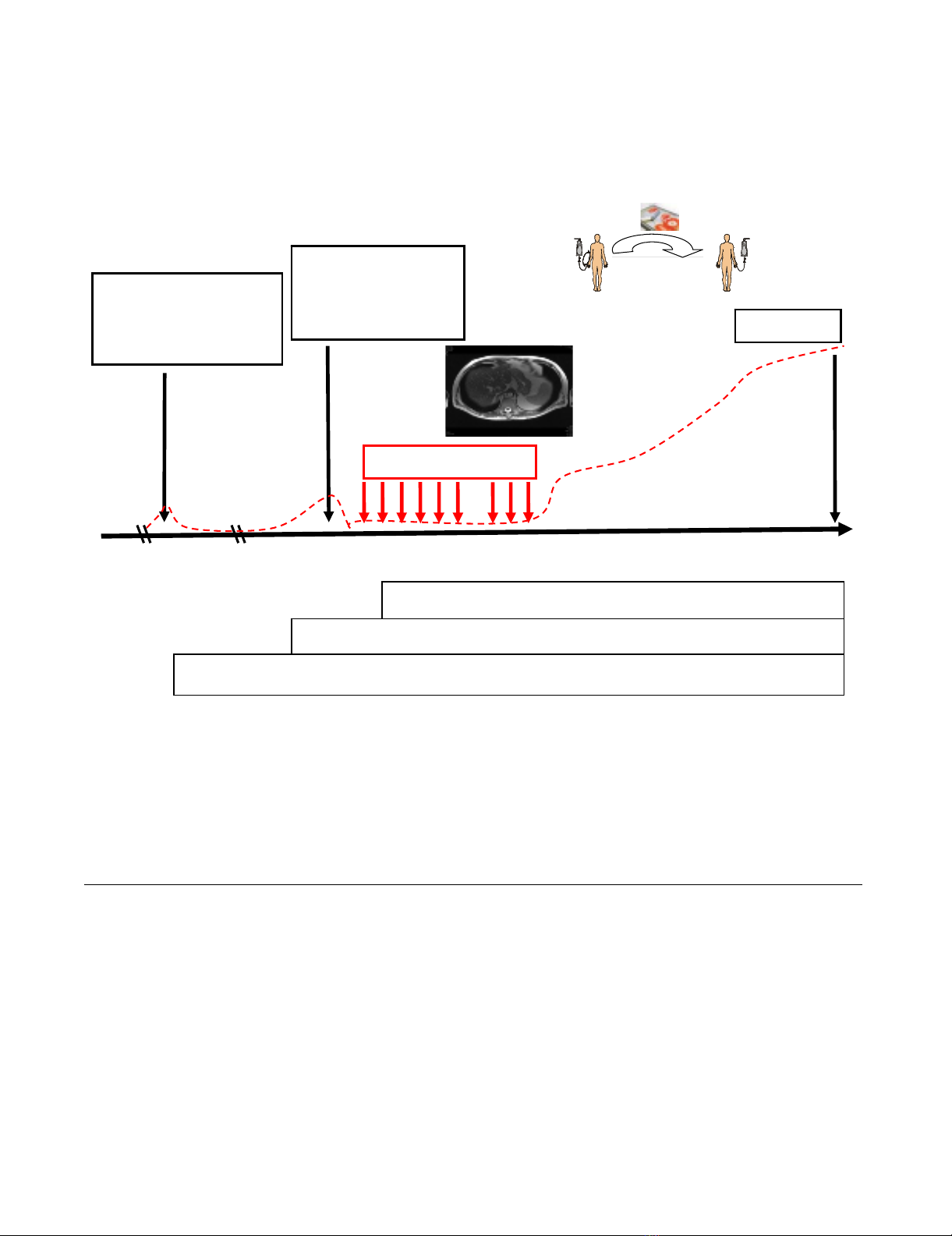
Study design upper panel) and clinical history of the patient (bottom panel)Figure 1
Study design upper panel) and clinical history of the patient (bottom panel). A 65 year old patient with an adenocar-
cinoma of the colon stage IIIc pT3, N2, M0, G2 (02/03) came to our attention at the time of an anastomotic relapse in 03/05.
After surgical resection of the colon carcinoma relapse in 06/05, a biopsy was provided to our laboratory for phenotypic char-
acterization. Two months later (08/05), the NK cell therapy was started. The patient received 6 sequential leukapheresis/re-
infusion cycles of autologous, ex vivo TKD/IL-2-activated PBMNC on a monthly basis. After a 3-month break, the patient
received another 3 cell re-infusions. The patient did not show any signs of metastases at the end of the NK cell therapy, as
determined by CT scan. The time interval between the beginning of the NK cell therapy and death was 27 months. Survival fol-
lowing recurrence and overall survival after first diagnosis was 32 and 58 months, respectively.
NK therapy-death: 27 months
Survival following recurrence: 32 months
Survival following primary tumor: 58 months
Study design
Clinical history
02/03 06/05 08/05 06/06 11/07
Su r ger y
adenocarcinoma
colon stage IIIc
pT3, pN2, M0, G2
Sur ger y
anastom otic
relapse
rpT3, rpN0, M0, G2
Death
CEA
NK cell therapy
Reinfusion
TKD/IL-2
stimulation
Leukapheresis

Journal of Translational Medicine 2009, 7:50 http://www.translational-medicine.com/content/7/1/50
Page 5 of 18
(page number not for citation purposes)
Hsp70 protein and Hsp70 antibody ELISA
The concentrations of Hsp70 protein and Hsp70 antibody
were measured in the patient's serum which was taken
before leukapheresis L7, L8, and L9 using a sandwich
ELSA kit (Duo Set IC; R&D Systems), according to the
manufacturer's instructions.
Generation of a tumor cell line
A 0.5 cm3 tumor specimen from the patient's anastomotic
relapse was obtained from the Department of Pathology.
After washing, the tumor tissue was mechanically minced
in RPMI 1640 medium supplemented with 10% v/v fetal
calf serum (FCS), 1 mM sodium pyruvate, antibiotics (all
from Gibco-BRL, Eggenstein, Germany) and 2 mM L-
glutamine (PAN Systems, Aidenbach, Germany) and the
homogenate was passed through a sterile mesh. An aliq-
uot of the single cell suspension was immediately used for
flow cytometry analysis, and the other was seeded into T-
25 culture flasks in supplemented RPMI 1640 medium.
After 2 weeks, adherent cells were trypsinized (trypsin/
EDTA, Gibco-BRL), counted and 0.5 × 10⁶ viable cells
were resuspended in 5 ml fresh medium for further flow
cytometric analyses. Aliquots of the established tumor cell
line from the first 5 cell passages were stored in liquid
nitrogen.
Flow cytometric analysis of tumor and effector cells
For flow cytometry of tumor cells, 2 × 10⁵ propidium
iodide negative (viable) cells were incubated for 30 min at
4°C in the dark with the following monoclonal antibod-
ies (mAbs): anti-fibroblast (ASO2-PE, Dianova, Ham-
burg, Germany), anti-MHC class I (W6/32-FITC, IgG2a;
Cymbus Biotechnology, Eastleigh, UK), anti HLA-E
(MEM-E/06-PE, IgG1; Biozol Diagnostica, Eching, Ger-
many), anti-MICA/B (BAMO1, IgG1; BAMO2, IgG2a,
Bamomab, Munich, Germany, kindly provided by Dr.
Alexander Steinle, Tübingen), anti-ULBP-1,2,3 (AUMO2,
IgG2a; BUMO1, IgG1; CUMO1, IgG1; all purchased from
Bamomab), anti-human Hsp70 (cmHsp70.1-FITC,
mouse IgG1, multimmune GmbH, Munich, Germany).
The cmHsp70.1 mAb recognizes the sequence NLLGRFEL
(aa 454–461) in the C-terminal domain of Hsp70 which
is exposed to the extracellular milieu of tumor cells [5].
This sequence acts as a recognition structure for NK cells
that have been stimulated either with full length Hsp70
protein or with the 14-mer Hsp70 peptide TKDNNLL-
GRFELSG (aa 450–463) when combined with low dose
IL-2 [11,18,19]. The phenotypic characterization of the
tumor was performed at the Klinikum rechts der Isar,
Technische Universität München.
Unstimulated and stimulated PBMNC harvested from
leukapheresis products and from the peripheral blood
were incubated with the following mAbs as described
above: anti-CD3 and anti-CD16/56-tricolor-conjugated
(Caltag, Hamburg, Germany), anti-CD94-FITC (HP-3D9,
IgG1; Becton Dickinson Pharmingen, Heidelberg, Ger-
many) and anti-CD94-PE (Ancell Bayport, Minneapolis,
MN, USA); anti-CD56-FITC (Becton Dickinson), anti-
NKG2D-PE (149810, IgG1, R&D Systems, Minneapolis,
MN, USA). FITC and PE labeled IgG1 and IgG2a immuno-
blobulins were used as isotype-matched non-specific
binding controls (Caltag, Hamburg, Germany). Differen-
tial counts and determination of lymphocyte subpopula-
tions in leukapheresis products was done with a dual-
color lyse and wash method (Sumlset, BD). Flow cytomet-
ric analysis of unstimualted leukapheresis products were
performed at the Klinikum rechts der Isar, Technische
Universität München and at the LMU, the agreement of
the results between both laboratories was verified apply-
ing Rainbow Calibration Particles (BD). Stimulated effec-
tor cells were only analyzed by flow cytometry at the
Klinkum rechts der Isar, Technische Universität München.
After 2 washing steps in PBS containing 2% v/v FCS (PBS/
FCS) and the addition of propidium iodide (PI, Sigma-
Aldrich, Deisenhofen, Germany, stock solution 1 μg/ml),
the cells were immediately analyzed by flow cytometry
using a FACSCalibur™ instrument (Becton Dickinson,
Heidelberg, Germany). The cell population was identified
on the basis of their forward (FSC) and right angle light
scatter properties (FSC vs SSC) and the fluorescence char-
acteristics of 5,000 to 10,000 gated events were deter-
mined. Data acquisition and analysis were performed
using CellQuest™ Pro software (Becton Dickinson).
Measurement of phenotype and cytolytic activity of
patient-derived PBMNC
For the in vitro analysis of stimulated cell populations,
sterile aliquots of the leukapheresis products were incu-
bated under identical culture conditions as the sample
which was to be re-infused. The cytolytic activity of
patient-derived PBMNC, without any further enrichment
for NK cells, against the classical NK target cell line K562
and the autologous, Hsp70 membrane-positive tumor
before and after in vitro stimulation with TKD/IL-2, and of
freshly isolated, non-cultured patient-derived PBMNC
before and after re-infusion in vivo was assessed using a
standard 4-hour granzyme B ELISPOT assay and a
51chromium release assay. As the lysis of Hsp70 mem-
brane-positive tumors by NK cells has previously been
identified as being perforin-independent, granzyme B
mediated apoptosis [13], this assay is suitable to deter-
mine the Hsp70-reactivity of NK cells.
For the ELISPOT assay, 96-well ELISPOT plates (Millipore
GmbH, Schwalbach, Germany) were coated with capture
antibody by overnight incubation at 4°C, after which they
were blocked using 10% v/v FCS. The effector and target
cells (3 × 10³) were added at different E/T ratios ranging


![Vaccine và ứng dụng: Bài tiểu luận [chuẩn SEO]](https://cdn.tailieu.vn/images/document/thumbnail/2016/20160519/3008140018/135x160/652005293.jpg)


