M I N I R E V I E W

Gonadotropin-releasing hormone: GnRH receptor signaling in extrapituitary tissues Lydia W. T. Cheung and Alice S. T. Wong

School of Biological Sciences, University of Hong Kong, China

Keywords cross-talk; extrapituitary; GnRH; GnRH receptor; MAPK; metastasis; pituitary; receptor tyrosine kinase; signaling; tumor

Correspondence A. S. T. Wong, School of Biological Sciences, University of Hong Kong, 4S-14 Kadoorie Biological Sciences Building, Pokfulam Road, Hong Kong, China Fax: +852 2559 9114 Tel: +852 2299 0865 E-mail: awong1@hku.hk

(Received 14 April 2008, revised 28 May 2008, accepted 11 June 2008)

doi:10.1111/j.1742-4658.2008.06677.x

Gonadotropin-releasing hormone (GnRH) has historically been known as in the past few years, a pituitary hormone; however, interest has been raised in locally produced, extrapituitary GnRH. GnRH receptor (GnRHR) was found to be expressed in normal human reproductive tissues (e.g. breast, endometrium, ovary, and prostate) and tumors derived from these tissues. Numerous studies have provided evidence for a role of GnRH in cell proliferation. More recently, we and others have reported a novel role for GnRH in other aspects of tumor progression, such as metastasis and angiogenesis. The multiple actions of GnRH could be linked to the divergence of signaling pathways that are activated by GnRHR. Recent observations also demonstrate cross-talk between GnRHR and growth fac- the classical Gaq–11-phospholipase C signal tor receptors. Intriguingly, transduction pathway, known to function in pituitary gonadotropes, is not involved in GnRH actions at nonpituitary targets. Herein, we review the key findings on the role of GnRH in the control of tumor growth, progres- sion, and dissemination. The emerging role of GnRHR in actin cytoskele- ton remodeling (small Rho GTPases), expression and ⁄ or activity of adhesion molecules (integrins), proteolytic enzymes (matrix metalloprotein- ases) and angiogenic factors is explored. The signal transduction mecha- nisms of GnRHR in mediating these activities is described. Finally, we discuss how a common GnRHR may mediate different, even opposite, responses to GnRH in the same tissue ⁄ cell type and whether an additional receptor(s) for GnRH exists.

Introduction

is

to GnRH-I, GnRH-II

in contrast

The hypothalamic gonadotropin-releasing hormone (GnRH) is a decapeptide that plays a critical role in the regulation of reproduction. GnRH-I (pGlu-His- Trp-Ser-Tyr-Gly-Leu-Arg-Pro-Gly-NH2) the first GnRH isoform discovered in mammalian brain. Its to stimulate pituitary secretion of major

role is

gonadotropins, luteinizing hormone and follicle-stimu- lating hormone, which in turn stimulate the gonads for steroid production. Subsequently, a second iso- form of GnRH (His5, Trp7, Tyr8) (GnRH-II) has been isolated from chicken brain. It is also highly conserved among vertebrates, including mammals [1]. However, is expressed at significantly higher levels outside the

Abbreviations EGF, epidermal growth factor; EGFR, epidermal growth factor receptor; ERK, extracellular signal-related kinase; FAK, focal adhesion kinase; FGF, fibroblast growth factor; GnRH, gonadotropin-releasing hormone; GnRHR, gonadotropin-releasing hormone receptor; JNK, Jun N-terminal kinase; MAPK, mitogen-activated protein kinase; MMP, matrix metalloproteinase; NF-jB, nuclear factor kappa B; PI3K, phosphatidylinositol 3-kinase; PKC, protein kinase C; Pyk2, proline-rich tyrosine kinase 2; RTK, receptor tyrosine kinase; uPA, urokinase-type plasminogen activator; VEGF, vascular endothelial growth factor.

FEBS Journal 275 (2008) 5479–5495 ª 2008 The Authors Journal compilation ª 2008 FEBS

5479

L. W. T. Cheung and A. S. T. Wong

GnRH receptor signaling

its

their tissue of origin, ovarian surface epithelium [9,10]. Interestingly, levels of GnRHR seem to be associated with cancer grading and have been reported to be elevated in advanced stage (stages III and IV) as compared to early stage (stages I and II) ovarian carcinomas [11]. Our recent findings that GnRH can promote the motility and invasiveness of ovarian can- cer cells further corroborate the view that GnRH may play a crucial role in tumor progression ⁄ metastasis [12,13], and these findings will be discussed in a later section.

specimens

in the kidney, brain and is particularly abundant bone marrow, and prostate [2]. This leads to the speculation that GnRH-II may have distinct physio- logical functions from those of GnRH-I. In line with this is the observation that although GnRH-II can stimulate gonadotropin secretion, efficiency is much lower than that of GnRH-I (only about 2% of that of GnRH-I) [3]. This suggests that the primary role of GnRH-II is not in the regulation of gonado- tropin secretion. Instead, this peptide has been shown to act as a neuromodulator [4]. The exact actions of GnRH-II in peripheral tissues are not entirely under- stood, but this is certainly an important topic for investigation which may offer an opportunity to eluci- date the undisclosed complexity of GnRH.

studied thus

Using [125I][d-Trp6]GnRH, specific receptor binding has been detected in membranes from 24 of 31 (77%) endometrial carcinomas and from three of 13 (23.1%) nonmalignant human endometrial [14]. GnRHR mRNA has been clearly detected in surgical endometrial carcinoma specimens and endometrial carcinoma cell lines [15,16]. As with normal myome- trium, most benign neoplasms far, including uterine leiomyoma, also possess GnRHR [17].

receptor

layers

cell

In this minireview, we will focus on recent progress in understanding the roles of GnRH-I and GnRH-II in particular its emerging in extrapituitary tissues, role in tumor growth, invasion, and metastasis. We will also describe the molecular mechanisms underlying these effects, focusing on the roles of proteolysis, adhesion, and signaling, as well as our still-emerging cross-talk with other understanding of pathways. Finally, we will discuss two important outstanding questions in the field regarding what might distinguish the different responses to the same ligand (GnRH) and whether an additional receptor(s) for GnRH exists in humans.

Early studies showed that the human placenta con- tains specific binding sites for GnRH that interact with GnRH agonists and antagonists [18]. Later on, GnRHR was localized to the cytotrophoblast and [19,20]. Temporal syncytiotrophoblast expression of GnRHR in the placental cells at different weeks of gestation has been observed, in parallel with the time-course of chorionic gonadotropin secretion during pregnancy [21], suggesting that the expression of the receptor is a function of pregnancy stage.

Localization of GnRH receptor (GnRHR) in peripheral reproductive tissues

lines [5]. Soon after this, a functional

The presence of GnRHR has been demonstrated in numerous human breast cancer cell lines and tumor biopsy specimens [22–24]. GnRHR was immunolocal- ized in the cytoplasm in 37 of 58 (64%) invasive ductal carcinoma cases [23]. The expression of GnRHR in normal human breast tissue is still controversial, but the sample size may have been too small to allow any definite conclusion [22,25].

The initial interest in extrapituitary GnRHR stemmed primarily from observations in the 1980s that GnRH analogs can inhibit the growth of nonpituitary tumor cell type I GnRHR was demonstrated in a variety of normal human reproductive tissues (e.g. breast, endometrium, ovary, and prostate) and tumors derived from these tissues.

GnRHR is also present in prostate cancer cells, as shown by radioligand-binding studies, PCR, and western blotting analysis [26,27]. GnRHR immunore- activity is localized to the luminal and basal epithelial cells in benign and malignant prostate tissues. In this study, the relative GnRHR mRNA levels showed a wide range of individual differences that were unrelated to the histological grades of the 16 cases [27]. There does, however, appear to be significantly higher expression of GnRHR in hormone-refractory prostate carcinoma than in other types of prostate tumor (n = 80) [28].

In the ovary, GnRHR mRNAs are expressed in granulosa-luteal cells, and increased expression of GnRHR correlates with follicular growth and develop- ment [6]. GnRHR binding has been demonstrated in luteinized granulosa cells, late follicles and developing corpora lutea, but not in primordial, early antral and preovulatory follicles [7,8]. This stage-specific expres- sion of GnRHR in the human granulosa and luteal cells suggests a role for GnRH in the regulation of ovarian physiology, particularly ovulation, follicular atresia and luteolysis. The presence of GnRHR protein and mRNA has also been demonstrated in human ovarian tumor specimens, ovarian cancer cell lines and

Although these extrapituitary GnRHRs share the same cDNA nucleotide sequence and encode tran- scripts and proteins of the same size as the pituitary

FEBS Journal 275 (2008) 5479–5495 ª 2008 The Authors Journal compilation ª 2008 FEBS

5480

L. W. T. Cheung and A. S. T. Wong

GnRH receptor signaling

GnRH-II has antiproliferative effects on ovarian cancer cells [41–43]. Although it has been suggested that this effect of GnRH-II is mediated through the type I GnRHR [43], there are other findings implicating a type I GnRHR-independent action [41,42].

It is interesting to note that although both GnRH-I agonists and antagonists exert antiproliferative effects, the effects of GnRH-I antagonists are stronger than those of the agonists [44]. This difference has also been seen in an in vivo model, which demonstrates a signifi- cant inhibition of tumor growth by GnRH-I antago- nists but not GnRH-I agonists [45]. The advantage of GnRH antagonists over the agonistic peptides is prob- ably due to the fact that they inhibit the secretion of gonadotropins and reduce sex steroid levels immedi- ately after application, thus achieving rapid therapeutic effects, whereas repeated exposure to agonistic agents is required to induce functional desensitization of the gonadotropes [46].

GnRHR [20,26,29], they also differ in several ways. First, cell surface receptor expression in extrapituitary sites is low as compared to that of the pituitary [15,27]. This may underlie the greater effect of the GnRHR ligands on the gonadotropes. Second, there are at least two classes of GnRHR: one has high affin- ity [with nanomolar dissociation constants (Kd)] for GnRH, and one has low affinity (with micromolar Kd values) for GnRH. The high-affinity GnRH-binding sites are commonly regarded as being the same as the GnRHR of the pituitary gland. Whereas in most of the reported cases, both the low-affinity and high-affin- ity GnRHR have been found in extrapituitary tissues [30–33], in some cases, only low-affinity GnRHR could be detected [10,18,34], and in others, e.g. in endome- trial cancers and nonmalignant endometrial specimens, only the high-affinity GnRHR has been demonstrated [14]. The exact role of each of these receptors and the implications of differential levels of expression remain to be elucidated.

Functions of GnRH-I and GnRH-II in cancers

Tumor growth

Treatment of human endometrial cancer cells (cell line Ishikawa) with the GnRH-I antagonist SB-75 results in growth inhibition, mainly due to the Fas ⁄ Fas ligand-mediated apoptotic pathway, whereas GnRH-I agonists have no effect on the same cell line [15,47,48]. Another endometrial carcinoma cell line, HEC-1A, also exhibits differential responses to different GnRH agon- ists and antagonists [15,30,36,48]. GnRH-II has been shown to have antiproliferative effects on endometrial carcinoma cells [41]. The effects of GnRH-I are abrogated after type I GnRHR knockout [36], whereas those of the GnRH-I antagonist cetrorelix and of GnRH-II persist [41]. These findings suggest that the antiproliferative effects of cetrorelix and GnRH-II are not mediated through the type I GnRHR.

Over the last two decades, both GnRH agonists and antagonists have been widely used as therapeutics in treating sex steroid-dependent tumors. The majority these GnRH analogs, when given continuously, of inhibit gonadotropin synthesis and secretion via downregulation of the pituitary GnRHRs. This indi- rect mechanism of action has provided the rationale for the use of GnRH analogs in the treatment of hor- mone-dependent tumors for many years. Only since the detection of GnRHR in extrapituitary tissues has there been increasing interest in its direct action on tumor cells.

independent

GnRH-I has been demonstrated to have antiprolifer- ative effects on prostate cancer cells [49–51], except in one in vivo study [52]. This antiproliferative effect appears to be independent of the androgen receptor status of the prostate carcinoma cells, as both andro- gen-sensitive LNCaP cells and androgen-resistant DU-145 cells remain sensitive to GnRH [49,50]. Acti- vation of GnRHR may mediate these effects via direct induction of apoptosis through caspase activation [53]. Compatible with a role for GnRH in survival at low doses, an enhancing effect of GnRH was observed when cells were treated with a low concentration (100 pm) of GnRH-I agonist [54]. GnRH-II was shown to have an antiproliferative effect on DU-145 cells and growth-stimulatory effect on TSU-Pr1 cells, but the type I GnRHR was not involved [55].

GnRH-I analogs have direct antiproliferative effects on ovarian cancer cells, which is linked to the disrup- tion of the cell cycle at G0 ⁄ G1 [31,35,36]. On the other in vitro studies failed to hand, several demonstrate significant growth inhibition by GnRH-I agonists, even at fairly high concentrations (micromolar range) [37,38]. In fact, a biphasic impact of GnRH-I agonists on growth has been reported: whereas GnRH-I agonists at high dose (1 lm) inhibit cell proliferation in vitro, cells treated with agonists at low dose (10 nm) show significant growth stimulation [39]. Further studies demonstrated that nanomolar concentrations of GnRH-I agonists also increase cell survival under multiple stress conditions, including DNA replication- cytotoxic agents and UV radiation [40]. specific

The influence of GnRH on the growth of human breast cancer cells was first studied with MCF-7 cells [56], and both in vitro and in vivo proliferation of

FEBS Journal 275 (2008) 5479–5495 ª 2008 The Authors Journal compilation ª 2008 FEBS

5481

L. W. T. Cheung and A. S. T. Wong

GnRH receptor signaling

GnRH-I and GnRH-II on cell invasion supports the view that the same receptor, type I GnRHR, is essen- tial for the effects of GnRH-I and GnRH-II in ovarian cancer cells.

breast cancer cells could be inhibited by both agonistic and antagonistic analogs of GnRH [57,58]. However, higher efficiency of GnRH antagonists in growth inhi- bition than that of GnRH agonists has been reported [24,58].

Invasion and metastasis

expression of

several

the

inhibitor

The observation that GnRH controls tumor growth suggests a regulatory role for this peptide in the meta- static behavior of cancer cells. This hypothesis is sup- ported by studies showing that GnRH-I and GnRH-II extracellular can affect matrix-degrading enzymes in human extravillous cyto- trophoblasts and decidual stromal cells to facilitate implantation [59,60]. However, its potential role in cancer metastasis has just begun to be revealed.

The decrease in uPA activity of cytosol from Dun- ning R3327H rat prostate tumors after treatment with GnRH-I analogs suggests that GnRH may be an important factor in reducing the invasiveness of pros- tate cancer [63]. High doses of GnRH-I agonists and antagonists have been reported to attenuate the invading capacity of both androgen-dependent and androgen-independent prostate cancer cells by modu- lating E-cadherin-mediated cell–cell contacts and pro- duction of uPA and its (plasminogen activator inhibitor-1) [64–66]. GnRH has also been shown to regulate cell motility through its interaction with the small GTPases Rac1, Cdc42, and RhoA, which are involved in the regulation of actin polymer- ization [67].

several

(uPA)

Metastasis is a complex phenomenon that requires several specific steps, such as decreased adhesion, increased motility, and proteolysis. The effects of GnRH in tumor metastasis are mediated through the regulation of adhesion molecules, Rho GTPases, and two families of metastasis-related proteinases, the matrix metallopro- teinases (MMPs) and the urokinase-type plasminogen activator levels: mRNA system, at transcription, secretion, and proenzyme activation.

treatment of

triptorelin,

the ability of melanoma cells

Up to now, there has been only one study, by Von Alten et al., investigating the role of GnRH in breast cancer metastasis, using a coculture system with human osteosarcoma cells to analyze tumor cell invasion to bone [68]. The consequences of GnRHR activation are complex and appear to be cell context cells with the dependent: whereas GnRH-I agonist the GnRH-II agonist [d-Lys6]GnRH-II and the GnRH-I antagonist cetrorelix decreases the invasion rate in most breast cancer cell lines, these agents have no significant effect in the GnRHR-positive MDA-MB-435 cells [68]. Further investigations are required to elucidate the reason why the MDA-MB-435 cell line reacts differently.

The ability of GnRH to regulate metastasis was first reported in melanoma cells [61]. High doses of GnRH-I significantly analog, at micromolar concentrations, to invade reduces and migrate [61]. Preliminary data (R. M. Moretti, M. Monagnani Marelli, J. C. van Groeninghen, M. Motta & P. Limonta, unpublished results, 2003) indicate that this inhibitory action is due to the effects of integrins and MMPs [62].

Organ-specific homing and colonization of cancer cells are important and interesting features of metasta- sis. A role for GnRH has also been suggested in the regulation of the immune response and metastasis. GnRH-I and GnRH-II are expressed in human normal and cancerous T-cells. GnRH triggers laminin receptor gene expression, adhesion to laminin, in vitro chemo- taxis, and in vivo homing to specific organs [69].

Angiogenesis

such as

We were the first to report possible metastatic activ- ity of GnRH-I in tumors of the female reproductive tract [12]. GnRH-I exerts a biphasic effect on cellular migration and invasion: whereas lower (nanomolar) concentrations of the GnRH-I agonist stimulate cellu- lar migration and invasion in a dose-dependent man- ner, high (micromolar) concentrations are not as efficient. This proinvasive effect is mediated through activation of metastasis-related proteinases, in particu- lar MMP-2 and MMP-9 [12]. Moreover, GnRH-I is able to transactivate the MMP-2 and MMP-9 promot- ers, which means that GnRH can be considered to be a new member of MMP-2 and MMP-9 transcriptional modulators. Like GnRH-I, native GnRH-II and its synthetic analog also induce a similar biphasic regula- tion of ovarian cancer invasion [13]. The finding that small interfering RNA-mediated downregulation of type I GnRHR completely reversed the effects of both

Angiogenesis is crucial to a number of physiological and pathological processes, reproduction, development, and tissue repair, as well as tumor growth and metastasis. Vascular endothelial growth factor (VEGF) is implicated as the most important angiogenesis inducer, because of its potency in a variety of normal and tumor cells. Other angiogenic factors include fibroblast growth factor (FGF), plate- let-derived growth factor and the angiopoietin family.

FEBS Journal 275 (2008) 5479–5495 ª 2008 The Authors Journal compilation ª 2008 FEBS

5482

L. W. T. Cheung and A. S. T. Wong

GnRH receptor signaling

very interesting to determine whether GnRH also plays a key role in tumor angiogenesis.

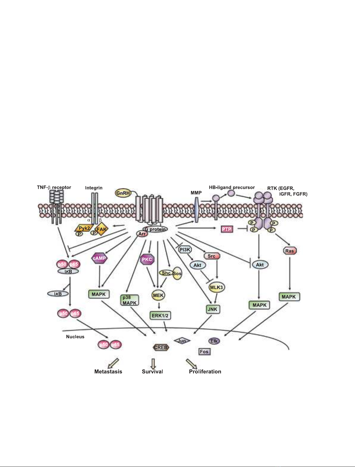
Intracellular signal transduction

Upon GnRH binding, GnRHR undergoes a conforma- tional change and stimulates a unique G-protein. Inter- estingly, the classical Gaq–11-phospholipase C signal transduction pathway, which is known to operate in the pituitary, is not involved in the antitumor activity of GnRH analogs. Rather, GnRHRs couple to Gai in these tumors and result in the activation of several downstream signaling cascades [73,74], such as mito- gen-activated protein kinase (MAPK), phosphatidyl- inositol-3-kinase (PI3K), and nuclear factor kappa B (NF-jB) signaling. The GnRH-induced signaling path- ways in extrapituitary tissues are shown schematically in Fig. 1.

The effect of GnRH on angiogenesis in the ovary, in which this neovascularization is necessary for follicular and luteal function, has been demonstrated. A recent in vivo study using rats revealed that an application of the GnRH-I agonist leuprolide acetate decreases the protein expression of VEGF and angiopoietin-1 and their receptors in ovarian follicles, and that this can be reversed by coinjection of the GnRH antagonist antide [70]. A similar inhibitory effect on angiogenesis can be observed in marmosets injected with the GnRH-I antagonist antarelix [71]. However, VEGF mRNA expression is unaffected by the treatment. The clinical response of uterine shrinkage after GnRH analog treatment and a pathological role of FGF-2, VEGF and platelet-derived growth factor in uterine leiomy- oma growth and vascularization has also been sug- is an gested [72]. Considering that angiogenesis important process in many human cancers, it would be

Fig. 1. Schematic representation of GnRHR signaling in extrapituitary tissues. Binding of GnRH to GnRHR triggers several intracellular signal- ing cascades and cross-talk with mitogenic signaling, depending on the cell context. Some of these signaling modules can transduce extra- cellular signals to the nucleus and thereby regulate genes that are involved in cell growth, metastasis, or survival. Arr, b-arrestin; CREB, cAMP response element-binding protein; FGFR, fibroblast growth factor receptor; HB-EGF, heparin-binding EGF; IjB, inhibitory factor kap- pa B; IGFR, IGF receptor; MEK, mitogen-activated protein kinase kinase; MLK3, mixed-lineage kinase 3; PTP, protein tyrosine phosphatase; Sos, son of sevenless; TNF-a, tumor necrosis factor alpha.

FEBS Journal 275 (2008) 5479–5495 ª 2008 The Authors Journal compilation ª 2008 FEBS

5483

L. W. T. Cheung and A. S. T. Wong

GnRH receptor signaling

MAPK

receptor-interacting proteins

(Fig. 1).

implicated in nerve growth factor-mediated neuronal differentiation of PC12 cells, whereas a rapid and transient activation is associated with growth factor- mediated proliferation of PC12 cells [80]. Thus, the duration of kinase activation seems to be a major determinant of signal outcome. We have shown differ- ential regulation of ERK1 ⁄ 2, p38 MAPK, and JNK by GnRH-I with sustained signaling through the JNK pathway in ovarian cancer cells [12]. Consistently, GnRH-stimulated MMP-2 and MMP-9 expression, secretion and cell invasion were attenuated by specific inhibition of JNK but not of ERK1 ⁄ 2 and p38 MAPK, suggesting that prolonged activation of JNK may contribute to a more invasive phenotype. Strong and sustained activation of MAPK has been reported to be necessary for its cytoplasm-to-nucleus transloca- tion, and thereby contributes to the regulation of gene expression [79,81]. It will be interesting to see whether sustained activation of JNK leads to its nuclear trans- location, which is required for GnRH-stimulated cell invasion. The JNK pathway targets multiple transcrip- including c-Jun, c-Fos, ATF and PEA, tion factors, and putative binding sites for these DNA-binding pro- teins are present in the MMP promoters [82]. Whether these putative regulatory elements participate in the the MMP-2 and GnRH-dependent activation of MMP-9 genes remains to be determined.

Cross-talk with mitogenic signaling

is now receiving further

to activate ERK1 ⁄ 2,

Cross-talk between cell surface receptors, which has been recognized as a mechanism capable of generating signal diversity, interest. Figure 1 illustrates the cross-talk between GnRHR and receptor tyrosine kinases (RTKs). For instance, GnRH causes transactivation of RTKs, such as EGFR [75,78,83]. MMP-2 and MMP-9 seem to be essential for GnRH-induced EGFR activation by cleavage of the heparin-binding epidermal growth factor (EGF) precursor [84]. Transactivation of EGFR has been shown as GnRH-induced ERK1 ⁄ 2 phosphorylation can be abolished in the pres- ence of the EGFR inhibitor AG1478 [53,78]. However, the biological importance of ERK1 ⁄ 2 activation in response to this cross-talk still remains elusive.

The major MAPK cascades include extracellular sig- nal-regulated kinase (ERK), Jun N-terminal kinase (JNK), and p38 MAPK. Many studies have shown that the MAPK pathway is critical for GnRH activi- ties, which provides an important link for the trans- mission of signals from the cell surface to the nucleus. Activation of MAPK by GnRH involves distinct upstream pathways in generating tissue-specific and cell-specific signaling (Fig. 1). This can occur at differ- ent levels via different mechanisms: (a) second messen- [26]; [protein kinase C (PKC) and cAMP] gers (b) such as Src and b-zarrestin [53,75]; and (c) upstream kinases such as MAPK ⁄ ERK kinase and PI3K [53,76]. For example, GnRH-I induces apoptosis in DU-145 prostate cancer cells via JNK, which is activated through two indepen- dent mechanisms [53]. Activation of the pathway is dependent on c-Src with concomitant decrease in Akt activity, and the combination of these two events relieves the inhibition of the upstream activator of JNK, MLK3 [53] Interestingly, although ERK1 ⁄ 2 is phosphorylated through epidermal growth factor receptor (EGFR) under the same conditions, this pathway is not involved in the apoptotic effects. These findings demonstrate that activation of the two MAPKs, which lead to distinct physiological out- comes, is separated already at the upstream levels. In the ovarian cancer cell line CaOV-3, prolonged stimu- lation of ERK1 ⁄ 2 through Shc and son of sevenless is required for GnRH-I-mediated growth inhibition [76]. Consistent with the fact that sustained activation of ERK1 ⁄ 2 is often correlated with cell cycle progression, GnRH-I-induced growth inhibition is attributed to G1 arrest [76]. Moreover, the signaling cascade was shown to be initiated by Gbc, supporting the notion that the post-GnRHR signaling cascade in extrapituitary cells is different from that in pituitary cells. GnRH-induced MAPK activation has also been shown in another ovarian cancer cell line, OVCAR-3. Both ERK1 ⁄ 2 and p38 MAPK mediate the antiproliferative effects of GnRH-I and GnRH-II in a PKC-dependent manner [43,77]. GnRH-II induces the activation of activator protein-1 transcription factor via p38 MAPK, suggest- ing a potential role of activator protein-1 in ovarian cancer cell growth [77]. The JNK pathway also drives tumor invasion and migration in ovarian cancer cells [12], but the activation mechanism(s) remains to be elucidated.

signaling

adhesive

Negative cross-talk between GnRHR and growth factor receptors has also been described. For instance, the antiproliferative effects of GnRH-I and GnRH-II agonists are mediated through attenuation of EGFR signaling in many reproductive tumor cells [57,66,85– 87]. In prostate cancer cells, cetrorelix is able to coun- ter EGFR-dependent through a PKC-dependent mechanism [66]. Activation of

Temporal and spatial differences in cellular signaling may have significant phenotypic manifestations [78,79]. For example, sustained activation of ERK1 ⁄ 2 has been

FEBS Journal 275 (2008) 5479–5495 ª 2008 The Authors Journal compilation ª 2008 FEBS

5484

L. W. T. Cheung and A. S. T. Wong

GnRH receptor signaling

[95]. These data suggest

tor has no effect on the activation of NF-jB [94]. It has also been shown that GnRH-I suppresses interleu- kin-8 expression via attenuation of tumor necrosis fac- tor alpha-induced NF-jB signaling in endometriotic that stromal cells (Fig. 1) modulation of cytokine signal transduction by GnRH may be one of the mechanisms contributing to its growth-inhibitory effect.

reorganization.

GnRHR appears to mediate these effects via activation of phosphotyrosine phosphatase, thereby reducing EGF-induced EGFR autophosphorylation, resulting in downregulation of mitogenic signal transduction and cell proliferation [85,86,88]. A negative regulatory interaction between GnRHR and mitogenic signaling pathways has also been reported in human prostate cancer cells via insulin-like growth factor. GnRH-I agonists inhibit expression of the insulin-like growth factor receptor, receptor tyrosine phosphorylation, and the subsequent downstream activation of Akt [89–91]. Another example is FGF-2. GnRH analog treatment has been shown to block cell proliferation and inva- sion induced by FGF-2 stimulation [65].

PI3K

The non-RTKs focal adhesion kinase (FAK) and proline-rich tyrosine kinase 2 (Pyk2) are the predomi- nant mediators of integrin signaling. GnRHR has been shown to signal through these molecules, suggesting a In role for GnRH in cytoskeletal human endometrial cancer cells (HEC-1A), b3-integrin- dependent activation of FAK is associated with the inhibitory effects of GnRH-I and GnRH-II on growth [96]. Leiomyoma regression induced by GnRH-I agon- ists has been suggested to be mediated, at least in part, through a mechanism involving suppression of FAK [97]. Maudsley et al. demonstrated a novel signaling cascade of GnRHR that functionally antagonizes the testosterone and inhibits prostate tumor actions of growth [98]. GnRH controls the tyrosine phosphoryla- tion status of the focal adhesion proteins Pyk2 and Hic-5. This alteration of the focal adhesion dynamics then results in nuclear translocation of the androgen receptor, which renders it transcriptionally inactive [98].

The PI3K signaling pathway and its downstream target Akt (also named protein kinase B) has been implicated in promoting cell survival, proliferation, and invasion. In uterine leiomyomas, the GnRH-I ago- nist leuprolide acetate causes a significant reduction in PI3K ⁄ Akt activity and inhibits the expression of the antiapoptotic proteins (c-FLIP and PED ⁄ PEA15), thereby inducing apoptosis [92]. In the SKOV-3 ovar- ian cancer cell line, GnRH-I and GnRH-II interfere with activation of the PI3K ⁄ Akt cascade, and this is is associated with the inhibitory effects of GnRH on cell invasion [13].

Mechanisms underlying the diverse responses to GnRH action

in prostate

cells,

[12,13,39]. Moreover,

Although PI3K ⁄ Akt and MAPK are two parallel pathways in some cell types, they are two related path- ways in the mediation of GnRH actions, as inhibition of PI3K ⁄ Akt can alter the activation of MAPK. For stimulation of cancer instance, PI3K ⁄ Akt releases mixed-lineage kinase 3, which in turn activates the JNK pathway, and this positive reg- ulation is important for the proapoptotic effect of GnRH-I (Fig. 1) [53]. PI3K ⁄ Akt is also an upstream kinase of ERK1 ⁄ 2, and EGFR transactivation by GnRH-I may be required for the activation of this cascade [75,93].

Other signaling pathways

signaling pathways

studied,

Activation of NF-jB is important for the protection against apoptosis in ovarian tumors induced by the GnRH-I agonist tiptorelin [94]. The effect is probably mediated by the Gai-coupled GnRHR, and receptor activation causes nuclear translocation of NF-jB [94]. Unlike the other the GnRH-I-induced NF-jB activation appears to be inde- pendent of the cross-talk between GnRHR and growth factor signaling, as treatment with phosphatase inhibi-

As discussed earlier, GnRH and its agonists have a dual and biphasic action: whereas low concentrations (0.1–10 nm) of GnRH stimulate cell proliferation, migration and invasion in a dose-dependent manner, high concentrations (100 nm to 1 lm) these inhibit the same dose of functions GnRH can elicit completely opposite responses in cells derived from the same tissue. We demonstrated that in two human ovarian cancer cell lines, OVCAR-3 and SKOV-3, GnRH-I and GnRH-II induce invasion of OVCAR-3 cells, but inhibit the invasiveness of SKOV- 3 cells [13]. A similar difference has been found in the effects of GnRH on cell proliferation and cell migra- tion in the prostate carcinoma cell lines TSU-Pr1 and DU-145 [67]. Whereas GnRH-I and GnRH-II stimu- lated cell proliferation, induced actin cytoskeleton remodeling and promoted migration in TSU-Pr1 cells, they were inhibitory in DU-145 cells [67]. The observa- tion that GnRH-I and GnRH-II have no significant effect in cell lines with type I GnRHR depletion indi- cates that the type I GnRHR is indispensable for the

FEBS Journal 275 (2008) 5479–5495 ª 2008 The Authors Journal compilation ª 2008 FEBS

5485

L. W. T. Cheung and A. S. T. Wong

GnRH receptor signaling

Table 1. Potential mechanisms that underlie the diverse responses to GnRH.

Determinants

References

[99–101]

for dual

Treatment conditions, e.g. duration and dose Different Ga subtypes Different G-protein subunits GnRHR expression level Ligand selectivity Presence of GnRHR splice variants Intrinsic cellular properties

[73,74,101,103,104] [105–109] [6,110–112] [114,115] [118,119] [61,62]

Third, multiple GnRH-dependent signaling path- ways may occur via different subunits of a single G-protein [105]. After ligand-induced dissociation, both the a-subunit and bc-subunits are capable of activating various effectors, such as adenylyl cyclase, phospholipase C, and ion channels, thereby conferring on the receptor signaling the potential [106,107]. The effector pathway to be activated is instance, to the upstream subunits. For specific whereas the a-subunit of Gi inhibits adenylyl cyclase activity, the bc-subunits may stimulate the activities of some adenylyl cyclase subtypes [108,109].

The receptor expression level is also known to be a determinant for different signal outcomes [6,110,111]. In gonadotropes, different cell surface densities of GnRHR result in the differential regulation of luteiniz- ing hormone and follicle-stimulating hormone subunit gene expression by GnRH-I [112]. We and others have previously shown that low doses of GnRH upregulate the expression of its receptor, whereas high doses decrease it [12,111,113]. This difference in regulation suggests that high levels of GnRHR expression may enhance the cellular response to GnRH stimulation, presumably due to more efficient signal amplification or altered signaling through coupling to different G-proteins.

Moreover,

effects of both GnRH-I and GnRH-II [13,67]. Thus, one intriguing question is how a common GnRHR may mediate different, even opposite, responses to GnRH in the same cell type ⁄ tissue. The reasons are unknown, but several possibilities (as summarized in Table 1) can be envisaged. First, the treatment condi- tion may be one determinant of the outcome. The pulsatility of GnRH release is necessary for the hor- mone to stimulate pituitary gonadotropes. On the other hand, sustained administration of the peptide brings about a short initial stimulation that is rapidly followed by a decrease in gonadotropin synthesis and secretion [99]. In support of this, the signal response is different at different doses. It has been shown that pul- satile GnRH stimulates more sustained ERK activity (more than 8 h), whereas continuous infusion of aT3-1 cells with GnRH stimulates short-term (2 h) ERK activity [100]. There is also evidence that GnRH treat- ment stimulates cAMP production at nanomolar con- centrations, but has an inhibitory effect at micromolar concentrations [101]. It should be pointed out that the nanomolar concentration (0.01–1 nm) corresponds to the physiological circulating level, and the effects caused by this dose range may represent the physiolog- ical functions of GnRH [54,102].

ligand selectivity has been proposed to explain the opposite (stimulatory and inhibitory) effects of GnRH. For instance, in positively respond- lines, GnRH-I is more ing prostate carcinoma cell effective than GnRH-II. On the other hand, in nega- tively responding cell lines, GnRH-II is much more effective than GnRH-I. Given the short plasma half- life of GnRH, efforts have been made to obtain GnRH analogs, to resist degradation and to increase potency. However, the different GnRH agonists may selectively stabilize different receptor-active conforma- tions and therefore different ligand-induced selective signaling pathways [114]. In this regard, it has been shown that the highly variable amino acid at posi- tion 8 of GnRH plays a discriminating role in selecting the receptor conformational state [115].

Second, GnRH action has been shown to be medi- ated by coupling to different Ga-proteins, depending on the time and dose of exposure [101,103]. In general, Gaq and Gas are associated with a stimulatory effect [103], whereas Gai often mediates the antiproliferative and proapoptotic effects of GnRH [73,74]. Low GnRH concentrations promote the coupling of GnRHR to [101]. High concentrations of GnRH cause a Gas switch in receptor coupling from Gas to Gai [101]. Moreover, stimulation of cAMP production by GnRH is through coupling to Gas, whereas inhibition of cAMP production at high concentrations of GnRH is through coupling to Gai [101,104]. These findings sug- gest that the intracellular milieu in different tissues results in differential coupling and different phenotypic effects.

The presence of splice variants of the GnRHR tran- script may be another possible reason for the different or opposite responses to GnRH. To date, variant tran- scripts of GnRHR have been isolated in many species, e.g. chicken [116], mouse [117] and human [118]. Although these splice variants are totally incapable of ligand binding or signal transduction, they have been implicated in the functional regulation of the wild-type receptor. Previous studies have reported their inhibi- tory activity on full-length GnRHR function [119]. This inhibition is specific, augmented by increasing

FEBS Journal 275 (2008) 5479–5495 ª 2008 The Authors Journal compilation ª 2008 FEBS

5486

L. W. T. Cheung and A. S. T. Wong

GnRH receptor signaling

s e c n e r e f e R

] 2 3 1 , 8 2 1 , 7 2 1 , 0 2 1 , 7 6 , 5 5 , 0 5 [

] 6 2 1 , 5 2 1 , 6 7 , 2 1 [

] 1 3 1 – 9 2 1 , 8 4 , 6 3 , 0 3 , 5 1 [

] 1 3 1 – 9 2 1 , 8 4 , 7 4 , 5 1 [

] 8 2 1 , 7 2 1 , 0 2 1 , 9 4 [

] 5 3 1 , 4 2 1 – 2 2 1 , 8 6 , 6 5 [

] 5 3 1 – 3 3 1 , 1 2 1 , 8 6 [

] 5 2 1 , 3 4 , 3 1 [

] 7 2 1 , 0 2 1 , 7 6 , 5 5 , 4 5 [

] 7 3 1 , 6 3 1 , 3 4 , 3 1 , 2 1 [

H R n G

f o

i

i

n o s a v n

t c e f f E

y t i l i t o m n o

i

?

?

?

. d e n m r e t e d n u

f o

, ?

;

t c e f f E

n o H R n G

h t w o r g

– ; fl

– ; fl

– ; fl

– ; fl

; t c e f f e

o n

, –

y t i l i

w o

l

i

o r t i v

i

i

i

i

i

n

h g H

w o L

b a p a c

e v s a v n I

i

w o L

h g H

h g H

h g H

w o L

y r e V

w o L

h g H

; e s a e r c n

i

,

f o

w o

n

l

i

i

i

i

i

i

i

e c n e s b a

n e g o r t s e

; e s a e r c e d

t n e d n e p e d n

h g H

w o L

h g H

i

- e g a r o h c n A

h t w o r g

h g H

w o L

h g H

h g H

y r e V

w o L

h g H

,

. s e n

l

i l

l l

i

i

e c

i

n e g o r t s e

e b a v e h c a

h t i

l

e c m e d u n

r e c n a c

w

d e d d a

n

s e Y

s e Y

y t i c n e g i r o m u T

i

s e Y

s e Y

y n O

s e Y

s e Y

s e Y

s e Y

s e Y

n o m m o c

l

l

l

l

l

i

l l

l l

a n g i r

i

d e t a i t n e r e f f i d

d e t a i t n e r e f f i d

d e t a i t n e r e f f i d

d e t a i t n e r e f f i d

d e t a i t n e r e f f i d

y l r o o P

s u o r e S

s u o r e S

y g o o t s h

O

y e t a r e d o M

e W

s u o r e S

e W

a t c u D

a t c u D

y l r o o P

f o H R n G o t

i

i

l

l

i

i

i

s e s n o p s e r

i

l

y r a m

y r a m

y r a m

l

l

a m o n c r a c

a m o n c r a c

a m o n c r a c

e d o n

n o s u f f e

n o s u f f e

e d o n

d n a

n o i t a o s I

i r P

i r P

a r u e P

n a r B

i r P

h p m y L

a r u e P

h p m y L

s e t i c s A

s e t i c s A

i

s c i t s i r e t c a r a h c

l

n g i r

i

O

t s a e r B

m u i r t e m o d n E

e t a t s o r P

m u i r t e m o d n E

y r a v O

e t a t s o r P

t s a e r B

e t a t s o r P

y r a v O

y r a v O

l

i

a c g o o s y h P

. 2

e n

-

i l

-

i

l l

e C

e l b a T

A 1 - C E H

5 4 1 - U D

a w a k h s I

3 - V O a C

P a C N L

5 3 4 - B M A D M

7 - F C M

1 r P U S T

3 - V O K S

3 - R A C V O

FEBS Journal 275 (2008) 5479–5495 ª 2008 The Authors Journal compilation ª 2008 FEBS

5487

L. W. T. Cheung and A. S. T. Wong

GnRH receptor signaling

amounts of the cotransfected splice variant cDNA and possibly by preventing or diverting the normal process- ing of GnRHR or enhancing GnRHR degradation [118].

it

Finally,

that differences

is also possible

invasive effect

tor that is not rapidly desensitized or internalized. There is evidence that GnRH-II may act through the type I GnRHR. In monkey pituitary cultures, in which the type II GnRHR is functional, GnRH-II has been found to stimulate gonadotropin secretion exclusively through the type I GnRHR [146]. In contrast, other evidence suggests that the neuromodulatory action of GnRH-II on mammalian behavior is not mediated via it the type I receptor in musk shrews [147]. Thus, appears that GnRH-II may selectively interact with different GnRHRs to mediate its different actions, pre- sumably due to the structural differences between the two GnRHR subtypes.

in response may be ascribed to the intrinsic properties of the cells. The physiological characteristics of the human cancer cell lines mentioned in this minireview are summarized in Table 2. For example, in contrast to SKOV-3 cells, OVCAR-3 cells have low invasive potential. Thus, whereas low doses of GnRH-I and GnRH-II can exert a significant in OVCAR-3 cells, they fail to stimulate SKOV-3 maxi- mally [12,13]. Both GnRH-I and GnRH-II only exert inhibitory effects on SKOV-3 cells at high doses [13].

Novel receptor(s) for GnRH in humans?

functional,

Alternatively, it is possible that the human type II GnRHR may be encoded by a different gene that has yet to be identified. Database searches have revealed the presence of more than two other GnRHR genes in the human genome apart from the conventional type I receptor gene [148]. These genes are located on sepa- rate chromosomes. Whether full-length transcripts can be produced from these receptor-like genes remains to be determined. Recently, a novel GnRH-II-binding protein, in addition to a conven- tional GnRHR, has been identified by using photo- affinity labeling with an azidobenzoyl-conjugated GnRH-II in prostate cancer cells [149]. Taken together, these observations thus suggest the potential existence of novel receptors for GnRH-I and GnRH-II.

Concluding remarks

(positive

even opposite

studies

further

cross-talk. Clearly,

An important issue that remains unresolved in this field is whether one or more other GnRHR subtypes exist in humans. The discovery of GnRH-II has stimu- lated the search for a cognate type II GnRHR. Molec- ular cloning of the type II GnRHR in goldfish, marmoset and monkey has shown that the type II receptor is structurally and functionally distinct from the type I receptor [138–140]. In humans, a type II GnRHR has not been found. However, a search of the revealed a putative human genome database has type II GnRHR gene chromosome 1q21.1 on [140,141]. Expression of this type II GnRHR mRNA has been shown in many human tissues, including endometrium, ovary, placenta, and prostate cancer cells [42,55,140–142]. Although these findings suggest that the human type II receptor gene is transcription- ally active, the mRNA is disrupted by a frameshift in coding exon 1 and a premature stop codon in exon 2, suggesting that a conventional seven-transmembrane receptor cannot be translated from this gene. The gene also overlaps two flanking genes and displays alterna- tive splicing [143]. Thus, the functionality of these human type II GnRHR splice variants and their involvement in transmitting signals from GnRH-II are still in question.

One noteworthy feature of

This overview shows that GnRH modulates a variety of cellular functions in extrapituitary tissues, such as cell growth, invasion, and angiogenesis. However, the effects of GnRH are complex and appear to be cell context dependent. The ability of GnRH to elicit very different, and negative), responses in extrapituitary tissues may arise from dif- ferential usage of signal transduction pathways and receptor are required to unravel this complex signaling network and the coordinated regulatory roles of different factors in specific cellular events during tumorigenesis. High-throughput gene profiling and bioinformatics approaches should be helpful to expand this area of research. The information may also serve as a basis for investigators in the field to explore the signaling mechanisms of other G-protein-coupled receptors. Most studies thus far have only been conducted in cellular models, but in vivo approaches will be essential for a complete understanding of the specific role of each GnRH isoform, including the putative GnRH-III isolated from the human brain [150]. Given the clinical

the primate type II GnRHR is that, unlike the type I receptor, it possesses a C-terminal tail, which is responsible for the recep- tor’s susceptibility to rapid desensitization and inter- nalization [138,144,145]. Finch et al. showed that GnRH was able to efficiently inhibit the proliferation of breast cancer cells when engineered with sheep type I GnRHR, but not with Xenopus type II GnRHR [145]. This clearly implies that the antiproliferative effect of GnRH is mediated most efficiently by a recep-

FEBS Journal 275 (2008) 5479–5495 ª 2008 The Authors Journal compilation ª 2008 FEBS

5488

L. W. T. Cheung and A. S. T. Wong

GnRH receptor signaling

utility of GnRH analogs in hormone-dependent dis- eases, better characterization of GnRH actions and its key molecular effectors is a necessary first step to more effective and perhaps new therapeutic strategies for improving clinical outcome.

mone binding sites in human epithelial ovarian carci- nomata. Eur J Cancer Clin Oncol 25, 215–221. 11 Chien CH, Chen CH, Lee CY, Chang TC, Chen RJ &

Chow SN (2004) Detection of gonadotropin-releasing hormone receptor and its mRNA in primary human epithelial ovarian cancers. Int J Gynecol Cancer 14, 451–458.

Acknowledgements

This work was supported by the Hong Kong Research Grant Council grant 778108 to A. S. T. Wong.

12 Cheung LW, Leung PC & Wong AS (2006) Gona-

dotropin-releasing hormone promotes ovarian cancer cell invasiveness through c-Jun NH2-terminal kinase- mediated activation of matrix metalloproteinase (MMP)-2 and MMP-9. Cancer Res 66, 10902– 10910.

References

1 Chen A, Yahalom D, Ben-Aroya N, Kaganovsky E,

Okon E & Koch Y (1998) A second isoform of gona- dotropin-releasing hormone is present in the brain of human and rodents. FEBS Lett 435, 199–203. 13 Chen CL, Cheung LW, Lau MT, Choi JH, Auersperg N, Wang HS, Wong AS & Leung PC (2007) Differen- tial role of gonadotropin-releasing hormone on human ovarian epithelial cancer cell invasion. Endocrine 31, 311–320. 14 Srkalovic G, Wittliff JL & Schally AV (1990)

2 White RB, Eisen JA, Kasten TL & Fernald RD (1998) Second gene for gonadotropin-releasing hormone in humans. Proc Natl Acad Sci USA 95, 305–309.

Detection and partial characterization of receptors for [D-Trp6]-luteinizing hormone-releasing hormone and epidermal growth factor in human endometrial carcinoma. Cancer Res 50, 1841–1846.

3 Densmore VS & Urbanski HF (2003) Relative effect of gonadotropin-releasing hormone (GnRH)-I and GnRH-II on gonadotropin release. J Clin Endocrinol Metab 88, 2126–2134. 4 Kauffman AS (2004) Emerging functions of gonado-

tropin-releasing hormone II in mammalian physiology and behaviour. J Neuroendocrinol 16, 794–806. 5 Schally AV, Comaru-Schally AM & Redding TW 15 Chatzaki E, Bax CM, Eidne KA, Anderson L, Grudz- inskas JG & Gallagher CJ (1996) The expression of gonadotropin-releasing hormone and its receptor in endometrial cancer, and its relevance as an autocrine growth factor. Cancer Res 56, 2059–2065. 16 Imai A, Ohno T, Iida K, Fuseya T, Furui T &

(1984) Antitumor effects of analogs of hypothalamic hormones in endocrine-dependent cancers. Proc Soc Exp Biol Med 175, 259–281.

Tamaya T (1994) Presence of gonadotropin-releasing hormone receptor and its messenger ribonucleic acid in endometrial carcinoma and endometrium. Gynecol Oncol 55, 144–148. 17 Marinaccio M, Reshkin S, Pinto V & Paradiso A

6 Peng C, Fan NC, Ligier M, Vaananen J & Leung PC (1994) Expression and regulation of gonadotropin- releasing hormone (GnRH) and GnRH receptor mes- senger ribonucleic acids in human granulosa-luteal cells. Endocrinology 135, 1740–1746. (1994) The estimation of LHRH receptors in the tissue of human leiomyoma, myometrium and endometrium. Minerva Ginecol 46, 519–526.

18 Iwashita M, Evans MI & Catt KJ (1986) Characteriza- tion of a gonadotropin-releasing hormone receptor site in term placenta and chorionic villi. J Clin Endocrinol Metab 62, 127–133. 19 Casan EM, Raga F & Polan ML (1999) GnRH 7 Brus L, Lambalk CB, de Koning J, Helder MN, Jans- sens RM & Schoemaker J (1997) Specific gonadotro- phin-releasing hormone analogue binding predominantly in human luteinized follicular aspirates and not in human pre-ovulatory follicles. Hum Reprod 12, 769–773.

mRNA and protein expression in human preimplanta- tion embryos. Mol Hum Reprod 5, 234–239.

8 Choi JH, Gilks CB, Auersperg N & Leung PC (2006) Immunolocalization of gonadotropin-releasing hor- mone (GnRH)-I, GnRH-II, and type I GnRH receptor during follicular development in the human ovary. J Clin Endocrinol Metab 91, 4562–4570.

20 Boyle TA, Belt-Davis DI & Duello TM (1998) Nucleo- tide sequence analyses predict that human pituitary and human placental gonadotropin-releasing hormone receptors have identical primary structures. Endocrine 9, 281–287. 21 Lin LS, Roberts VJ & Yen SS (1995) Expression of

9 Choi KC, Auersperg N & Leung PC (2001) Expression and antiproliferative effect of a second form of gona- dotropin-releasing hormone in normal and neoplastic ovarian surface epithelial cells. J Clin Endocrinol Metab 86, 5075–5078.

FEBS Journal 275 (2008) 5479–5495 ª 2008 The Authors Journal compilation ª 2008 FEBS

5489

human gonadotropin-releasing hormone receptor gene in the placenta and its functional relationship to human chorionic gonadotropin secretion. J Clin Endo- crinol Metab 80, 580–585. 10 Emons G, Pahwa GS, Brack C, Sturm R, Oberheuser F & Knuppen R (1989) Gonadotropin releasing hor-

L. W. T. Cheung and A. S. T. Wong

GnRH receptor signaling

through specific tumour receptors. Br J Cancer 62, 96–99.

22 Kottler ML, Starzec A, Carre MC, Lagarde JP, Mar- tin A & Counis R (1997) The genes for gonadotropin- releasing hormone and its receptor are expressed in human breast with fibrocystic disease and cancer. Int J Cancer 71, 595–599.

34 Pahwa GS, Kullander S, Vollmer G, Oberheuser F, Knuppen R & Emons G (1991) Specific low affinity binding sites for gonadotropin-releasing hormone in human endometrial carcinomata. Eur J Obstet Gynecol Reprod Biol 41, 135–142.

23 Moriya T, Suzuki T, Pilichowska M, Ariga N, Kimura N, Ouchi N, Nagura H & Sasano H (2001) Immuno- histochemical expression of gonadotropin releasing hormone receptor in human breast carcinoma. Pathol Int 51, 333–337. 24 Eidne KA, Flanagan CA, Harris NS & Millar RP 35 Thompson MA, Adelson MD & Kaufman LM (1991) Lupron retards proliferation of ovarian epithelial tumor cells cultured in serum-free medium. J Clin Endocrinol Metab 72, 1036–1041. 36 Volker P, Grundker C, Schmidt O, Schulz KD &

(1987) Gonadotropin-releasing hormone (GnRH)-bind- ing sites in human breast cancer cell lines and inhibi- tory effects of GnRH antagonists. J Clin Endocrinol Metab 64, 425–432.

25 Eidne KA, Flanagan CA & Millar RP (1985) Gonado- tropin-releasing hormone binding sites in human breast carcinoma. Science 229, 989–991. Emons G (2002) Expression of receptors for luteinizing hormone-releasing hormone in human ovarian and endometrial cancers: frequency, autoregulation, and correlation with direct antiproliferative activity of luteinizing hormone-releasing hormone analogues. Am J Obstet Gynecol 186, 171–179.

37 Connor JP, Buller RE & Conn PM (1994) Effects of GnRH analogs on six ovarian cancer cell lines in cul- ture. Gynecol Oncol 54, 80–86. 38 Slotman BJ, Poels LG & Rao BR (1989) A direct

26 Limonta P, Moretti RM, Marelli MM, Dondi D, Par- enti M & Motta M (1999) The luteinizing hormone- releasing hormone receptor in human prostate cancer cells: messenger ribonucleic acid expression, molecular size, and signal transduction pathway. Endocrinology 140, 5250–5256. LHRH-agonist action on cancer cells is unlikely to be the cause of response to LHRH-agonist treatment. Anticancer Res 9, 77–80. 39 Arencibia JM & Schally AV (2000) Luteinizing hor-

mone-releasing hormone as an autocrine growth factor in ES-2 ovarian cancer cell line. Int J Oncol 16, 1009– 1013.

40 Gunthert AR, Grundker C, Bottcher B & Emons G (2004) Luteinizing hormone-releasing hormone (LHRH) inhibits apoptosis induced by cytotoxic agent and UV-light but not apoptosis mediated through CD95 in human ovarian and endometrial cancer cells. Anticancer Res 24, 1727–1732. 27 Tieva A, Stattin P, Wikstrom P, Bergh A & Damber JE (2001) Gonadotropin-releasing hormone receptor expression in the human prostate. Prostate 47, 276–284. 28 Straub B, Muller M, Krause H, Schrader M, Goessl C, Heicappell R & Miller K (2001) Increased incidence of luteinizing hormone-releasing hormone receptor gene messenger RNA expression in hormone-refractory human prostate cancers. Clin Cancer Res 7, 2340–2343. 29 Kakar SS, Grizzle WE & Neill JD (1994) The nucleo- tide sequences of human GnRH receptors in breast and ovarian tumors are identical with that found in pituitary. Mol Cell Endocrinol 106, 145–149. 30 Emons G, Schroder B, Ortmann O, Westphalen S,

41 Grundker C, Schlotawa L, Viereck V, Eicke N, Horst A, Kairies B & Emons G (2004) Antiproliferative effects of the GnRH antagonist cetrorelix and of GnRH-II on human endometrial and ovarian cancer cells are not mediated through the GnRH type I recep- tor. Eur J Endocrinol 151, 141–149. Schulz KD & Schally AV (1993) High affinity binding and direct antiproliferative effects of luteinizing hor- mone-releasing hormone analogs in human endome- trial cancer cell lines. J Clin Endocrinol Metab 77, 1458–1464. 31 Emons G, Ortmann O, Becker M, Irmer G, Springer

42 Grundker C, Gunthert AR, Millar RP & Emons G (2002) Expression of gonadotropin-releasing hor- mone II (GnRH-II) receptor in human endometrial and ovarian cancer cells and effects of GnRH-II on tumor cell proliferation. J Clin Endocrinol Metab 87, 1427–1430. B, Laun R, Holzel F, Schulz KD & Schally AV (1993) High affinity binding and direct antiproliferative effects of LHRH analogues in human ovarian cancer cell lines. Cancer Res 53, 5439–5446. 43 Kim KY, Choi KC, Auersperg N & Leung PC (2006)

32 Segal-Abramson T, Kitroser H, Levy J, Schally AV & Sharoni Y (1992) Direct effects of luteinizing hor- mone-releasing hormone agonists and antagonists on MCF-7 mammary cancer cells. Proc Natl Acad Sci USA 89, 2336–2339. Mechanism of gonadotropin-releasing hormone (GnRH)-I and -II-induced cell growth inhibition in ovarian cancer cells: role of the GnRH-I receptor and protein kinase C pathway. Endocr Relat Cancer 13, 211–220.

FEBS Journal 275 (2008) 5479–5495 ª 2008 The Authors Journal compilation ª 2008 FEBS

5490

44 Yano T, Pinski J, Radulovic S & Schally AV (1994) Inhibition of human epithelial ovarian cancer cell 33 Qayum A, Gullick W, Clayton RC, Sikora K & Wax- man J (1990) The effects of gonadotrophin releasing hormone analogues in prostate cancer are mediated

L. W. T. Cheung and A. S. T. Wong

GnRH receptor signaling

55 Enomoto M, Endo D, Kawashima S & Park MK

growth in vitro by agonistic and antagonistic analogues of luteinizing hormone-releasing hormone. Proc Natl Acad Sci USA 91, 1701–1705. (2004) Human type II GnRH receptor mediates effects of GnRH on cell proliferation. Zool Sci 21, 763–770. 56 Miller WR, Scott WN, Morris R, Fraser HM &

Sharpe RM (1985) Growth of human breast cancer cells inhibited by a luteinizing hormone-releasing hor- mone agonist. Nature 313, 231–233.

45 Yano T, Pinski J, Halmos G, Szepeshazi K, Groot K & Schally AV (1994) Inhibition of growth of OV-1063 human epithelial ovarian cancer xenografts in nude mice by treatment with luteinizing hormone-releasing hormone antagonist SB-75. Proc Natl Acad Sci USA 91, 7090–7094.

46 Schally AV (1999) Luteinizing hormone-releasing hor- mone analogs: their impact on the control of tumori- genesis. Peptides 20, 1247–1262.

57 Gunthert AR, Grundker C, Olota A, Lasche J, Eicke N & Emons G (2005) Analogs of GnRH-I and GnRH-II inhibit epidermal growth factor-induced signal trans- duction and resensitize resistant human breast cancer cells to 4OH-tamoxifen. Eur J Endocrinol 153, 613–625. 58 Sharoni Y, Bosin E, Miinster A, Levy J & Schally AV (1989) Inhibition of growth of human mammary tumor cells by potent antagonists of luteinizing hor- mone-releasing hormone. Proc Natl Acad Sci USA 86, 1648–1651. 47 Imai A, Takagi A, Horibe S, Takagi H & Tamaya T (1998) Fas and Fas ligand system may mediate anti- proliferative activity of gonadotropin-releasing hor- mone receptor in endometrial cancer cells. Int J Oncol 13, 97–100. 59 Raga F, Casan EM, Wen Y, Huang HY, Bonilla-

48 Kleinman D, Douvdevani A, Schally AV, Levy J & Sharoni Y (1994) Direct growth inhibition of human endometrial cancer cells by the gonadotropin-releasing hormone antagonist SB-75: role of apoptosis. Am J Obstet Gynecol 170, 96–102.

Musoles F & Polan ML (1999) Independent regulation of matrix metalloproteinase-9, tissue inhibitor of metalloproteinase-1 (TIMP-1), and TIMP-3 in human endometrial stromal cells by gonadotropin-releasing hormone: implications in early human implantation. J Clin Endocrinol Metab 84, 636–642.

49 Limonta P, Dondi D, Moretti RM, Maggi R & Motta M (1992) Antiproliferative effects of luteinizing hor- mone-releasing hormone agonists on the human pros- tatic cancer cell line LNCaP. J Clin Endocrinol Metab 75, 207–212.

60 Chou CS, Zhu H, Shalev E, MacCalman CD & Leung PC (2002) The effects of gonadotropin-releasing hor- mone (GnRH) I and GnRH II on the urokinase-type plasminogen activator ⁄ plasminogen activator inhibitor system in human extravillous cytotrophoblasts in vitro. J Clin Endocrinol Metab 87, 5594–5603.

50 Dondi D, Limonta P, Moretti RM, Marelli MM, Ga- rattini E & Motta M (1994) Antiproliferative effects of luteinizing hormone-releasing hormone (LHRH) agon- ists on human androgen-independent prostate cancer cell line DU 145: evidence for an autocrine-inhibitory LHRH loop. Cancer Res 54, 4091–4095.

61 Moretti RM, Montagnani Marelli M, Van Groening- hen JC & Limonta P (2002) Locally expressed LHRH receptors mediate the oncostatic and antimetastatic activity of LHRH agonists on melanoma cells. J Clin Endocrinol Metab 87, 3791–3797.

51 Montagnani Marelli M, Moretti RM, Dondi D, Limonta P & Motta M (1997) Effects of LHRH agonists on the growth of human prostatic tumor cells: ‘in vitro’ and ‘in vivo’ studies. Arch Ital Urol Androl 69, 257–263. 52 Jungwirth A, Pinski J, Galvan G, Halmos G, 62 Moretti RM, Monagnani Marelli M, van Groeninghen JC, Motta M & Limonta P (2003) Inhibitory activity of luteinizing hormone-releasing hormone on tumor growth and progression. Endocr Relat Cancer 10, 161– 167.

63 Hierowski MT, Wettler O & Schally AV (1984) Plas- minogen activator and nuclear androgen receptor in rat prostate tumors after treatment with D-Trp-6-LH- RH. Biomed Pharmacother 38, 297–303. Szepeshazi K, Cai RZ, Groot K, Vadillo-Buenfil M & Schally AV (1997) Inhibition of growth of androgen- independent DU-145 prostate cancer in vivo by lutein- ising hormone-releasing hormone antagonist Cetrorelix and bombesin antagonists RC-3940-II and RC-3950- II. Eur J Cancer 33, 1141–1148.

64 Dondi D, Festuccia C, Piccolella M, Bologna M & Motta M (2006) GnRH agonists and antagonists decrease the metastatic progression of human prostate cancer cell lines by inhibiting the plasminogen activa- tor system. Oncol Rep 15, 393–400.

53 Kraus S, Levy G, Hanoch T, Naor Z & Seger R (2004) Gonadotropin-releasing hormone induces apoptosis of prostate cancer cells: role of c-Jun NH2- terminal kinase, protein kinase B, and extracellular signal-regulated kinase pathways. Cancer Res 64, 5736–5744.

FEBS Journal 275 (2008) 5479–5495 ª 2008 The Authors Journal compilation ª 2008 FEBS

5491

65 Gnanapragasam VJ, Darby S, Khan MM, Lock WG, Robson CN & Leung HY (2005) Evidence that pros- tate gonadotropin-releasing hormone receptors mediate an anti-tumourigenic response to analogue therapy in hormone refractory prostate cancer. J Pathol 206, 205–213. 54 Enomoto M, Seong JY, Kawashima S & Park MK (2004) Proliferation of TSU-Pr1, a human prostatic carcinoma cell line is stimulated by gonadotropin- releasing hormone. Life Sci 74, 3141–3152.

L. W. T. Cheung and A. S. T. Wong

GnRH receptor signaling

kinase and apoptosis in ovarian cancer cells. J Clin Endocrinol Metab 89, 3020–3026. 78 Shah BH, Farshori MP, Jambusaria A & Catt KJ

66 Yates C, Wells A & Turner T (2005) Luteinising hor- mone-releasing hormone analogue reverses the cell adhesion profile of EGFR overexpressing DU-145 human prostate carcinoma subline. Br J Cancer 92, 366–375.

(2003) Roles of Src and epidermal growth factor recep- tor transactivation in transient and sustained ERK1 ⁄ 2 responses to gonadotropin-releasing hormone receptor activation. J Biol Chem 278, 19118–19126. 79 Marshall CJ (1995) Specificity of receptor tyrosine

67 Enomoto M, Utsumi M & Park MK (2006) Gonado- tropin-releasing hormone induces actin cytoskeleton remodeling and affects cell migration in a cell-type-spe- cific manner in TSU-Pr1 and DU145 cells. Endocrinol- ogy 147, 530–542. kinase signaling: transient versus sustained extracellu- lar signal-regulated kinase activation. Cell 80, 179–185. 68 von Alten J, Fister S, Schulz H, Viereck V, Frosch

KH, Emons G & Grundker C (2006) GnRH analogs reduce invasiveness of human breast cancer cells. Breast Cancer Res Treat 100, 13–21. 80 Kao S, Jaiswal RK, Kolch W & Landreth GE (2001) Identification of the mechanisms regulating the differ- ential activation of the mapk cascade by epidermal growth factor and nerve growth factor in PC12 cells. J Biol Chem 276, 18169–18177. 81 Pouyssegur J, Volmat V & Lenormand P (2002) Fidel-

69 Chen A, Ganor Y, Rahimipour S, Ben-Aroya N, Koch Y & Levite M (2002) The neuropeptides GnRH-II and GnRH-I are produced by human T cells and trigger laminin receptor gene expression, adhesion, chemotaxis and homing to specific organs. Nat Med 8, 1421–1426. 70 Parborell F, Irusta G, Rodriguez Celin A & Tesone M (2008) Regulation of ovarian angiogenesis and apoptosis by GnRH-I analogs. Mol Reprod Dev 75, 623–631.

ity and spatio-temporal control in MAP kinase (ERKs) signalling. Biochem Pharmacol 64, 755–763. 82 Chakraborti S, Mandal M, Das S, Mandal A & Chak- raborti T (2003) Regulation of matrix metalloprotein- ases: an overview. Mol Cell Biochem 253, 269–285. 83 Shah BH, Soh JW & Catt KJ (2003) Dependence of gonadotropin-releasing hormone-induced neuronal MAPK signaling on epidermal growth factor receptor transactivation. J Biol Chem 278, 2866–2875.

71 Taylor PD, Hillier SG & Fraser HM (2004) Effects of GnRH antagonist treatment on follicular development and angiogenesis in the primate ovary. J Endocrinol 183, 1–17. 72 Di Lieto A, De Falco M, Pollio F, Mansueto G,

84 Roelle S, Grosse R, Aigner A, Krell HW, Czubayko F & Gudermann T (2003) Matrix metalloproteinases 2 and 9 mediate epidermal growth factor receptor trans- activation by gonadotropin-releasing hormone. J Biol Chem 278, 47307–47318. 85 Moretti RM, Marelli MM, Dondi D, Poletti A,

Salvatore G, Somma P, Ciociola F, De Rosa G & Staibano S (2005) Clinical response, vascular change, and angiogenesis in gonadotropin-releasing hormone analogue-treated women with uterine myomas. J Soc Gynecol Investig 12, 123–128. 73 Kraus S, Naor Z & Seger R (2006) Gonadotropin-

releasing hormone in apoptosis of prostate cancer cells. Cancer Lett 234, 109–123. 74 Imai A, Horibe S, Takagi A & Tamaya T (1997) Gi

Martini L, Motta M & Limonta P (1996) Luteinizing hormone-releasing hormone agonists interfere with the stimulatory actions of epidermal growth factor in human prostatic cancer cell lines, LNCaP and DU 145. J Clin Endocrinol Metab 81, 3930–3937. 86 Grundker C, Volker P & Emons G (2001) Antiprolif- erative signaling of luteinizing hormone-releasing hor- mone in human endometrial and ovarian cancer cells through G protein alpha(I)-mediated activation of phosphotyrosine phosphatase. Endocrinology 142, 2369–2380. 87 Eicke N, Gunthert AR, Emons G & Grundker C

protein activation of gonadotropin-releasing hormone- mediated protein dephosphorylation in human endo- metrial carcinoma. Am J Obstet Gynecol 176, 371–376. 75 Kraus S, Benard O, Naor Z & Seger R (2003) c-Src is activated by the epidermal growth factor receptor in a pathway that mediates JNK and ERK activation by gonadotropin-releasing hormone in COS7 cells. J Biol Chem 278, 32618–32630. 76 Kimura A, Ohmichi M, Kurachi H, Ikegami H, (2006) GnRH-II agonist [D-Lys6]GnRH-II inhibits the EGF-induced mitogenic signal transduction in human endometrial and ovarian cancer cells. Int J Oncol 29, 1223–1229. 88 Lamharzi N, Halmos G, Jungwirth A & Schally AV

Hayakawa J, Tasaka K, Kanda Y, Nishio Y, Jikihara H, Matsuura N et al. (1999) Role of mitogen-activated protein kinase ⁄ extracellular signal-regulated kinase cascade in gonadotropin-releasing hormone-induced growth inhibition of a human ovarian cancer cell line. Cancer Res 59, 5133–5142.

FEBS Journal 275 (2008) 5479–5495 ª 2008 The Authors Journal compilation ª 2008 FEBS

5492

(1998) Decrease in the level and mRNA expression of LH-RH and EGF receptors after treatment with LH- RH antagonist cetrorelix in DU-145 prostate tumor xenografts in nude mice. Int J Oncol 13, 429–435. 89 Montagnani Marelli M, Moretti RM, Mai S, Procacci P & Limonta P (2007) Gonadotropin-releasing hor- mone agonists reduce the migratory and the invasive 77 Kim KY, Choi KC, Park SH, Chou CS, Auersperg N & Leung PC (2004) Type II gonadotropin-releasing hormone stimulates p38 mitogen-activated protein

L. W. T. Cheung and A. S. T. Wong

GnRH receptor signaling

99 Kaiser UB, Conn PM & Chin WW (1997) Studies of

behavior of androgen-independent prostate cancer cells by interfering with the activity of IGF-I. Int J Oncol 30, 261–271. 90 Marelli MM, Moretti RM, Dondi D, Motta M & gonadotropin-releasing hormone (GnRH) action using GnRH receptor-expressing pituitary cell lines. Endocr Rev 18, 46–70.

Limonta P (1999) Luteinizing hormone-releasing hor- mone agonists interfere with the mitogenic activity of the insulin-like growth factor system in androgen-inde- pendent prostate cancer cells. Endocrinology 140, 329–334. 100 Haisenleder DJ, Cox ME, Parsons SJ & Marshall JC (1998) Gonadotropin-releasing hormone pulses are required to maintain activation of mitogen- activated protein kinase: role in stimulation of gonadotrope gene expression. Endocrinology 139, 3104–3111.

91 Lamharzi N, Schally AV & Koppan M (1998) Lutein- izing hormone-releasing hormone (LH-RH) antagonist Cetrorelix inhibits growth of DU-145 human andro- gen-independent prostate carcinoma in nude mice and suppresses the levels and mRNA expression of IGF-II in tumors. Regul Pept 77, 185–192. 101 Krsmanovic LZ, Mores N, Navarro CE, Arora KK & Catt KJ (2003) An agonist-induced switch in G protein coupling of the gonadotropin-releasing hormone recep- tor regulates pulsatile neuropeptide secretion. Proc Natl Acad Sci USA 100, 2969–2974. 102 Loumaye E & Catt KJ (1982) Homologous regulation

of gonadotropin-releasing hormone receptors in cul- tured pituitary cells. Science 215, 983–985. 103 Liu F, Usui I, Evans LG, Austin DA, Mellon PL, 92 Bifulco G, Miele C, Pellicano M, Trencia A, Ferraioli M, Paturzo F, Tommaselli GA, Beguinot F & Nappi C (2004) Molecular mechanisms involved in GnRH analogue-related apoptosis for uterine leiomyomas. Mol Hum Reprod 10, 43–48.

Olefsky JM & Webster NJ (2002) Involvement of both G(q ⁄ 11) and G(s) proteins in gonadotropin-releasing hormone receptor-mediated signaling in L beta T2 cells. J Biol Chem 277, 32099–32108.

93 Shah BH, Neithardt A, Chu DB, Shah FB & Catt KJ (2006) Role of EGF receptor transactivation in phos- phoinositide 3-kinase-dependent activation of MAP kinase by GPCRs. J Cell Physiol 206, 47–57. 94 Grundker C, Schulz K, Gunthert AR & Emons G

104 Krsmanovic LZ, Mores N, Navarro CE, Tomic M & Catt KJ (2001) Regulation of Ca2+-sensitive adenylyl cyclase in gonadotropin-releasing hormone neurons. Mol Endocrinol 15, 429–440. 105 Grosse R, Schmid A, Schoneberg T, Herrlich A, Muhn (2000) Luteinizing hormone-releasing hormone induces nuclear factor kappaB-activation and inhibits apopto- sis in ovarian cancer cells. J Clin Endocrinol Metab 85, 3815–3820.

P, Schultz G & Gudermann T (2000) Gonadotropin- releasing hormone receptor initiates multiple signaling pathways by exclusively coupling to G(q ⁄ 11) proteins. J Biol Chem 275, 9193–9200.

95 Sakamoto Y, Harada T, Horie S, Iba Y, Taniguchi F, Yoshida S, Iwabe T & Terakawa N (2003) Tumor necrosis factor-alpha-induced interleukin-8 (IL-8) expression in endometriotic stromal cells, probably through nuclear factor-kappa B activation: gonadotropin-releasing hormone agonist treatment reduced IL-8 expression. J Clin Endocrinol Metab 88, 730–735.

106 Guo CH, Janovick JA, Kuphal D & Conn PM (1995) Transient transfection of GGH3-1¢ cells [GH3 cells stably transfected with the gonadotropin-releasing hormone (GnRH) receptor complementary deoxyribo- nucleic acid] with the carboxyl-terminal of beta-adren- ergic receptor kinase 1 blocks prolactin release: evidence for a role of the G protein beta gamma-sub- unit complex in GnRH signal transduction. Endocri- nology 136, 3031–3036.

96 Choi JR, Park DW, Choi DS & Min CK (2006) Role of integrin, FAK (focal adhesion kinase) and ERK (extracellular signal regulated kinase) on the sup- pressed cell proliferation of endometrial cancer cells by GnRH (gonadotropin-releasing hormone). Korean J Fertil Steril 33, 115–123. 97 Chegini N & Kornberg L (2003) Gonadotropin releas- 107 Clapham DE & Neer EJ (1993) New roles for G-pro- tein beta gamma-dimers in transmembrane signalling. Nature 365, 403–406. 108 Federman AD, Conklin BR, Schrader KA, Reed RR

& Bourne HR (1992) Hormonal stimulation of ade- nylyl cyclase through Gi-protein beta gamma subunits. Nature 356, 159–161. ing hormone analogue therapy alters signal transduc- tion pathways involving mitogen-activated protein and focal adhesion kinases in leiomyoma. J Soc Gynecol Investig 10, 21–26. 109 Tang WJ & Gilman AG (1991) Type-specific regula-

tion of adenylyl cyclase by G protein beta gamma subunits. Science 254, 1500–1503.

FEBS Journal 275 (2008) 5479–5495 ª 2008 The Authors Journal compilation ª 2008 FEBS

5493

110 Kaiser UB, Jakubowiak A, Steinberger A & Chin WW (1993) Regulation of rat pituitary gonadotropin-releas- ing hormone receptor mRNA levels in vivo and in vitro. Endocrinology 133, 931–934. 98 Maudsley S, Davidson L, Pawson AJ, Freestone SH, Lopez de Maturana R, Thomson AA & Millar RP (2006) Gonadotropin-releasing hormone functionally antagonizes testosterone activation of the human androgen receptor in prostate cells through focal adhe- sion complexes involving Hic-5. Neuroendocrinology 84, 285–300.

L. W. T. Cheung and A. S. T. Wong

GnRH receptor signaling

lines from different laboratories. Breast Cancer Res Treat 9, 111–121.

111 Kang SK, Cheng KW, Nathwani PS, Choi KC & Leung PC (2000) Autocrine role of gonadotropin- releasing hormone and its receptor in ovarian cancer cell growth. Endocrine 13, 297–304. 112 Kaiser UB, Sabbagh E, Katzenellenbogen RA, Conn 123 Dickson RB, Bates SE, McManaway ME & Lippman ME (1986) Characterization of estrogen responsive transforming activity in human breast cancer cell lines. Cancer Res 46, 1707–1713.

PM & Chin WW (1995) A mechanism for the differen- tial regulation of gonadotropin subunit gene expres- sion by gonadotropin-releasing hormone. Proc Natl Acad Sci USA 92, 12280–12284.

124 Xie D, Miller CW, O’Kelly J, Nakachi K, Sakashita A, Said JW, Gornbein J & Koeffler HP (2001) Breast cancer. Cyr61 is overexpressed, estrogen-inducible, and associated with more advanced disease. J Biol Chem 276, 14187–14194. 125 Yao JJ, Liu Y, Lacorazza HD, Soslow RA, Scandura

113 McArdle CA, Gorospe WC, Huckle WR & Conn PM (1987) Homologous down-regulation of gonadotropin- releasing hormone receptors and desensitization of gonadotropes: lack of dependence on protein kinase C. Mol Endocrinol 1, 420–429. JM, Nimer SD & Hedvat CV (2007) Tumor promoting properties of the ETS protein MEF in ovarian cancer. Oncogene 26, 4032–4037. 126 Hongo A, Kuramoto H, Nakamura Y, Hasegawa K,

114 Lu ZL, Gallagher R, Sellar R, Coetsee M & Millar RP (2005) Mutations remote from the human gonadotro- pin-releasing hormone (GnRH) receptor-binding sites specifically increase binding affinity for GnRH II but not GnRH I: evidence for ligand-selective, receptor- active conformations. J Biol Chem 280, 29796–29803. 115 Lu ZL, Coetsee M, White CD & Millar RP (2007) Nakamura K, Kodama J & Hiramatsu Y (2003) Anti- tumor effects of a soluble insulin-like growth factor I receptor in human ovarian cancer cells: advantage of recombinant protein administration in vivo. Cancer Res 63, 7834–7839.

Structural determinants for ligand–receptor conforma- tional selection in a peptide G protein-coupled recep- tor. J Biol Chem 282, 17921–17929. 127 Webber MM, Bello D & Quader S (1997) Immortal- ized and tumorigenic adult human prostatic epithelial cell lines: characteristics and applications. Part 2. Tumorigenic cell lines. Prostate 30, 58–64. 128 Sobel RE & Sadar MD (2005) Cell lines used in pros-

tate cancer research: a compendium of old and new lines – part 1. J Urol 173, 342–359. 116 Shimizu M & Bedecarrats GY (2006) Identification of a novel pituitary-specific chicken gonadotropin-releas- ing hormone receptor and its splice variants. Biol Reprod 75, 800–808. 117 Zhou W & Sealfon SC (1994) Structure of the mouse

gonadotropin-releasing hormone receptor gene: variant transcripts generated by alternative processing. DNA Cell Biol 13, 605–614. 129 Kyo S, Nakamura M, Kiyono T, Maida Y, Kanaya T, Tanaka M, Yatabe N & Inoue M (2003) Successful immortalization of endometrial glandular cells with normal structural and functional characteristics. Am J Pathol 163, 2259–2269. 130 Tanaka R, Saito T, Ashihara K, Nishimura M,

118 Kottler ML, Bergametti F, Carre MC, Morice S, Dec- oret E, Lagarde JP, Starzec A & Counis R (1999) Tis- sue-specific pattern of variant transcripts of the human gonadotropin-releasing hormone receptor gene. Eur J Endocrinol 140, 561–569.

Mizumoto H & Kudo R (2003) Three-dimensional coculture of endometrial cancer cells and fibroblasts in human placenta derived collagen sponges and expres- sion matrix metalloproteinases in these cells. Gynecol Oncol 90, 297–304.

131 Sillem M, Prifti S, Koumouridis A & Runnebaum B (1999) Invasiveness corresponds to differentiation rather than to proteinase secretion in endometrial can- cer cell lines. Eur J Gynaecol Oncol 20, 367–370. 132 Stone KR, Mickey DD, Wunderli H, Mickey GH & Paulson DF (1978) Isolation of a human prostate carcinoma cell line (DU 145). Int J Cancer 21, 274– 281. 119 Grosse R, Schoneberg T, Schultz G & Gudermann T (1997) Inhibition of gonadotropin-releasing hormone receptor signaling by expression of a splice variant of the human receptor. Mol Endocrinol 11, 1305–1318. 120 Bahk JY, Hyun JS, Lee H, Kim MO, Cho GJ, Lee BH & Choi WS (1998) Expression of gonadotropin- releasing hormone (GnRH) and GnRH receptor mRNA in prostate cancer cells and effect of GnRH on the proliferation of prostate cancer cells. Urol Res 26, 259–264. 133 Honjo Y, Nangia-Makker P, Inohara H & Raz A 121 Chatzistamou I, Schally AV, Varga JL, Groot K,

FEBS Journal 275 (2008) 5479–5495 ª 2008 The Authors Journal compilation ª 2008 FEBS

5494

(2001) Down-regulation of galectin-3 suppresses tumorigenicity of human breast carcinoma cells. Clin Cancer Res 7, 661–668. 134 Mukhopadhyay R, Theriault RL & Price JE (1999) Busto R, Armatis P & Halmos G (2001) Inhibition of growth and metastases of MDA-MB-435 human estrogen-independent breast cancers by an antagonist of growth hormone-releasing hormone. Anticancer Drugs 12, 761–768. 122 Osborne CK, Hobbs K & Trent JM (1987) Biological differences among MCF-7 human breast cancer cell Increased levels of alpha6 integrins are associated with the metastatic phenotype of human breast cancer cells. Clin Exp Metastasis 17, 325–332.

L. W. T. Cheung and A. S. T. Wong

GnRH receptor signaling

135 Thompson EW, Paik S, Brunner N, Sommers CL,

Zugmaier G, Clarke R, Shima TB, Torri J, Donahue S, Lippman ME et al. (1992) Association of increased basement membrane invasiveness with absence of estrogen receptor and expression of vimentin in human breast cancer cell lines. J Cell Physiol 150, 534–544. 136 Hamilton TC, Young RC, McKoy WM, Grotzinger

143 Morgan K, Conklin D, Pawson AJ, Sellar R, Ott TR & Millar RP (2003) A transcriptionally active human type II gonadotropin-releasing hormone receptor gene homolog overlaps two genes in the antisense orientation on chromosome 1q.12. Endocrinology 144, 423–436. 144 Pawson AJ, Faccenda E, Maudsley S, Lu ZL, Naor Z & Millar RP (2007) Mammalian type I GnRH recep- tors undergo slow, constitutive, agonist-independent internalization. Endocrinology 149, 1415–1422. 145 Finch AR, Green L, Hislop JN, Kelly E & McArdle

KR, Green JA, Chu EW, Whang-Peng J, Rogan AM, Green WR & Ozols RF (1983) Characterization of a human ovarian carcinoma cell line (NIH:OVCAR-3) with androgen and estrogen receptors. Cancer Res 43, 5379–5389. 137 Maeda T, Tashiro H, Katabuchi H, Begum M, Ohtake CA (2004) Signaling and antiproliferative effects of type I and II gonadotropin-releasing hormone recep- tors in breast cancer cells. J Clin Endocrinol Metab 89, 1823–1832. 146 Okada Y, Murota-Kawano A, Kakar SS & Winters SJ

H, Kiyono T & Okamura H (2005) Establishment of an immortalised human ovarian surface epithelial cell line without chromosomal instability. Br J Cancer 93, 116–123.

(2003) Evidence that gonadotropin-releasing hormone (GnRH) II stimulates luteinizing hormone and follicle- stimulating hormone secretion from monkey pituitary cultures by activating the GnRH I receptor. Biol Reprod 69, 1356–1361. 147 Kauffman AS, Wills A, Millar RP & Rissman EF 138 Millar R, Lowe S, Conklin D, Pawson A, Maudsley S, Troskie B, Ott T, Millar M, Lincoln G, Sellar R et al. (2001) A novel mammalian receptor for the evolution- arily conserved type II GnRH. Proc Natl Acad Sci USA 98, 9636–9641. 139 Illing N, Troskie BE, Nahorniak CS, Hapgood JP,

Peter RE & Millar RP (1999) Two gonadotropin- releasing hormone receptor subtypes with distinct ligand selectivity and differential distribution in brain and pituitary in the goldfish (Carassius auratus). Proc Natl Acad Sci USA 96, 2526–2531. 140 Neill JD, Duck LW, Sellers JC & Musgrove LC (2001) (2005) Evidence that the type-2 gonadotrophin-releas- ing hormone (GnRH) receptor mediates the behaviour- al effects of GnRH-II on feeding and reproduction in musk shrews. J Neuroendocrinol 17, 489–497. 148 van Biljon W, Wykes S, Scherer S, Krawetz SA & Hapgood J (2002) Type II gonadotropin-releasing hormone receptor transcripts in human sperm. Biol Reprod 67, 1741–1749. 149 Maiti K, Oh DY, Moon JS, Acharjee S, Li JH, Bai

A gonadotropin-releasing hormone (GnRH) receptor specific for GnRH II in primates. Biochem Biophys Res Commun 282, 1012–1018.

DG, Park HS, Lee K, Lee YC, Jung NC et al. (2005) Differential effects of gonadotropin-releasing hormone (GnRH)-I and GnRH-II on prostate cancer cell signal- ing and death. J Clin Endocrinol Metab 90, 4287–4298. 141 Neill JD (2002) GnRH and GnRH receptor genes in the human genome. Endocrinology 143, 737–743. 142 Eicke N, Gunthert AR, Viereck V, Siebold D, Behe 150 Yahalom D, Chen A, Ben-Aroya N, Rahimipour S,

FEBS Journal 275 (2008) 5479–5495 ª 2008 The Authors Journal compilation ª 2008 FEBS

5495

M, Becker T, Emons G & Grundker C (2005) GnRH- II receptor-like antigenicity in human placenta and in cancers of the human reproductive organs. Eur J Endocrinol 153, 605–612. Kaganovsky E, Okon E, Fridkin M & Koch Y (1999) The gonadotropin-releasing hormone family of neuro- peptides in the brain of human, bovine and rat: identi- fication of a third isoform. FEBS Lett 463, 289–294.