
A novel tachykinin-related peptide receptor
Sequence, genomic organization, and functional analysis
Tsuyoshi Kawada
1
, Yasuo Furukawa
2
, Yoriko Shimizu
2
, Hiroyuki Minakata
1
, Kyosuke Nomoto
3
and Honoo Satake
1
1
Suntory Institute for Bioorganic Research, Osaka, Japan;
2
Department of Biological Science, Faculty of Science,
Hiroshima University, Japan;
3
Faculty of Life Sciences, Toyo University, Gunma, Japan
Structurally tachykinin-related peptides have been isolated
from various invertebrate species and shown to exhibit their
biological activities through a G-protein-coupled receptor
(GPCR) for a tachykinin-related peptide. In this paper, we
report the identification of a novel tachykinin-related pep-
tide receptor, the urechistachykinin receptor (UTKR) from
the echiuroid worm, Urechis unitinctus. The deduced UTKR
precursor includes seven transmembrane domains and typ-
ical sites for mammalian tachykinin receptors and inver-
tebrate tachykinin-related peptide receptors. A functional
analysis of the UTKR expressed in Xenopus oocytes dem-
onstrated that UTKR, like tachykinin receptors and
tachykinin-related peptide receptors, activates calcium-
dependent signal transduction upon binding to its endo-
genous ligands, urechistachykinins (Uru-TKs) IV and VII,
which were isolated as Urechis tachykinin-related peptides
from the nervous tissue of the Urechis unitinctus in our
previous study. UTKR responded to all Uru-TKs equival-
ently, showing that UTKR possesses no selective affinity
with Uru-TKs. In contrast, UTKR was not activated by
substance P or an Uru-TK analog containing a C-terminal
Met-NH
2
instead of Arg-NH
2
. Furthermore, the genomic
analysis revealed that the UTKR gene, like mammalian
tachykinin receptor genes, consists of five exons interrupted
by four introns, and all the intron-inserted positions are
completely compatible with those of mammalian tachykinin
receptor genes. These results suggest that mammalian
tachykinin receptors and invertebrate tachykinin-related
peptide receptors were evolved from a common ancestral
GPCR gene. This is the first identification of an invertebrate
tachykinin-related peptide receptor from other species than
insects and also of the genomic structure of a tachykinin-
related peptide receptor gene.
Keywords: tachykinin-related peptide; Uru-TK; UTKR;
Urechis unicinctus; G-protein-coupled receptor.
Tachykinins are vertebrate multifunctional brain/gut pep-
tides that play crucial roles not only in the various peripheral
activities but also in the functions of the central nervous
system including the processing of sensory information
[15]. The major mammalian tachykinin family peptides are
substance P (SP), neurokinin A (NKA), and neurokinin B
(NKB). Three mammalian tachykinin receptors, namely,
NK1, NK2, and NK3 receptors, have also been well
characterized. They belong to a G-protein-coupled receptor
(GPCR) superfamily, and their interaction with their
agonists causes the activation of phospholipase C (PLC)
inducing the production of inositol 1,4,5-triphosphate
(InsP
3
) and an increase of intracellular calcium as second
messengers [6].
Numerous structurally tachykinin-related peptides have
been characterized from various invertebrates since
locustatachykinins (Lom-TKs) I and II were purified [7].
Previously, we also identified urechistachykinins (Uru-TKs)
I and II from the ventral nervous cord of the echiuroid
worm Urechis unicinctus [8]. Furthermore, we cloned the
Uru-TKs cDNA as the first example of cDNA encoding an
invertebrate tachykinin-related peptide, showing that the
Uru-TK precursor polypeptide encodes five more Uru-TK
sequences (Uru-TKs IIIVII) as well as Uru-TKs I and II,
and that six of seven Uru-TKs (Uru-TKs IV and VII,
Table 1) are produced from this precursor [9,10]. Of
particular importance in tachykinin-related peptides is that
most tachykinin-related peptides share the C-terminal
common sequence Phe-X-Gly-Y-Arg-NH
2
, which is ana-
logous to the mammalian tachykinin consensus sequence
Phe-X-Gly-Leu-Met-NH
2
. In addition, no tachykinin-rela-
ted peptides containing the Phe-X-Gly-Y-Arg-NH
2
sequence have ever been isolated from vertebrates.
Some biochemical activities of tachykinin-related pep-
tides such as the contraction of cockroach hindgut and
oviduct as well as depolarization or hyperpolarization of
identified interneurons of locusts have been documented [7].
These bioactivities of tachykinin-related peptides are expec-
ted to be exerted upon interaction with their receptors. To
date, DTKR, NKD, and STKR have been cloned as
tachykinin-related peptide receptors or receptor candidates
Correspondence to H. Satake, Wakayamadai 1-1-1, Shimamoto-cho,
Mishima-gun, Osaka 6188503, Japan.
Fax: + 81 75 962 2115, Tel.: + 81 75 962 3743,
E-mail: Hono_Satake@suntory.co.jp
Abbreviations: GPCR, G-protein coupled receptor; InsP
3
, inositol
1,4,5-triphosphate; NKA, neurokinin A; NKB, neurokinin B; PLC,
phospholipase C; RACE, rapid amplification of cDNA ends; RT,
reverse transcriptase; SP, substance P; Uru-TK, urechistachykinin;
UTKR, Uru-TK receptor.
Note: cDNA and genomic DNA sequence data are available in the
DDBJ/EMBL/GenBank databases under accession numbers
AB050456 and AB081457, respectively.
(Received 26 April 2002, revised 8 July 2002,
accepted 11 July 2002)
Eur. J. Biochem. 269, 42384246 (2002) FEBS 2002 doi:10.1046/j.1432-1033.2002.03106.x

[11]. More recently, a partial sequence of another putative
tachykinin-related peptide receptor, LTKR was also iden-
tified from the cockroach Leucophaea maderae [12]. These
receptors or putative receptors show high amino-acid
sequence similarity to mammalian tachykinin receptors
[1114],andNKDandSTKR,whichwereclonedfromthe
fruitfly Drosophila melanogaster and the stable fly Stomoxys
calcitrans, respectively, were found to interact with some
tachykinin-related peptides [13,14]. Furthermore, recent
studies revealed that STKR, like mammalian tachykinin
receptors, activates the PLC-InsP
3
-calcium signal transduc-
tion cascade [15,16]. These findings imply that tachykinin-
related peptides are the invertebrate functional counterparts,
at least partially, for vertebrate tachykinin family peptides.
However, only a few tachykinin-related peptide receptors
have been characterized from several insects as mentioned
above. Furthermore, tachykinin-related peptides and their
receptors from different species have so far been employed
for studies of tachykinin-related peptide activity on insect
tachykinin-related peptide receptors. Therefore, the bio-
chemical characteristics of tachykinin-related peptides and
their receptors such as the binding selectivity still need to be
fully elucidated, and the interphyletic relationships and
molecular evolution of tachykinin-related peptide receptors
have not been investigated. To further study the biological
functions and evolutionary and phylogenetic relationship of
tachykinin-related peptide receptors and tachykinin recep-
tors, we identified a novel tachykinin-related peptide recep-
tor, UTKR from the echiuroid worm Urechis unicinctus.
In this paper, we present a UTKR sequence, an exon/intron
structure of the UTKR gene, and the response of the UTKR
to Uru-TKs. To the best of our knowledge, this is the first
characterization of a noninsect tachykinin-related peptide
receptor and the structural organization of the tachykinin-
related peptide receptor gene.
MATERIALS AND METHODS
Preparation of RNA from echiuroid worms
Echiuroid worms were purchased from a fishing-bait shop.
Total RNA was prepared from ventral nervous tissues using
TRIzol reagent (Gibco, Gaithersburg, MD, USA),
and mRNA was purified using OligotexTM-dT 30
(Daiichikagaku, Tokyo, Japan) according to the manufac-
turer’s instructions.
Oligonucleotide primers
All oligonucleotide primers were ordered from Kiko-
Technology (Osaka, Japan). The oligo-dT anchor primer
and the anchor primer were supplied in a 5¢/3¢RACE kit
(Roche Diagnostics, Basel, Switzerland).
Identification of the partial fragment of
UTKR
cDNA
All reverse transcription polymer chain reactions
(RT-PCRs) and rapid amplifications of cDNA ends were
performed using Taq
Ex
polymerase (Takara, Kyoto, Japan)
or rTaq DNA polymerase (Toyobo, Osaka, Japan) and a
thermal cycler (model GeneAmp PCR system 9600;
PE-Biosystems, Foster City, CA, USA). The mRNA
(0.5 lg) was reverse-transcribed to cDNA at 55 C for
60 min using the oligo-dT anchor primer and the AMV
reverse transcriptase supplied in the 5¢/3¢RACE kit
(Roche). The first-strand cDNA was amplified using the
degenerate primers 5¢-AI(A/C)GIATG(A/C)GIACIGTIA
CIAA(T/C)TA(T/C)TT-3¢(I represents an inosine residue)
and 5¢-CA(A/G)CA(A/G)TAIATIGG(A/G)TT(A/G)TA
CAT-3¢, corresponding to amino-acid sequences
RMRTVTNYF (at transmembrane domain II of mamma-
lian tachykinin receptors) and MYNPIIYC (at transmem-
brane domain VII), respectively. These PCR experiments
were performed with five cycles, consisting of 94 C for 30 s,
40 C for 30 s and 72 C for 3 min, followed by 35 cycles,
consisting of 94 C for 15 s, 50 C for 30 s, and 72 C for
3 min. The first-round PCR products were reamplified
using the degenerate primers 5¢-AI(A/C)GIATG(A/C)GIA
CIGTIACIAA(T/C)TA(T/C)TT-3¢and 5¢-TG(A/G)(A/T)
AIGGIA(A/G)CCA(A/G)CAIATIGC-3¢corresponding to
the sequences RMRTVTNYF and AICWLP(F/Y)H (trans-
membrane domains II and VI, respectively). The PCR
was performed with five cycles of 94 C for 30 s, 37 C for
1min,and72C for 2 min, followed by 15 cycles of a 94 C
for 30 s, 45 C for 30 s, and 72 C for 2 min and a final
extension at 72 C for 10 min. The resultant PCR product
was purified using the Qiaquick Gel Extraction kit (Qiagen,
Valencia, CA, USA) and subcloned into the pCR2.1 vector
using a TA cloning kit (Invitrogen, San Diego, CA, USA)
according to the manufacturer’s instructions. Subcloned
inserts were sequenced on an ABI PRISMTM 310 Genetic
Analyzer (PE-Biosystems) using a Big-Dye sequencing kit
(PE-Biosytems) and universal primers (M13 or T7 primers).
3¢RACE of
UTKR
cDNA
First-strand cDNA was amplified using the oligo-dT primer
and a gene-specific primer (5¢-CTTGGCCTGTGCGTATT
CGATGG-3¢, complementary to nucleotides 104163), and
the first-round PCR products were reamplified using the
anchor primer for 30 cycles of 94 C for 30 s, 55 C for 30 s,
and 72 C for 3 min (10 min for the last cycle). The
products were subcloned and sequenced as described above.
5¢RACE of
UTKR
cDNA
The template cDNA was synthesized using a primer
complementary to nucleotides 752730 (5¢-ACGGACGCT
GCAATAGTGCATGG-3¢), followed by dA-tailing of the
cDNA using dATP and terminal transferase (Roche). The
first cDNA was amplified using an oligo-dT anchor primer
and a gene-specific primer (5¢-GTGAACTTGCAGAATG
GTAGCTCG-3¢; complementary to nucleotides 716693),
and the first-round PCR products were amplified using the
Table 1. Amino-acid sequences of Uru-TK peptides. The conserved
amino acids are shown in bold.
Peptide Sequence
Uru-TK I LRQSQFVGAR-NH
2
Uru-TK II AAGMGFFGAR-NH
2
Uru-TK III AAPSGFFGAR-NH
2
Uru-TK IV AAYSGFFGAR-NH
2
Uru-TK V APSMGFFGAR-NH
2
Uru-TK VII APKMGFFGAR-NH
2
FEBS 2002 An Urechis tachykinin-related peptide receptor (Eur. J. Biochem. 269) 4239

PCR anchor primer and a primer (5¢-CGAACACCCAG
TGGTTATTCAAC-3¢, complementary to nucleotides
693672), followed by reamplification using the anchor
primer and a primer (5¢-GATATCAAAGCGTCAGCAA
CTGC-3¢, complementary to nucleotides 638616). PCRs
were performed as described for 3¢RACE, and the final
PCR products were subcloned and sequenced as described
above.
Determination of the exon/intron structure
of the
UTKR
gene
The genomic DNA of echiuroid worms was extracted using
the MagExtractor (Toyobo) and the UTKR gene was
amplified using the Genomic PCR with ExpandTM Long
Template PCR System (Roche). The reaction was per-
formed with primers corresponding to the 5¢-and
3¢-terminal regions of UTKR cDNA according to the
manufacturer’s instructions. The amplified products were
subcloned and sequenced using several gene-specific pri-
mers. To sequence intron 1, the subcloned PCR products
containing the full-length intron 1 were digested with
EcoRI, HindIII, HpaIandXhoI, and each fragment was
re-subcloned and sequenced.
Peptide synthesis and purification
Uru-TKs and their analogs were synthesized by a solid-
phase peptide synthesizer (Model 433 A, PE-Biosystems,
Tokyo, Japan) using the FastMocTM method and were
purified by a C18 reversed-phase HPLC column (Model
UG 80, 5 lm, size 20 mm ø ·250 mm, Shiseido, Tokyo,
Japan). The peptide sequences were confirmed by a peptide
sequencer (Model PSQ-1, Shimadzu, Kyoto, Japan).
Expression of UTKR in
Xenopus
oocytes
The ORF region of UTKR cDNA was amplified and
inserted into the Xenopus expression vector pSPUTK
(Stratagene, La Jolla, CA, USA). The plasmid was linea-
rized with HpaI, and cRNA was prepared using SP6 RNA
polymerase (Ambion, Texas, USA). 50 nL of the cRNA
solution (0.05 lgÆlL
)1
) were injected into oocytes. The
oocytes were incubated for 24daysat17C and trans-
ferredtoND96buffer[96m
M
NaCl, 2 m
M
KCl, 1.8 m
M
CaCl
2
,1m
M
MgCl
2
and 5 m
M
Hepes (pH 7.6)]. The
oocytes were voltage-clamped at )80 mV. The dose
response data and the EC50 values of the experiment were
analyzed using
ORIGIN
6.1 software (Microcal Software
Inc.).
RESULTS
Cloning of a Uru-TK receptor cDNA
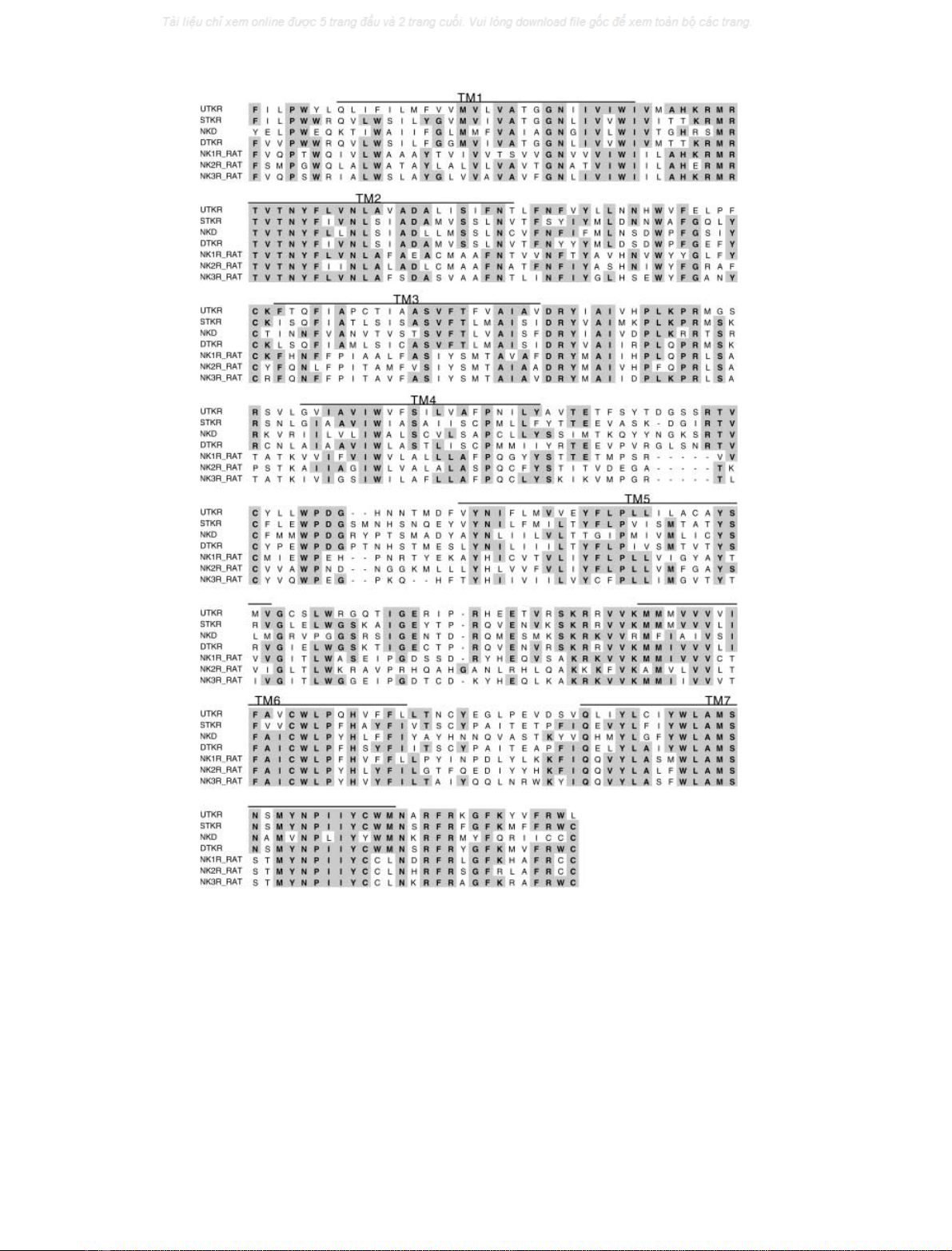
Comparative analysis of amino-acid sequences of mamma-
lian tachykinin receptors and insect tachykinin-related
peptide receptors showed that the second, sixth, and seventh
transmembrane domains are highly conserved among all
receptors. To identify a tachykinin-related peptide receptor
of the echiuroid worm, we first performed RT-PCR
experiments using degenerative primers corresponding to
the conserved regions (see Materials and methods). An
amplified cDNA product of 628 bp was subcloned and
sequenced. The putative amino-acid sequence was shown to
encode a partial transmembrane domain of a GPCR.
Moreover, we determined the full-length cDNA sequence
encoding the putative GPCR using the 5¢-and3¢RACE
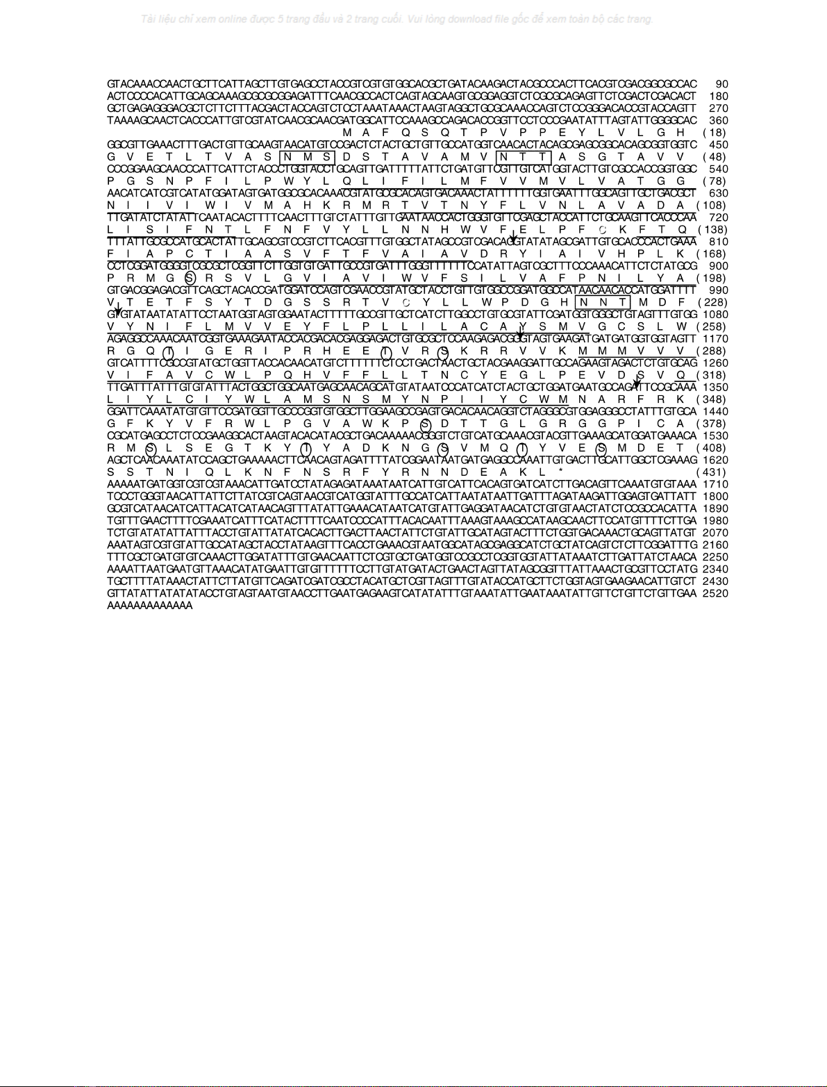
method. Figure 1A shows the 2533 bp putative receptor
cDNA containing a 1293 bp ORF flanked by a 306 bp
5¢-untranslatedregion(UTR)anda924bp3¢-UTR. The
ORF begins with the ATG codon at position 307, which is
supported by the Kozak rule [17], and terminates with a
TGA stop codon at position 1602. Only one potential
polyadenylation signal AATAAA was found to be located
19 bases upstream of a poly(A) tail.
The deduced receptor protein is composed of 431 amino-
acid residues (Fig. 1). The sequence showed the presence of
the seven hydrophobic transmembrane regions that are the
most typical characteristic of GPCRs. The common Cys
residues (Cys134 and Cys214) responsible for the disulfide
bridge between the first and second extracellular loops are
found at corresponding positions of known tachykinin
receptors. N-linked glycosylation sites (Asn-X-Ser/Thr,
Asn28, Asn39, and Asn223) are also located at the
N-terminal and second extracellular domains. The GPCR
sequence were also found to contain potential phosphory-
lation sites by protein kinase A (Arg/Lys-X-(X)-Ser/Thr,
Ser173, Thr262, Ser365, Ser381, Thr389, and Ser396), by
protein kinase C (Ser/Thr-X-Arg/Lys, Thr273 and Ser276),
and by casein kinase 2 (Ser/Thr-X-(X)-Asp/Glu, Thr262,
Ser381, Thr389, Thr400, and Ser404) in the second and
third intracellular loop and C-terminal region. Further-
more, the Asp/Glu-Arg-Tyr motif (Asp158Tyr160) in the
second intracellular loop and the Lys/Arg-Lys/Arg-X-X-
Lys/Arg motif(Arg278Lys282) in the third intracellular
loop which are often shown in most GPCRs are also present
(Fig. 1), whereas a cysteine residue utilized as a palmityla-
tion site in the C-terminal region was not found, given that
the Trp/Cys-Cys palmitylation site in tachykinin receptors
was replaced with Trp356Leu357 at the corresponding
positions of the putative Urechis GPCR (Fig. 1). The lack of
this site was not the result of a PCR error or an artifact, as
all clones obtained using different polymerases encoded the
identical sequence. Comparative study of amino-acid
sequences verified that the putative Urechis GPCR sequence
including the transmembrane domains and intracellular and
extracellular regions displayed high identity to those of
mammalian tachykinin receptors and insect tachykinin-
related peptide receptors (Fig. 2 and Table 2). In addition,
the sequence of this region was shown to be closer to those
of tachykinin-related peptide receptors than tachykinin
receptors (Table 2). Furthermore, the homology-searching
showed no significant similarity of UTKR to any other
GPCR. Taken together, these results revealed that the
putative Urechis GPCR possesses the essential properties of
tachykinin receptors and tachykinin-related peptide recep-
tors. Consequently, we concluded that this GPCR is a
putative Urechis tachykinin-related peptide receptor and
designated the receptor as the Uru-TK receptor, UTKR.
Functional expression of UTKR in
Xenopus
oocytes
It is well established that the binding of tachykinins and
tachykinin-related peptides to their receptors results in the
activation of PLC followed by the production of the
4240 T. Kawada et al. (Eur. J. Biochem. 269)FEBS 2002
intracellular second messengers, InsP
3
and calcium
[1316,1820]. In Xenopus oocytes, the interaction of an
agonist with its GPCR, inducing an elevation of intracel-
lular calcium, leads to the activation of a calcium-dependent
chloride channel, which is evaluated by direct observation of
the resultant inward chloride current. This system has been
employed for functional analyses of tachykinin receptors
and tachykinin-related peptide receptors [1821], and thus,
we examined whether the UTKR expressed in Xenopus
oocytes was activated by its putative endogenous ligands,
Uru-TKs.
After UTKR cRNA was injected into oocytes followed
by incubation at 17 C for 24 days, the receptor-expres-
sing oocytes were voltage-clamped at )80 mV. Subse-
quently, Uru-TK I was added to an oocyte every 20 min at
indicated concentrations in order to prevent desensitization
of the receptor. As shown in Fig. 3(A), application of Uru-
TK I to the UTKR-expressing Xenopus oocytes evoked a
clear response, whereas no signal was observed in the
absence of the UTKR cRNA (data not shown). A maximal
response was observed at more than 20 n
M
,andthehalf-
maximal response value (EC50) was calculated to be
approximately 1 n
M
by a doseresponse curve of current
shift (Fig. 3B). These results confirmed that UruTK I is
an endogenous ligand of UTKR.
In a previous study, we showed that six Uru-TK peptides
(Uru-TK IV and VII, as summarized in Table 1) were
yielded from the single Uru-TK precursor in the nervous
tissue of echiuroid worms [10]. To examine whether other
Uru-TKs are also endogenous agonists of UTKR, the
activities of Uru-TKs IIVandVIIonUTKRwere
observed by the voltage-clamp method. As shown in
Fig. 3B, all EC50 values of Uru TKs IIVandVIIwere
showntobe0.623.15 n
M
, demonstrating that the effects of
all Uru-TKs on UTKR were as potent as that of Uru-TK I.
These results indicate that Uru-TKs IIVandVIIalsoserve
as endogenous agonistic ligands of UTKR with equivalent
activity to Uru-TK I. Furthermore, no marked difference in
the activity of Uru-TKs on UTKR suggested that UTKR
possessed no significant selective affinity with any Uru-TK.
Fig. 1. A cDNA and deduced amino-acid sequence of Uru-TK receptor, UTKR. Seven putative transmembrane domains are underlined. The
conserved N-glycosylation sites (Asn28, Asn39, and Asn223) are boxed. Potentially phosphorylated serines or threonines (Ser173, Thr262, Thr273,
Ser276, Ser365, Ser381, Thr389, Ser396, Thr400, and Ser404) are marked by circles. Cysteines in a disulfide bridge (Cys134 and Cys214) are
indicated in black. The Asp-Arg-Tyr and Lys/Arg-Lys/Arg-X-X-Lys/Arg characteristic sequences in G-coupled receptors are written in italic
(Asp158-Tyr160 and Arg278-Lys282). Arrows indicate introns-inserted positions.
FEBS 2002 An Urechis tachykinin-related peptide receptor (Eur. J. Biochem. 269) 4241
Structureactivity relationships of Uru-TKs
and mammalian tachykinins
Most invertebrate tachykinin-related peptides contain a
common Phe-X-Gly-Y-Arg-NH
2
sequence at their
C-termini, whereas the C-terminal consensus motif of
vertebrate tachykinins is Phe-X-Gly-Leu-Met-NH
2
.More-
over, we demonstrated in our previous study that conver-
sion of Arg-NH
2
to Met-NH
2
in all Uru-TKs resulted in the
loss of the contractile activity of Uru-TKs on the cockroach
hindgut, although the peptides and tissues used in these
studies were derived from different species [10,22]. To
confirm whether the C-terminal Arg-NH
2
is critical for
activation of the UTKR, an Uru-TK I analog ([Met10]Uru-
TK I), in which the C-terminal Arg-NH
2
is replaced with
Met-NH
2
, was synthesized and applied in the voltage-clamp
experiment. As shown in Fig. 4A, the [Met10]Uru-TK I
analog exhibited no activity on UTKR at concentrations
comparable to those of Uru-TK I. This result clearly
showed that the Phe-X-Gly-Y-Arg-NH
2
is essential for
Fig. 2. Alignment of the amino-acid sequence of receptor core region. Four invertebrate tachykinin-related peptide receptors (UTKR, STKR, NKD
and DTKR) and three rat tachykinin receptors (NK13R) are aligned. Conserved residues are shadowed and shown in bold. Seven putative
transmembrane regions (TM1-7) are indicated above the corresponding sequence part.
4242 T. Kawada et al. (Eur. J. Biochem. 269)FEBS 2002


![Báo cáo seminar chuyên ngành Công nghệ hóa học và thực phẩm [Mới nhất]](https://cdn.tailieu.vn/images/document/thumbnail/2025/20250711/hienkelvinzoi@gmail.com/135x160/47051752458701.jpg)


