R E V I E W A R T I C L E
Structural recognition of DNA by poly(ADP-ribose)polymerase-like zinc finger families Stefania Petrucco and Riccardo Percudani
Department of Biochemistry and Molecular Biology, University of Parma, Italy
Keywords DNA binding; DNA damage; PARP; phylogenesis; zinc fingers
Correspondence S. Petrucco, Department of Biochemistry and Molecular Biology, Univesrity of Parma, Parco Area delle Scienze 23 ⁄ A, I-43100 Parma, Italy Fax: +39 0521 905151 Tel: +39 0521 905149 E-mail: petrucco@unipr.it
PARP-like zinc fingers (zf-PARPs) are protein domains apt to the recogni- tion of multiple DNA secondary structures. They were initially described as the DNA-binding, nick-sensor domains of poly(ADP-ribose)polymerases (PARPs). It now appears that zf-PARPs are evolutionary conserved in the eukaryotic lineage and associated with various enzymes implicated in nucleic acid transactions. In the present study, we discuss the functional and structural data of zf-PARPSs in the light of a comparative analysis of the protein family. Sequence and structural analyses allow the definition of the conserved features of the zf-PARP domain and the identification of five distinct phylogenetic groups. Differences among the groups accumulate on the putative DNA binding surface of the PARP zinc-finger fold. These observations suggest that different zf-PARP types have distinctive recogni- tion properties for DNA secondary structures. A comparison of various functional studies confirms that the different finger types can accomplish a selective recognition of DNA structures.
(Received 14 November 2007, revised 17 December 2007, accepted 24 December 2007)
doi:10.1111/j.1742-4658.2008.06259.x
Introduction
(zf-PARPs),
(PARPs)
zf-PARPs
represent
the
PARP-like zinc fingers (zf-PARP) are zinc coordi- nated protein domains that assist the DNA structure recognition of different eukaryotic enzymes, and owe their name to the proteins where these domains were identified for the first time, namely poly(ADP-ribose- [1,2]. Beyond PARPs, other polymerases enzymes involved in the DNA metabolism are also characterized by the presence of zf-PARPs and, among them, mammalian DNA ligases III and plant DNA 3¢ phosphatases have been studied in some detail.
PARPs
substrates
the most represented and studied member of the PARP family, is characterized by the presence of two unusually long zinc fingers that are 
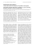
positioned upstream of the catalytic domain (Fig. 1). zf-PARPs mediate DNA recognition by PARP-1 and were initially termed as nick-sensors due to their spe- cific binding to nicked DNA [3]. It was subsequently demonstrated that zf-PARPs also recognize other DNA structures, including double-strand breaks, three- and four-way junctions, hairpins, bubbles, etc. [4–8]. Importantly, regulatory domain of PARP-1, and they are required for inducing enzyme activity upon DNA recognition [8–10]. The amount of activation depends upon the bound DNA structure as well as upon the relative concentrations of NAD+ and of ATP [11,12]. PARP-1 is its own best include histones, DNA substrate; other synthesis and repair enzymes, topoisomerases, tran- scription factors, centromeric proteins, etc. [13–18].
PARPs are a family of abundant eukaryotic enzymes that catalyse the reversible, NAD+-dependent poly ADP-ribosylation of protein substrates. PARP-1,
FEBS Journal 275 (2008) 883–893 ª 2008 FEBS. No claim to original Italian government works
883
Abbreviations FI, N-terminal finger of PARP; FII, second finger of PARP; G1–5, groups, 1 to 5; PARP, poly(ADP-ribose)polymerase; zf-PARP, PARP-like zinc fingers.
S. Petrucco and R. Percudani zf-PARP families
would even seem that, in the case of the ligase III, the zf-finger domain competes with the catalytic domain for nicked DNA binding. Leppard et al. [24] suggested that the DNA ligase III finger recognizes and interacts with single strand breaks, when DNA ligase III, and possibly the associated single strand DNA repair complex, is bound to negatively charged, auto modified PARP-1. Furthermore, it has been suggested that DNA ligase III finger stimulates rejoining of DNA strand breaks at sites of clustered damage [21]. Yet, a clear role for the DNA ligase finger in vivo has not emerged.
The nick sensing activity of zf-PARPs has sustained the general opinion that DNA breaks are the major sites of PARP-1 modifying activity. In recent studies, however, a new property of DNA recognition by PARP-1 has been described, which stresses the more general aptitude of PARP-1 to function as a chromatin modifier [16,19]. According to Kim et al. [16], PARP-1 is specifically bound to nucleosomes of nontranscribed, H1-histone-free chromatin domains. The authors pro- pose that PARP-1 structures a silent, but ready-to-be- open, chromatin conformation, where the activity of nucleosome-bound PARP-1 (but not of unbound PARP) is regulated by the relative concentrations of NAD and ATP.
Plant DNA 3¢ phosphatases are phosphoesterases that can restore functional 3¢ DNA ends by specifically removing 3¢ blocking phosphates. This is a necessary step in DNA repair pathways because DNA polyme- rases and DNA ligases can only process DNA that carries free 3¢ OH ends.
Mammalian DNA ligase III and plant DNA 3¢ phosphatases
Mammalian DNA ligases III and plant DNA 3¢ phos- phatases represent the other two types of enzymes for which zf-PARPs have been described [20–23].
Plant, but not animal, 3¢ DNA phosphatases, bear a unique N-terminal region comprising multiple copies of zf-PARPs. As for ligase III and PARP-1, fingers can bind to different DNA secondary structures but, similar to ligase III and in contrast to PARP-1, DNA the enzymatic activity of binding does not control plant DNA 3¢ phosphatases [22,23].
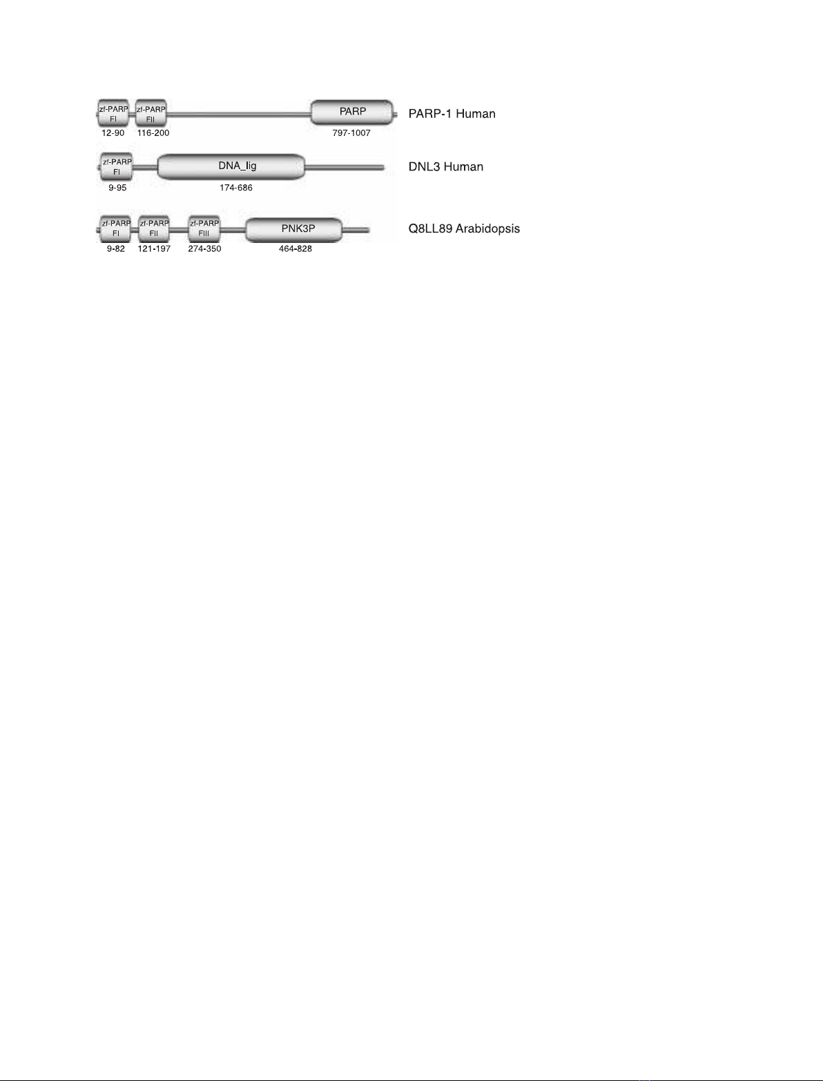
As shown in Fig. 1, DNA ligase III bears a single zf-PARP, whereas Arabidopsis DNA 3¢ phosphatase has three such fingers. Both enzymes are implicated in single-strand DNA repair processes, which are respon- sible for removing damage that has occurred on either thus anticipating the occurrence of DNA filament, much more dangerous double-strand DNA breaks.
zf-PARPs are thus components of a very abundant chromatin modifier, PARP-1, and they are necessary for binding at specific DNA sites. They are also found in DNA repair enzymes that do not require zf-PARPs to recognize DNA damages. DNA ligases and 3¢ DNA phosphatases can obviously bind damaged DNA (nicked and 3¢ blocked, respectively) via the active site of their catalytic domains.
DNA ligase III is an ATP dependent DNA ligase that appears to be a repair specific enzyme. The speci- ficity might originate from its interactions within a single-strand DNA repair complex, which restricts enzymatic activities to the damaged DNA position [20,21].
Here, we address questions and propose answers concerning the functional differences existing between zf-PARPs, which have been proposed to share binding specificity in different protein contexts [21,25].
Fig. 1. Domain architecture of the charac- terized zf-PARP proteins. Only zf-PARPs and associated catalytic domains are indicated, according to Pfam annotation: DNA_ligase, DNA ligase III; PNK3P, polynucleotide kinase 3 phosphatase. Proteins are drawn to scale and the limits of each domain are indi- cated by corresponding amino acid posi- tions.
The zf-PARP family
Searches in the DNA database for zf-PARP sequences immediately show that this protein module is not unique to the three enzymes mentioned above. More
The distinctive feature of DNA ligase III with respect to other DNA ligases is the presence of a zf-PARP at its N-terminus. Similar to PARP-1 fingers, the ligase finger also recognizes different DNA secondary struc- tures, such as nicks and cruciforms. In the case of ligase III, however, and in contrast to PARP-1, DNA recognition by the finger does not have an obvious it influence on enzymatic activity [20,21]. Curiously,
FEBS Journal 275 (2008) 883–893 ª 2008 FEBS. No claim to original Italian government works
884
types,
predicted
including
polypeptides protein encoded in the genomes of lower eukaryotes, also dis- play zf-PARPs (Table 1).
phosphatases. Finally, no member of the group five has yet been characterized. Future work might elicit insights regarding the specific functions of zf-PARPs of this group, which is the most heterogeneous in terms of protein domain architecture.
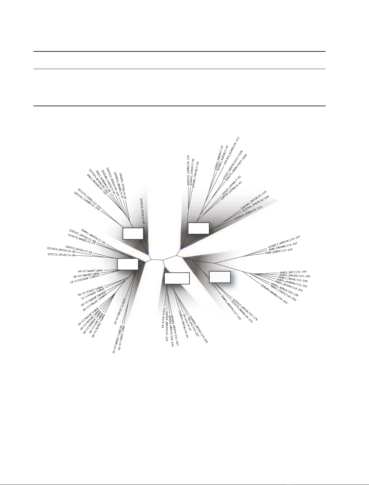
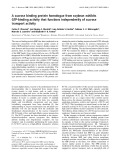
A sequence alignment of the complete family was used to obtain the phylogenetic tree shown in Fig. 2, which allows clustering zf-PARPs into five different groups. Group one (G1) comprises the first, N-termi- nal finger (FI) of PARPs. Group two (G2) comprises DNA ligase III fingers and only includes animal and mycetozoan sequences. Group three (G3) comprises the zf-PARPs that are exclusively found in plant DNA 3¢ phosphatases. Group four (G4) comprises the sec- ond (FII) fingers of animal and plant PARPs. Interest- ingly, all other eukaryotes appear to lack a second finger in their PARPs. Group five (G5) comprises fun- gal and protozoan sequences of putative DNA heli- cases, high mobility group proteins and RNA binding kinases. Furthermore, fingers of this group can be orphan of any catalytic domain and simply be associ- ated with low complexity protein regions. Given that low complexity regions often provide sites for protein– protein interactions, orphan fingers possibly provide DNA binding functions to interacting protein com- plexes.
When the sequences of different groups are com- pared, a number of aligned positions show strong amino acid conservation, allowing the definition of a the zf-PARP domain (Fig. 3). general signature of Beyond residues for zinc coordination, four hydropho- bic and four charged amino acid residues appear to be almost invariant in all finger types (Fig. 3A,B). Indeed, some of the invariant residues have been functionally tested in vitro and in vivo and shown to be essential for DNA binding [21,26]. However two regions, named region V1 and V2 in Fig. 3, are highly variable among zf-PARPs, both in length and sequence. A clear signa- ture of the group is only observable in the case of G2 and G4 fingers, but conserved features can also be noticed in the variable regions of other groups (Fig. 4). In particular, V1 in G1 fingers displays a prevalence of hydrophobic amino acid residues sepa- rated by a highly conserved aspartic residue. In the same region, G2 fingers display a predominance of hydrophobic and small amino acid residues, whereas G4 fingers have a prevalence of charged amino acids. V1 in G3 and G5 fingers shows poor conservation. V2 is mostly conserved within G2, 3 and 4 fingers, with a large majority of charged residues in G2 and a highly in G4. V2 is shorter in conserved RxELxF motif G3 fingers and characterized by conserved proline resi- dues. Functional divergence among proteins of G5 could also account for the sequence heterogeneity observed within this group.
In summary, the alignment of zf-PARPs suggests that, in a very much conserved backbone scaffold, two variable regions might be in charge of providing spe- cific properties to the different zf-PARP groups.
S. Petrucco and R. Percudani zf-PARP families
zf-PARP structure
Multiple studies provide direct evidence that isolated zf-PARP domains can recapitulate the binding proper- ties of full-length proteins. Thus, structural features of zf-PARPs are the basis of the DNA recognition. The recent addition of two zf-PARP structures deriving from structural genomics initiatives, corresponding to the FI (PDB 1v9x) and FII (PDB 2cs2) fingers of PARP-1, allows comparison with the published struc- ture of the ligase III finger [25]. As expected, a similar overall organization can be recognized within these three structures, which belong to the glucocorticoid receptor-like (DNA binding domain) super family. A
Overall, this group composition suggests an ancient origin for the zf-PARP module, which appears to be generally associated with domains involved in nucleic acid transactions. Because of the ancient separation of all zf-PARP groups, the branching order of the deepest nodes of the tree, and thus the relationships between the different groups, cannot be assessed with high con- fidence. In the case of PARP-1, however, it appears that the acquisition of a second finger predates animal and plant divergence. This would imply that FII fin- gers have been subsequently lost in some lineages (e.g. Caenorhabditis elegans). The presence of an FII finger in complex organisms might reflect additional roles acquired by PARP-1 as a chromatin modifier. It is interesting to note that FI and FII fingers of PARP-1 are clearly divergent and, indeed, PARP FI appears to be more related to DNA ligase III fingers than to PARP FII. In keeping with the phylogenetic analysis, it was previously shown that the DNA ligase III finger is specifically recognized by PARP FI, but not FII, anti-antibodies [20]. These observations may indicate that FI and FII fingers are not redundant and supply diverse functions in PARP-1 activity. By contrast, all DNA 3¢ phosphatase fingers cluster in G3 and show relatively recent duplications, which occurred after the monocots–dicots split. Also, the number of fingers associated with these enzymes is quite variable (from one in Citrus to three in Arabidopsis), thus suggesting that these fingers have redundant roles in DNA 3¢
FEBS Journal 275 (2008) 883–893 ª 2008 FEBS. No claim to original Italian government works
885
S. Petrucco and R. Percudani zf-PARP families
Table 1. List of the zf-PARP containing proteins considered in the present study. Sequences containing the zf-Parp domain were retrieved from the Pfam entry PF00645 (http://pfam.sanger.ac.uk) with the addition of Zea mays and Citrus clementina polynucleotide 3-phosphatases, which where deduced from EST assemblies. Sequences less than 90% identical were retained in the final set and utilized for phylogenetic analysis. Catalytic domains are indicated according to Pfam annotation: DNA_ligase, DNA ligase III; PNK3P, polynucleotide kinase 3 phospha- tase; PI3_PI4_kinase, phosphatidylinositol 3- and 4-kinase; Helicase_C, helicase conserved C-terminal domain.
IDa Protein description Organism name Taxon Catalytic domainsb Length Phylogenetic Groupa
NAD+ ADP-ribosyltransferase-1B Q8I7C5_DICDI Q5RHR0_BRARE Novel protein similar to 804 1013 PARP PARP Dictyostelium discoideum Mycetozoa Brachydanio rerio Metazoa G1 G1 + G2
vertebrate ADP- ribosyltransferase Q7QBC7_ANOGA ENSANGP00000014723 995 PARP Anopheles gambiae str. Metazoa G1 + G2 PEST
Poly(ADP-ribose) polymerase Q510C0 ENTHI_ NAD+ ADP-ribosyltransferase-1A Q7Z115_DICDI Q61WX1_CAEBR Hypothetical protein CBG04221 PME1_CAEEL Poly(ADP-ribose)polymerase 845 938 936 945 PARP PARP PARP PARP Entamoeba histolytica Dictyostelium discoideum Mycetozoa Caenorhabditis briggsae Caenorhabditis elegans Enthamoebidae G1 G1 G1 G1 Metazoa Metazoa pme-1
PARP_SARPE PARP_DROME PARP1_XENLA PARP1_RAT PARP1_MOUSE PARP1_HUMAN PARP1_CRIGR PARP1_CHICK PARP1_BOVIN PARP1_ARATH Q4KM23_BRARE Q5ZLW6_CHICK Q8UVU2_XENLA Q4SEP2_TETNG Poly(ADP-ribose)polymerase Poly(ADP-ribose)polymerase Poly(ADP-ribose)polymerase Poly(ADP-ribose)polymerase 1 Poly(ADP-ribose)polymerase 1 Poly(ADP-ribose)polymerase 1 Poly(ADP-ribose)polymerase 1 Poly(ADP-ribose)polymerase 1 Poly(ADP-ribose)polymerase 1 Poly(ADP-ribose)polymerase 1 Zgc:112973 Hypothetical protein DNA ligase III isoform alpha Chromosome undetermined 996 994 998 1014 1013 1014 1013 1011 1016 983 752 902 988 873 PARP PARP PARP PARP PARP PARP PARP PARP PARP PARP DNA_ligase DNA_ligase DNA_ligase DNA_ligase Metazoa Sarcophaga peregrina Drosophila melanogaster Metazoa Metazoa Xenopus laevis Metazoa Rattus norvegicus Metazoa Mus musculus Metazoa Homo sapiens Metazoa Cricetulus griseus Metazoa Gallus gallus Metazoa Bos taurus Viridiplantae Arabidopsis thaliana Metazoa Brachydanio rerio Metazoa Gallus gallus Metazoa Xenopus laevis Metazoa Tetraodon nigroviridis G1 + G2 G1 + G2 G1 + G2 G1 + G2 G1 + G2 G1 + G2 G1 + G2 G1 + G2 G1 + G2 G1 + G2 G4 G4 G4 G4 SCAF14615
Q2T9Y5_BOVIN DNL3_MOUSE DNL3_HUMAN Zm ESTb Citrus ESTc Q84JE8_ARATH Similar to DNA ligase III DNA ligase 3 DNA ligase 3 EST assembly EST assembly Putative DNA nick-sensor 943 1015 922 462 276 694 DNA_ligase DNA_ligase DNA_ligase PNK3P PNK3P PNK3P Bos taurus Mus musculus Homo sapiens Zea mays Citrus clementina Arabidopsis thaliana Metazoa Metazoa Metazoa Viridiplantae Viridiplantae Viridiplantae G4 G4 G4 G3 G3 G3 protein Oryza sativa
463 2729 3409 Viridiplantae Fungi Fungi G3 G5 G5 Q5JND9_ORYSA Q4I275_GIBZE Q7SI27_NEUCR Putative phosphoesterase Hypothetical protein Hypothetical protein PNK3P PI3_PI4_kinase Gibberella zeae PI3_PI4_kinase Neurospora crassa NCU00625.1
Helicase_C Helicase_C Helicase_C
Q4QA20_LEIMA Q387H5_9TRYP Q4E4N3_TRYCR Q4RR05_TETNG Q4Q1U1_LEIMA Q21275_CAEEL Q5BY75_SCHJA Q54E19_DICDI 1092 984 983 233 285 493 165 895 Leishmania major Trypanosoma brucei Trypanosoma cruzi Tetraodon nigroviridis Leishmania major Caenorhabditis elegans Schistosoma japonicum Dictyostelium Euglenozoa Euglenozoa Euglenozoa Metazoa Euglenozoa Metazoa Metazoa Mycetozoa G5 G5 G5 G1 G5 G5 G1 G5 DNA repair protein, putative DNA repair protein, putative DNA repair protein, putative Chromosome 14 SCAF15003 Hypothetical protein Hypothetical protein SJCHGC03951 protein SMAD ⁄ FHA domain-containing protein discoideum AX4
FEBS Journal 275 (2008) 883–893 ª 2008 FEBS. No claim to original Italian government works
886
Q61C61_CAEBR Q38AV1_9TRYP Q4E4B2_TRYCR Q5KJS7_CRYNE Hypothetical protein CBG13063 Hypothetical protein Hypothetical protein Hypothetical protein 467 240 230 254 Metazoa Euglenozoa Euglenozoa Fungi G5 G5 G5 G5 Caenorhabditis briggsae Trypanosoma brucei Trypanosoma cruzi Cryptococcus neoformans
S. Petrucco and R. Percudani zf-PARP families
Table 1. (Continued).
IDa Protein description Length Organism name Catalytic domainsb Phylogenetic Groupa Taxon
Q9Y7K9_SCHPO SPBC2A9.07c protein 274 Fungi G5
a Present study. b Deduced by the assembly of EST sequences DR813175, DN226524, DV517124, DR813176, DT649995. c Deduced by the assembly of EST sequences DY292829, DY289144, DY300019.
Q4PF94_USTMA Q5B8J3_EMENI Q2UBX7_ASPOR Hypothetical protein Hypothetical protein Predicted protein 546 279 143 Schizosaccharomyces pombe Ustilago maydis Emericella nidulans Aspergillus oryzae Fungi Fungi Fungi G5 G5 G5
G5 : fungi protozoa
G2 : DNA Ligase
G1 : PARP Finger I
G4 : PARP Finger II
G3 : PNK3P Fingers I/II/III
schematic view of the zf-PARP fold is shown in Fig. 5. A three-stranded antiparallel b sheet characterizes the N-terminal half of the domain (b1, b2 and b3 in Figs 3A and 5), with a long loop connecting b1 and b2
and containing two of the cysteine residues involved in is coordinating the zinc ion. The C-terminal half mainly a helical (a1 and a2 in Figs 3A and 5), with the third and the forth zinc-chelating residues
FEBS Journal 275 (2008) 883–893 ª 2008 FEBS. No claim to original Italian government works
887
Fig. 2. Phylogenetic relationships in the zf-PARP family. Alignment of the zf-Parp domains was carried out with the family Hidden Markov model of Pfam using programs of the HMMER package [33]. Maximum-likelihood phylogeny was obtained with the PHYML program [34]. The resulting unrooted maximum-likelihood tree was visualized with branch length adjustment for visibility enhancement using TREE ILLUSTRATOR. Branches leading to the main phylogenetic groups are shadowed in gray and labelled according to the group composition. Sequenced are indicated with identifiers (for details, see Table 1), followed by the sequence interval considered in the analysis.
S. Petrucco and R. Percudani zf-PARP families
A
B
FEBS Journal 275 (2008) 883–893 ª 2008 FEBS. No claim to original Italian government works
888
Fig. 3. Sequence features of the zf-PARP family. V1 and V2 indicate regions that are not conserved in sequence and length among the differ- ent groups of the zf-PARP family. (A) Sequence alignment of representative members of the zf-PARP family groups. Numbers preceding sequence identifiers indicate phylogenetic groups as designated in Fig. 2. Identical residues are shown in white on a red background; residues with equivalent physical-chemical features and conserved among 90% of the sequences are shown in red and boxed. Secondary structure features deduced from PDB coordinates of the Arabidopsis PAPR-1 FI finger (blue), of the human PARP-1 FII (orange) and of the human DNA ligase III finger (green) are drawn above the aligned sequences. The alignment of zf-PARPs, decorated with secondary structure elements, was visualized using ESPRIPT (http://espript.ibcp.fr/ESPript). (B) Sequence logo of the zf-PARP family. The logo from the complete alignment of the family was obtained at http://weblogo.berkeley.edu/logo.cgi. Amino acids are coloured according to their physical-chemical features.
S. Petrucco and R. Percudani zf-PARP families
it
in this case,
the entry of (histidine and cysteine) exposed at helix a1. Zinc coordination orients the long loop between sheets b1 and b2, toward the a1 helix. Vari- able region V1 identified in the sequence alignment (Fig. 3B) comprises the loop between sheets b2 and b3, whereas variable region V2 comprises the C-termi- nal side of helix 1 and the loop between helices a1 and a2. It thus follows that helix 1 has different length amongst the different structures.
in the FII finger
Superposition of the three structures shows that the zinc-coordinating residues, as well as all other highly conserved amino acid residues, occupy quasi-identical positions in the three finger types (Fig. 5B). Side chains of conserved hydrophobic residues are buried and appear to be mainly responsible for the folding of the finger structure. Side chains of conserved residues that are polar or charged are exposed at the surface and contribute to the solvent-exposed electrostatic variable potential of
the fingers. As
expected,
regions V1 and V2 display different shapes in the three finger types. NMR studies show that much of region V1 is disordered in all three finger types, most likely due to internal flexibility (demonstrated experimentally for the case of the ligase finger), whereas the V2 region is partially disordered only in the case of the ligase structure, although, is not known whether this reflects dynamic processes or not. Differ- ences in the C-terminal side of helix 1, and thus in the length of this helix, are possibly an important determi- nant for the specific structural recognition. Indeed Gradwohl et al. [5] have demonstrated that substitut- ing helix 1 of PARP FII with helix 1 of FI induces FI-binding properties [5]. Loops including regions V1 and V2 are part of the protein– DNA interface and, accordingly, display positive elec- in the ligase III finger [25]. Fig. 6 trostatic potential shows that the charge distribution on the solvent- accessible surface is only partially conserved among
FEBS Journal 275 (2008) 883–893 ª 2008 FEBS. No claim to original Italian government works
889
Fig. 4. Within-group conservation of zf-PARP variable regions. Sequence logos of the variable regions V1 (left panels) and V2 (right panels) were obtained from sequence alignments of distinct zf-PARP groups. Groups (G1–G5) are designated as in Fig. 2.
S. Petrucco and R. Percudani zf-PARP families
A
B
Fig. 5. Structural features of the PARP-like finger family. (A) Sche- matic representation of the zf-PARP fold. The most conserved sec- ondary structure elements (a1, a2, b1, b2, b3) and the two variable regions (V1, V2) are indicated; the coordinated zinc ion is repre- sented as a blue sphere. (B) Stereo view of the C-alpha trace com- parison among the Arabidopsis PARP-1 FI (PDB 1v9x, conformer 1, amino acids 11–89; cyan), the human PARP-1 FII (PDB 2cs2, best representative conformer 1, amino acids 18–103; magenta) and the human DNA ligase III (PDB 1uw0, best representative conformer 1, amino acids 6–93; black) zf-PARPs. Structures were aligned and visualized using the PYMOL software (http://www.pymol.org/). Con- served amino acids in the complete alignment of zf-PARPs (bits score ‡ 3; see Fig. 3B) are represented as spheres.
immunological
analyses
and
the three finger types. When comparing corresponding views of the proteins, the charge distribution on the PARP-1 FI finger appears to be more similar to the DNA ligase finger than to the PARP-1 FII finger. Phylogenetic also suggested a closer relationship of PARP FI with the ligase III finger than with PARP FII.
binding surface of the different finger types might have different shapes and sizes.
It can thus be assumed that the DNA binding sur- face of zf-PARPs depends upon sequence and struc- tural features of variable regions 1 and 2 and that, due to loop mobility, the DNA binding surface can accommodate various DNA structures. Obviously, these analyses do not take into account the contribu- tion of unstructured portions of zf-PARPs observed in the NMR studies that are known to be important for DNA binding [25]. Still, they indicate that the DNA
Based on the known finger structures and on the sequence conservation among zf-PARPs, it is possible to obtain predictions about the structure of fingers for which data are not yet available. As expected, major differences are observed in the shape and length of variable regions 1 and 2 (not shown), and these differ- ences may provide insights into DNA binding specifici- ties. However, given the very poor conservation of V1 and V2, structural predictions in those regions are
FEBS Journal 275 (2008) 883–893 ª 2008 FEBS. No claim to original Italian government works
890
Fig. 6. Electrostatic potential on solvent accessible surfaces of Ara- bidopsis PARP-1 FI, human PARP-1 FII and human DNA ligase III zf-PARPs. The electrostatic potentials of the zf-PARPs were calcu- lated using APBS Poisson–Boltzmann solver software [35]; zinc ions were not included in the calculation. The left panel shows fin- ger surfaces oriented as in Fig. 5; the right panel shows corre- sponding surfaces following a 180(cid:2) rotation about the y-axis. Surfaces were coloured by potential (red negative, blue positive) using an arbitrary scale, as indicated. The same aligned conformers were used as in Fig. 5; however the results were stable with respect to choice of ensemble member.
expected to be inaccurate when distantly related zf- PARPs are compared. Experimental structural and functional data are thus mandatory to evaluate any assumptions concerning zf-PARPs that are not closely related to proteins already characterized.
S. Petrucco and R. Percudani zf-PARP families
zf-PARP binding specificity and function
that
including gaps, hairpins,
all bind to gapped DNA). Such observations would imply that common features among nonB DNA struc- tures could be recognized by the different zf-PARPs. The available experimental evidence indeed suggests that exposed DNA ends in a double-helix context and that helical flexibility might represent the recognition target for structural DNA recognition [29–32]. As shown in Table 1, however, different zf-PARPs do not always bind to same DNA structures and, in fact, a very specific recognition can be produced (e.g. 3¢ DNA phosphatase fingers recognized bulged, but not, nicked DNA; the opposite is true for PARP-1 fingers). This being the case, rather than relying on general DNA flexibility or DNA ends exposure, zf-PARPs must use more sophisticated tools for targeting DNA. These possibly include the monitoring of fine differences in DNA flexibility as well as transitions between single and double strands in the DNA. Such recognition skills would distinguish between nicks, gaps, hairpins, etc., which all represent possible different signals on the DNA molecule. More generally, structural specific recognition of the DNA could provide complex regula- tory potential within the cell.
When zf-PARPs were initially described, they were named nick-sensors, due to their ability to bind nicked DNA. When DNA ligase III and 3¢ DNA phosphatase fingers were identified, they were also named nick-sen- sors due to their sequence similarity with PARP-1 fin- gers and their ability to bind strand-breaks. It became rapidly clear zf-PARP recognition was not restricted to nicked DNA, but many different second- ary structures, three- and four-way cruciforms, double-strand breaks, recessed DNA ends, as well sequences that are prone to gener- ate altered secondary structures, could be specifically recognized by this new type of zinc finger. The specific role of such structural recognition in living cells has not been clarified and very little data are available in vivo. The difficulties in obtaining in vivo data come from the very limited experimental approaches for gen- erating predictable secondary DNA structures in living cells, thus explaining why the functional properties of zf-PARPs have been mostly investigated in vitro.
The fact that the different fingers are prone to a dis- tinctive DNA recognition was anticipated by the struc- tural information provided above. However, a simple correlation between binding specificity and finger type cannot be easily drawn. It must be emphasized that binding to a very limited number of DNA templates has been tested in a straight comparison and that quantitative evaluations are still lacking. More exten- sive analyses with more templates under identical experimental conditions, both in vivo and in vitro, would be required to decipher the DNA selectivity of the different finger types. Nevertheless, we propose that functional and structural analyses provide a clear
A partial summary of these studies is provided in Table 2. From these data, it can be inferred that each finger type recognizes several DNA structures (e.g. the DNA ligase III finger binds to nick and gapped DNA, and to double-strand DNA breaks). Also, different fin- ger types can recognize the same DNA structures (e.g. PARP-1, DNA ligase and 3¢ DNA phosphatase fingers
Table 2. Summary of the DNA binding properties of zf-PARP fingers. Only results obtained with DNA templates that are recognized by more than one zf-PARP type are shown. F indicates zf-PARP fingers numbered as in Fig. 1; zf-PARP finger groups are designated as in Fig. 2. Presence (+) and absence ()) of DNA binding activity; + ⁄ ), poorly detected binding; ND, not determined.
DNA ligase III PARP-1 3¢ DNA phosphatasea
FI–FII (G1–G4) FI (G1) FII (G4) FI–FII–FIII (G3) FI–FII (G3) FII–FIII (G3) FI (G3) DNA template
+ ⁄ )g )g + ⁄ ) ) + ⁄ ) ) + ⁄ ) )
a Reference [23] and Fig. S1. b References [5,26,27] and Fig. S1. c References [4,5,27] and Fig. S1. d Reference [24] and Fig. S1. e Refer- ence [28]. f Fig. S1. g References [5,6,26]. The results were obtained by analysing mutants of PARP-1 FI–FII, in which one zinc finger was render inactive by mutating one of its zinc binding sites. h References [20,21,24,25]. i References [20,21]. j References [21,24].
FEBS Journal 275 (2008) 883–893 ª 2008 FEBS. No claim to original Italian government works
891
+ ) ) +b +c +d +e )e )f Double-strand DNA breaks Nicked DNA Gapped DNA 5¢ Recessed DNA ends 3¢ Recessed DNA ends Bulged DNA ND ND ND ND + ⁄ )g +g ND ND ND ND + ND ND + + ⁄ )i +h +j ND ND ND + + ND ND +
stranded breaks in DNA. Proc Natl Acad Sci USA 87, 2990–2994.
6 Ikejima M, Noguchi S, Yamashita R, Ogura T,
indication that zf-PARPs can generate versatile and specific recognition properties of DNA secondary structure.
S. Petrucco and R. Percudani zf-PARP families
Conclusion
Sugimura T, Gill DM & Miwa M (1990) The zinc fingers of human poly(ADP-ribose) polymerase are differentially required for the recognition of DNA breaks and nicks and the consequent enzyme activation. Other structures recognize intact DNA. J Biol Chem 265, 21907–21913.
7 Sastry SS & Kun E (1990) The interaction of adenosine
diphosphoribosyl transferase (ADPRT) with a cruciform DNA. Biochem Biophys Res Commun 167, 842–847. 8 Lonskaya I, Potaman VN, Shlyakhtenko LS, Oussat- cheva EA, Lyubchenko YL & Soldatenkov VA (2005) Regulation of poly(ADP-ribose) polymerase-1 by DNA structure-specific binding. J Biol Chem 280, 17076– 17083.
9 D’Amours D, Desnoyers S, D’Silva I & Poirier GG
zf-PARPs constitute a family of protein modules bear- ing specific recognition properties for nonB nucleic acid structures. The family is evolutionary conserved in eukaryotes, suggesting that structural recognition of DNA by zf-PARPs is mostly important in chromatin templates. Beyond the conserved amino acids that are required to preserve the finger folding, two much less conserved regions could account for structural changes in the DNA binding surface. A higher degree of con- servation in such variable regions is observed in some of the zf-PARP groups identified by phylogenetic anal- ysis. Functional data show that zf-PARPs belonging to different phylogenetic groups can have distinct DNA recognition properties. We thus suggest that proteins containing different zf-PARPs can selectively distin- guish signals embedded in DNA structures.
(1999) Poly(ADP-ribosyl)ation reactions in the regula- tion of nuclear functions. Biochem J 342, 249–268. 10 Burkle A (2001) Poly(APD-ribosyl)ation, a DNA dam- age-driven protein modification and regulator of geno- mic instability. Cancer Lett 163, 1–5.
Acknowledgement
11 Kim MY, Mauro S, Gevry N, Lis JT & Kraus WL (2004) NAD+-dependent modulation of chromatin structure and transcription by nucleosome binding properties of PARP-1. Cell 119, 803– 814.
S. Petrucco is grateful to D. Neuhaus for helpful dis- cussion. This work was supported by the FIL grants of Universita` di Parma.
References
12 Kun E, Kirsten E, Mendeleyev J & Ordahl CP (2004) Regulation of the enzymatic catalysis of poly(ADP- ribose) polymerase by dsDNA, polyamines, Mg2+, Ca2+, histones H1 and H3, and ATP. Biochemistry 43, 210–216.
13 Schreiber V, Hunting D, Trucco C, Gowans B,
1 Cherney BW, McBride OW, Chen DF, Alkhatib H, Bhatia K, Hensley P & Smulson ME (1987) cDNA sequence, protein structure, and chromosomal location of the human gene for poly(ADP-ribose) polymerase. Proc Natl Acad Sci USA 84, 8370–8374.
Grunwald D, De Murcia G & De Murcia JM (1995) A dominant-negative mutant of human poly(ADP-ribose) polymerase affects cell recovery, apoptosis, and sister chromatid exchange following DNA damage. Proc Natl Acad Sci USA 92, 4753–4757.
2 Suzuki H, Uchida K, Shima H, Sato T, Okamoto T, Kimura T & Miwa M (1987) Molecular cloning of cDNA for human poly(ADP-ribose) polymerase and expression of its gene during HL-60 cell differentiation. Biochem Biophys Res Commun 146, 403–409.
3 de Murcia G & Menissier de Murcia J (1994) Poly(ADP-
14 Saxena A, Saffery R, Wong LH, Kalitsis P & Choo KH (2002) Centromere proteins Cenpa, Cenpb, and Bub3 interact with poly(ADP-ribose) polymerase-1 pro- tein and are poly(ADP-ribosyl)ated. J Biol Chem 277, 26921–26926.
15 Ame JC, Spenlehauer C & de Murcia G (2004) The
ribose) polymerase: a molecular nick-sensor. Trends Biochem Sci 19, 172–176.
PARP superfamily. Bioessays 26, 882–893.
4 Menissier-de Murcia J, Molinete M, Gradwohl G,
16 Kim MY, Zhang T & Kraus WL (2005) Poly(ADP-
ribosyl)ation by PARP-1: ‘PAR-laying’ NAD+ into a nuclear signal. Genes Dev 19, 1951–1967.
Simonin F & de Murcia G (1989) Zinc-binding domain of poly(ADP-ribose)polymerase participates in the recognition of single strand breaks on DNA. J Mol Biol 210, 229–233.
5 Gradwohl G, Menissier de Murcia JM, Molinete M,
17 Heale JT, Ball AR Jr, Schmiesing JA, Kim JS, Kong X, Zhou S, Hudson DF, Earnshaw WC & Yokomori K (2006) Condensin I interacts with the PARP-1-XRCC1 complex and functions in DNA single-strand break repair. Mol Cell 21, 837–848.
Simonin F, Koken M, Hoeijmakers JH & de Murcia G (1990) The second zinc-finger domain of poly(ADP- ribose) polymerase determines specificity for single-
FEBS Journal 275 (2008) 883–893 ª 2008 FEBS. No claim to original Italian government works
892
28 Pion E, Bombarda E, Stiegler P, Ullmann GM, Mely
18 Schreiber V, Dantzer F, Ame JC & de Murcia G (2006) Poly(ADP-ribose): novel functions for an old molecule. Nat Rev Mol Cell Biol 7, 517–528.
Y, de Murcia G & Gerard D (2003) Poly(ADP-ribose) polymerase-1 dimerizes at a 5’ recessed DNA end in vitro: a fluorescence study. Biochemistry 42, 12409– 12417.
29 Spiro C, Bazett-Jones DP, Wu X & McMurray CT
19 Nusinow DA, Hernandez-Munoz I, Fazzio TG, Shah GM, Kraus WL & Panning B (2007) Poly(ADP- ribose) polymerase 1 is inhibited by a histone H2A variant, MacroH2A, and contributes to silencing of the inactive X chromosome. J Biol Chem 282, 12851– 12859.
20 Mackey ZB, Niedergang C, Murcia JM, Leppard J,
(1995) DNA structure determines protein binding and transcriptional efficiency of the proenkephalin cAMP-responsive enhancer. J Biol Chem 270, 27702– 27710.
30 Morrison C, Smith GC, Stingl L, Jackson SP, Wagner EF & Wang ZQ (1997) Genetic interaction between PARP and DNA-PK in V(D)J recombination and tumorigenesis. Nat Genet 17, 479–482.
Au K, Chen J, de Murcia G & Tomkinson AE (1999) DNA ligase III is recruited to DNA strand breaks by a zinc finger motif homologous to that of poly(ADP- ribose) polymerase. Identification of two functionally distinct DNA binding regions within DNA ligase III. J Biol Chem 274, 21679–21687.
31 Mizukoshi T, Tanaka T, Arai K, Kohda D & Masai H (2003) A critical role of the 3’ terminus of nascent DNA chains in recognition of stalled replication forks. J Biol Chem 278, 42234–42239.
21 Taylor RM, Whitehouse CJ & Caldecott KW (2000) The DNA ligase III zinc finger stimulates binding to DNA secondary structure and promotes end joining. Nucleic Acids Res 28, 3558–3563.
32 Nishikawa J, Amano M, Fukue Y, Tanaka S, Kishi H, Hirota Y, Yoda K & Ohyama T (2003) Left-handedly curved DNA regulates accessibility to cis-DNA ele- ments in chromatin. Nucleic Acids Res 31, 6651–6662.
33 Eddy SR (1998) Profile hidden Markov models.
22 Petrucco S, Volpi G, Bolchi A, Rivetti C & Ottonello S (2002) A nick-sensing DNA 3’-repair enzyme from. Arabidopsis J Biol Chem 277, 23675–23683.
23 Petrucco S. (2003) Sensing DNA damage by PARP-like
Bioinformatics 14, 755–763.
fingers. Nucleic Acids Res 31, 6689–6699.
34 Guindon S & Gascuel O (2003) A simple, fast, and accurate algorithm to estimate large phylogenies by maximum likelihood. Syst Biol 52, 696–704.
24 Leppard JB, Dong Z, Mackey ZB & Tomkinson AE (2003) Physical and functional interaction between DNA ligase III alpha and poly(ADP-ribose) polymerase 1 in DNA single-strand break repair. Mol Cell Biol 23, 5919–5927.
25 Kulczyk AW, Yang JC & Neuhaus D (2004) Solution
35 Baker NA, Sept D, Joseph S, Holst MJ & McCammon JA (2001) Electrostatics of nanosystems: application to microtubules and the ribosome. Proc Natl Acad Sci USA 98, 10037–10041.
S. Petrucco and R. Percudani zf-PARP families
Supplementary material
structure and DNA binding of the zinc-finger domain from DNA ligase IIIalpha. J Mol Biol 341, 723–738.
26 Molinete M, Vermeulen W, Burkle A, Menissier-de
is available
The following supplementary material online: Fig. S1. Differential affinity of zf-PARPs for DNA secondary structures.
This material is available as part of the online article
Murcia J, Kupper JH, Hoeijmakers JH & de Murcia G (1993) Overproduction of the poly(ADP-ribose) poly- merase DNA-binding domain blocks alkylation-induced DNA repair synthesis in mammalian cells. EMBO J 12, 2109–2117.
from http://www.blackwell-synergy.com
27 Le Cam E, Fack F, Menissier-de Murcia J, Cognet JA,
Please note: Blackwell Publishing are not responsible for the content or functionality of any supplementary materials supplied by the authors. Any queries (other than missing material) should be directed to the corre- sponding author for the article.
Barbin A, Sarantoglou V, Revet B, Delain E & de Murcia G (1994) Conformational analysis of a 139 base-pair DNA fragment containing a single-stranded break and its interaction with human poly(ADP-ribose) polymerase. J Mol Biol 235, 1062–1071.
FEBS Journal 275 (2008) 883–893 ª 2008 FEBS. No claim to original Italian government works
893