Granule-bound starch synthase I
A major enzyme involved in the biogenesis of B-crystallites in starch granules
Fabrice Wattebled
1
, Alain Bule
´on
2
, Brigitte Bouchet
2
, Jean-Philippe Ral
1
, Luc Lie
´nard
1
, David Delvalle
´
1
,
Kim Binderup
1
, David Dauville
´e
1
, Steven Ball
1
and Christophe D’Hulst
1
1
Unite
´de Glycobiologie Structurale et Fonctionnelle, Unite
´Mixte de Recherche CNRS/USTL n8576, Unite
´Sous Contrat de l’INRA,
Universite
´des Sciences et Technologies de Lille, Villeneuve d’Ascq, France;
2
Institut National de la Recherche Agronomique,
Centre de Recherches Agroalimentaires, Nantes, France
Starch defines a semicrystalline polymer made of two
different polysaccharide fractions. The A- and B-type
crystalline lattices define the distinct structures reported in
cereal and tuber starches, respectively. Amylopectin, the
major fraction of starch, is thought to be chiefly respon-
sible for this semicrystalline organization while amylose is
generally considered as an amorphous polymer with little
or no impact on the overall crystalline organization. STA2
represents a Chlamydomonas reinhardtii gene required for
both amylose biosynthesis and the presence of significant
granule-bound starch synthase I (GBSSI) activity. We
show that this locus encodes a 69 kDa starch synthase
and report the organization of the corresponding STA2
locus. This enzyme displays a specific activity an order of
magnitude higher than those reported for most vascular
plants. This property enables us to report a detailed
characterization of amylose synthesis both in vivo and
in vitro. We show that GBSSI is capable of synthesizing a
significant number of crystalline structures within starch.
Quantifications of amount and type of crystals synthesized
under these conditions show that GBSSI induces the
formation of B-type crystals either in close association
with pre-existing amorphous amylopectin or by crystalli-
zation of entirely de novo synthesized material.
Keywords: starch; amylose synthesis; granule-bound starch
synthase; Chlamydomonas reinhardtii;in vitro synthesis.
Starch accumulates in plants as a complex granular
mixture of a-glucans (a-1,4-linked and a-1,6-branched)
consisting chiefly of amylopectin and amylose. In amylo-
pectin, the major fraction is composed of small-size a-1,4-
linked chains that are clustered together by the presence of
5% a-1,6 linkages [1] (starch structure reviewed in [2] and
[3]; starch metabolism reviewed in [4]). Amylose is
composed of longer chains with less than 1% a-1,6
branches. Plant starch can be further distinguished from
glycogen by the presence of highly ordered parallel arrays
of double helical glucans (reviewed in [5]). The origin of
these arrays resides in the close packing of the a-1,6
linkages at the root of the unit amylopectin cluster. The
9 nm size of each repetitive unit or cluster is conserved
throughout the plant kingdom [6]. Two major types of
crystalline organization have been documented so far in
native starch granules. A-type powder diffraction patterns
can be recovered from most cereal endosperm and
Chlamydomonas reinhardtii starches while B-type struc-
tures were reported for tuber starches or high amylose
starches from mutants of algae and cereals. It is generally
assumed that amylopectin plays a major role in establish-
ing the crystalline organization of starch. Indeed, amylose-
defective mutants or antisense constructs of maize and
potato accumulate normal amounts of starch with the
same A- or B-type granule organization and similar
crystallinities to the corresponding wild-type references. In
addition, starches with elevated amylose content are
generally less crystalline suggesting that most, if not all,
of the amylose remains amorphous within the granule.
Amylose synthesis has been known since the foundation
work laid by Nelson & Rines [7], to depend on the
presence of granule-bound starch synthase I (GBSSI), an
enzyme identified by de Fekete et al.[8],asassociatedwith
starch granules. GBSSI was first reported to use non-
physiological concentrations of UDP-glucose [9] while
ADP-glucose was shortly discovered thereafter as the
preferred donor substrate [10]. Mutations leading to
defectsforGBSSIhavebeenisolatedinanever-increasing
number of species including waxy (wx) maize [11], wx rice
[12], wx barley [13], wx wheat [14], amylose-free (amf)
potato [15], low amylose (lam) pea [16], wx amaranth [17]
and sta2 C. reinhardtii [18]. A number of studies
approaching the synthesis of amylose in vitro [9,19–21],
Correspondence to C. D’Hulst, Unite
´de Glycobiologie
Structurale et Fonctionnelle, Unite
´Mixte de Recherche
CNRS/USTL n8576, Unite
´Sous Contrat de l’INRA,
Universite
´des Sciences et Technologies de Lille, 59655 Villeneuve
d’Ascq, Cedex France.
Fax: + 33 3 20436555, Tel.: + 33 3 20434881,
E-mail: christophe.dhulst@univ-lille1.fr
Abbreviations: GBSSI, granule-bound starch synthase I; RFLP,
restriction fragment length polymorphism.
Enzymes: soluble and granule-bound starch synthases:
ADPglucose:1,4-a-
D
-glucan 4-a-
D
-glucosyltransferases (EC 2.4.1.21);
ADP-glucose pyrophosphorylase: ADP:a-
D
-glucose-1-phosphate
adenylyltransferase (EC 2.7.7.27).
Note: a web site is available at http://www.univ-lille1.fr/ugsf/
(Received 11 January 2002, revised 21 June 2002,
accepted 25 June 2002)
Eur. J. Biochem. 269, 3810–3820 (2002) FEBS 2002 doi:10.1046/j.1432-1033.2002.03072.x

establish that GBSSI incorporates glucose both in amy-
lopectin and amylose according to the conditions used.
Leloir et al. [9] originally noted a stimulation of GBSSI by
high concentrations of malto-oligosaccharides and found
incorporation of radioactive glucose into both starch
fractions. In a recent study, Denyer et al. [21] showed that
in the absence of these oligosaccharides, the labelled
product synthesized in vitro by GBSSI was confined to the
amylopectin fraction. However in the presence of high
malto-oligosaccharide concentrations, GBSSI incorporated
glucose massively into amylose-like glucans. In vivo
evidence supporting the involvement of GBSSI in amylo-
pectin synthesis was produced in Chlamydomonas by
Maddelein et al. [22]. Additional in vitro synthesis experi-
ments performed with starch granules isolated from
C. reinhardtii show that amylose synthesis can occur in
the absence of malto-oligosaccharide priming by extension
and cleavage of a nonreducing end available on an
amylopectin molecule [23]. It has recently been shown that
thismechanismalsoappearstobeatworkinthestarches
extracted from higher plants [24]. However the total
amount of GBSSI activity measured in Chlamydomonas
starch appeared 10- to 50-fold higher than that measured
in vascular plant starches [24].
We now report the cloning and characterization of
cDNAs and gDNAs corresponding to a granule-bound
starch synthase from C. reinhardtii. We show that this
sequence corresponds to the previously characterized STA2
gene required for amylose synthesis. We show that this
69 kDa enzyme contains an extra 11.4 kDa at the
C-terminus that is not found in the higher plant enzymes.
Detailed in vivo investigations performed during the course of
storage starch synthesis show that amylopectin and amylose
synthesis are partly disconnected and that amylose synthesis
persists when the rate of polysaccharide and amylopectin
synthesis become minimal.Invitrosynthesis experiments
performed using wild-type Chlamydomonas starch with this
high specific activity enzyme establish that GBSSI induces
the formation of B-type crystalline structures.
EXPERIMENTAL PROCEDURES
Materials
ADP[U-
14
C]glucose and a[
32
P]dCTP were purchased from
Amersham (Amersham, Buckinghamshire, UK). ADP-
glucose was obtained from Sigma. CL-2B Sepharose
column and Percollwere obtained from Amersham
Pharmacia Biotech. Starch assay kit was obtained from
Roche (Germany).
Chlamydomonas
strains, growth conditions and media
The reference strains of C. reinhardtii used in this study are
137C (mt-nit1 nit2)and330(mt+ nit1 nit2 arg7-7 cw15).
CS9 (mt+) is a wild-type strain of Chlamydomonas smithii.
Both C. smithii and C. reinhardtii are interfertile ecotypes
that give rise to a fertile progeny. The GBSSI-defective strain
BAFR1 (mt+ nit1 nit2 sta2–29::ARG7) contains a disrup-
tion of the STA2 gene that was generated through random
integration of the pARG7 plasmid in the nuclear DNA of
C. reinhardtii [18]. Strain IJ2 has been already described
elsewhere [22] and contains mutations at both the STA2 and
STA3 loci. Mutation in the latter leads to the complete
disappearance of the major soluble starch synthase enzyme.
Strain 18B (mt-nit1 nit2 sta2-1) displays a mutation at the
STA2 locus which leads to synthesis of a truncated GBSSI
(58 kDa) [18]. The adequate strain for phenotypic comple-
mentation is TERBD20 (sta2-1nit1nit2cw15arg7-7)andis
a descendant from a cross involving strains 330 and 18B.
Finally, strain I7 has been described by van den Koornhuyse
et al. [25] and carries a mutation at locus STA1 encoding the
small subunit of ADP-glucose pyrophosphorylase. I7 accu-
mulates less than 5% of normal starch quantity. Standard
media are fully detailed in [26] while growth conditions and
nitrogen-starved media are described in [18,27–29].
Determination of starch levels, starch purification and
spectral properties of the iodine–starch complex
A full account of amyloglucosidase assays, starch purifica-
tion on Percoll gradients, starch granule-bound proteins
solubilization and k
max
(maximal absorbance wavelength of
the iodine polysaccharide complex) measures can be found
in [18].
In vitro
synthesis of amylose
Starch (13.9 mg) was incubated with 3.2 m
M
ADP-
glucose in the presence of 50 m
M
glycine (pH 9.0),
100 m
M
(NH
4
)
2
SO
4
, 0.4% 2-mercaptoethanol, 5 m
M
MgCl
2
and 0.05% BSA in a total volume of 52 mL at
30 C for 4, 14, 24 and 48 h incubation and in a total
volume of 78 mL for 72 h incubation. After incubation,
the suspension was centrifuged at 4000 gfor 10 min and
the supernatant discarded. The starch pellet was then
washed three times in 50 mL of sterile milliQ water. After
the last wash, the starch pellet was stored at 4 C
awaiting further analysis.
Separation of starch polysaccharides by gel permeation
chromatography
Starch (0.5–1.0 mg) dissolved in 10 m
M
NaOH (500 lL)
was applied to a column (0.5 cm internal diameter ·65 cm)
of Sepharose CL-2B, which was equilibrated and eluted
with 10 m
M
NaOH. Fractions of 300–320 lL were collected
at a rate of one fraction per 1.5 min. Glucans in the
fractions were detected by their reaction with iodine and the
levels of amylopectin and amylose were determined by
amyloglucosidase assays (Roche).
In vitro
assay of GBSSI activity
This assay is fully described in both [18] and [22]. Briefly,
50 lg of fresh starch granules were incubated at 30 Cfor
30 min in 100 lL of the following buffer: Glygly (NaOH),
pH 9, 50 m
M
;(NH
4
)
2
SO
4
,100m
M
; 2-mercaptoethanol,
5m
M
;MgCl
2
,5m
M
;BSA,0.25gÆL
)1
;ADP-glucose
3.2 m
M
;and[U
14
C]ADP-glucose (336 mCiÆm
M
)1
),
0.75 n
M
. The reaction was stopped by addition of 2 mL
of 70% ethanol. The resulting precipitate was subsequently
filtered on a glass-fibre filter (Whatmann GF/C), rinsed
with 15 mL of 70% ethanol, dried for 30 min at room
temperature and finally counted in a liquid scintillation
counter.
FEBS 2002 In vitro synthesis of amylose (Eur. J. Biochem. 269) 3811

Antibodies directed against whole starch-bound
proteins: Western blots
To produce antisera raised against whole starch-bound
proteins, native starch granules purified from strains IJ2 and
137C were applied to rabbits (New Zealand albinos) in three
successive intramuscular injections of 20 mg spaced by
3 weeks. Before injection, one volume of complete Freund
adjuvant (Difco, Detroit, MI, USA) was added to the
starch-granule suspension. Antisera were then prepared
from 20 to 50 mL of blood from immunized rabbit. After
blood coagulation, clots were removed by centrifugation at
13 000 gfor 15 min at 4 C and the resulting supernatant
(antiserum) was subsequently aliquoted into 1-mL samples
and could be kept at )80 C for several months.
Proteins bound to the starch granule were separated by
electrophoresis on classical SDS/PAGE gel (7.5% acryl-
amide and 0.1% SDS; methods to extract starch granule-
bound proteins are fully described in [18]). Before blotting
proteins onto nitrocellulose membrane (Protean BA,
Schleicher & Schuell), the gels were incubated for 15 min
in a Western blot buffer [48 m
M
Tris, 39 m
M
glycine,
0.0375% (w/v) SDS and 20% methanol]. The transfer was
carried out using the Mini Trans-Blot Cell (Biorad,
Hercules, CA, USA) for 45 min at 250 mA with the same
Western blot buffer. After blocking for 4 h in a 3% BSA
solution made in Tris/NaCl/Tween buffer (Tris base,
20 m
M
; NaCl, 137 m
M
;0.1%Tween20;pH7.6with1
M
HCl), membranes were incubated overnight at 4 Cwiththe
specific antiserum diluted in Tris/NaCl buffer (Tris base,
20 m
M
; NaCl, 137 m
M
;pH7.6with1
M
HCl). After
incubation, membranes were rinsed several times in Tris/
NaCl/Tween buffer at room temperature before immuno-
detection with a biotin and streptavidin/alkaline phospha-
tase kit (Sigma) following the supplier’s instructions.
Cloning of the full-length GBSSI cDNA
A partial cDNA clone corresponding to algal GBSSI was
isolated as follows. Approximately 500 000 lysis plaques of
aChlamydomonas kZAP II cDNA library were screened
with antisera SA137C and PA55 as described by Sambrook
et al. [30]. A cDNA clone (named CD142) with an insert of
1696 bp was isolated and fully sequenced on both strands
and submitted to GenBank (accession number AF026420).
To obtain more information about the 5¢end of this cDNA,
an RT-PCR amplification was done using a specific primer
5¢-CGCAAACACCTCGCTGGCAC and a degenerated
primer 5¢-AAGACSGGYGGYCT corresponding to the
highly conserved KTGGL sequence found at the
N-terminal part of all GBSSIs cloned to date. An amplified
fragment of 1380 bp (named CD142#A) was cloned in
pBluescriptII SK+ and fully sequenced on both strands. To
obtain the 5¢end of the GBSSI cDNA a RACE-PCR
protocol was used (Life Technologies) following the suppli-
er’s instructions. A total fraction of RNA from the wild-
type strain was reverse transcribed using the specific primer
5¢-CACGCGGGCAGCCTCAATAG. A first PCR ampli-
fication of the subsequently produced cDNA was done
using the specific primer 5¢-CGAAGCGCTTGTGG
TTGTC while the nested PCR amplification was carried
out with the following specific primer 5¢-CGTAGC
GAGGGGCAATGGTC. The complete cDNA obtained
was submitted to GenBank under the same previous
accession number (AF026420). Total RNA was extracted
from the wild-type strain 330 with RNeasy Plant Mini Kit
(Qiagen) following the supplier’s instructions.
Cloning of the full-length GBSSI gDNA
To isolate a genomic copy of the structural gene of
Chlamydomonas GBSSI, 11280 Escherichia coli clones from
a cosmid library [31] were screened using the CD142 insert
as a radiolabelled probe. This genomic library is indexed in
120 microtitration plates and the corresponding E. coli
clones were transferred onto nylon filters and consequently
treated as described by Sambrook et al.[30]beforehybrid-
ization with the specific nucleotide probe. From a total of 16
positives clones, three were selected for further analysis
because of their strong hybridization with probe CD142
(GB911, GB1114 and GB1411). Only GB911 gave pheno-
typic complementation of the sta2-1 mutant strain (see
Results). This prompted us to use this cosmid for complete
sequencing of the STA2 gene.
Complementation of the sta2-1 mutation
Strain TERBD20 was cotransformed with both GB911
cosmid clone and the plasmid pASL [32]. Approximately
10
8
cells were transformed by the glass bead method with
1lgofpASLmixedwith4lg of cosmid GB911 as
described by Kindle et al. [33]. Transformant clones were
selected and purified on minimal medium (high salt acetate)
prior to their analysis.
Restriction fragment length polymorphism (RFLP) analysis
Standard protocols for molecular biology as described by
Sambrook et al. [30] were used for RFLP analysis, including
gDNA restriction and subsequent electrophoresis on aga-
rose gel, transfer onto nylon membranes and hybridization
with a specific probe. Chlamydomonas gDNA was prepared
as described in [34]. Approximately 10 lg of gDNA was
digested with 50 units of restriction enzyme. Restriction
fragments were then separated on 0.8% agarose gel and
transferred onto a nylon membrane (Porablot, NY Amp,
Macherey-Nagel). Hybridization was performed overnight
at 65 C in the following hybridization buffer: 5 ·NaCl/
Cit, 5 ·Denhardt’s, 0.1% SDS, 0.1 gÆmL
)1
denatured
salmon sperm DNA where 1 ·NaCl/Cit is 0.15
M
NaCl,
0.015
M
sodium citrate and 1 ·Denhardt’s is 0.2 gÆL
)1
Ficoll 400, 0.2 gÆL
)1
PVP40 and 0.2 gÆL
)1
BSA. Probes
were radiolabelled by random primers method as described
by supplier’s instruction (Amersham Life Science). Mem-
branes were typically washed twice in 2 ·NaCl/Cit, 0.1%
SDS at 65 C for 10 min and twice in 0.5 ·NaCl/Cit, 0.1%
SDS at 65 C for 10 min before exposure to X-ray film.
Scanning electron microscopy
Scanning electron microscopy experiments were performed
as already described in [35]. Starch granules were stuck onto
brass stubs with double-sided carbon-conductive adhesive
tape and covered with a 30 nm gold layer using an 1100 ion-
sputtering device (Jeol). Samples were then examined with a
840-A scanning electron microscope (Jeol) operating at an
3812 F. Wattebled et al. (Eur. J. Biochem. 269)FEBS 2002

accelerating voltage of 5 keV with a current probe of 0.1
nA. The working distance was 15 mm.
X-ray diffraction measurements
Samples (10 mg) were sealed between two aluminium foils
to prevent any significant change in water content during
the measurement. Diffraction diagrams were recorded using
Inel (Orleans, France) X-ray equipment operating at 40 kV
and 30 mA. CuK
a1
radiation (k¼0.15405 nm) was select-
ed using a quartz monochromator. A curved position-
sensitive detector (Inel CPS120) was used to monitor the
diffracted intensities using 2 h exposure periods. Relative
crystallinity was determined, after bringing all recorded
diagrams to the same scale using normalization of the total
scattering between 3and 30(2h) following a method
derived from Wakelin et al. [36]. Dry extruded starch and
spherolitic crystals of amylose were used as amorphous and
crystalline standards, respectively.
RESULTS
Molecular cloning of cDNA encoding a protein
recognized by an antibody directed against
granule-associated proteins
Starch was purified from nitrogen-supplied cultures of both
the wild-type 137C reference and a mutant strain carrying a
gene disruption in the STA2 locus of C. reinhardtii (strain
IJ2). This sta2-29::ARG7 mutation induces the simulta-
neous loss of GBSSI activity and of the major protein
associated with starch. The latter migrates as a 76 kDa band
on SDS/PAGE gels [18]. The sta2-1 mutation was previ-
ously described as leading to the production of a truncated
58 kDa GBSSI protein. Microsequencing of both sta2-1
and wild-type GBSSI have shown that both N-termini were
strictly identical [18]. Moreover, several mass spectrometry
analyses recently conducted on mutant and wild-type
proteins showed the specific disappearance of C-terminal
peptides in the truncated protein. Whereas all peptides
upstream of the sequence EGLLEEV VYGKG (positions
502–513 on the mature protein) are present in both proteins,
peptides downstream of the sequence IPGDLPA
VSYAPNTLKPVSASVEGNGAAAPK (positions 531–
561) are selectively absent in the sta2-1 mutant polypeptide.
The absence of the C-terminal tail in sta2-1 mutants
correlates with an increase in the ADP-glucose K
m
from 4 to
over 20 m
M
ADP-glucose [18].
Whole wild-type native starch granules were injected
intramuscularly into rabbits (to give a total of 60 mg).
Antisera were prepared from these animals as detailed in
Experimental procedures. These antisera were analysed by
Western blotting against starch-bound proteins isolated
from the aforementioned wild-type and mutant Chlamydo-
monas strains. The blots gave results identical to those
generated by the PA55 antibody directed against a synthetic
peptide conserved at the C-terminal of all starch synthases
examined to date [37]. This prompted us to use both the
PA55 and the SA137C antibodies to screen for expression of
corresponding epitopes within a kZAP II cDNA library.
From a total of 25, we found one and four phage plaques
reacting against PA55 and SA137C, respectively, and their
sequences showed high similarities to GBSSI already cloned
in higher plants. These sequences covered a total of 1696 bp
an were deposited in GenBank as CD142 (accession number
AF026420).
Characterization of the GBSSI cDNA sequences
To obtain additional GBSSI sequences, we used RT-PCR
and amplified a 1380-bp fragment that covers the
N-terminal part of the protein. This was performed by
selecting oligonucleotide primers derived from the con-
served KTGGL sequence found towards the N-terminus of
all GBSSI proteins studied to date. Finally, to generate the
full GBSSI cDNA sequence we used RACE-PCR (as
described in Experimental procedures) to generate an
additional fragment of 435 bp. Three independent RACE-
PCR experiments were performed in order to determine the
+1 nucleotide for transcription. N-Terminal sequencing of
the GBSSI protein solubilized from wild-type granules [18]
established the transit peptide cleavage site at position 57.
The full GBSSI protein contains an extra 11.4 kDa
C-terminal tail with no significant homology to any
previously published starch or glycogen-synthase sequence.
The predicted mass of the mature protein appeared to be
7 kDa smaller than that inferred by the SDS/PAGE
measurements (i.e. 69 and not 76 kDa). The sequence
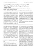
comparisons displayed in Fig. 1 using the
CLUSTALW
method with PAM (percent accepted mutation) series
residue weight matrix (gap penalty ¼10; gap length penalty
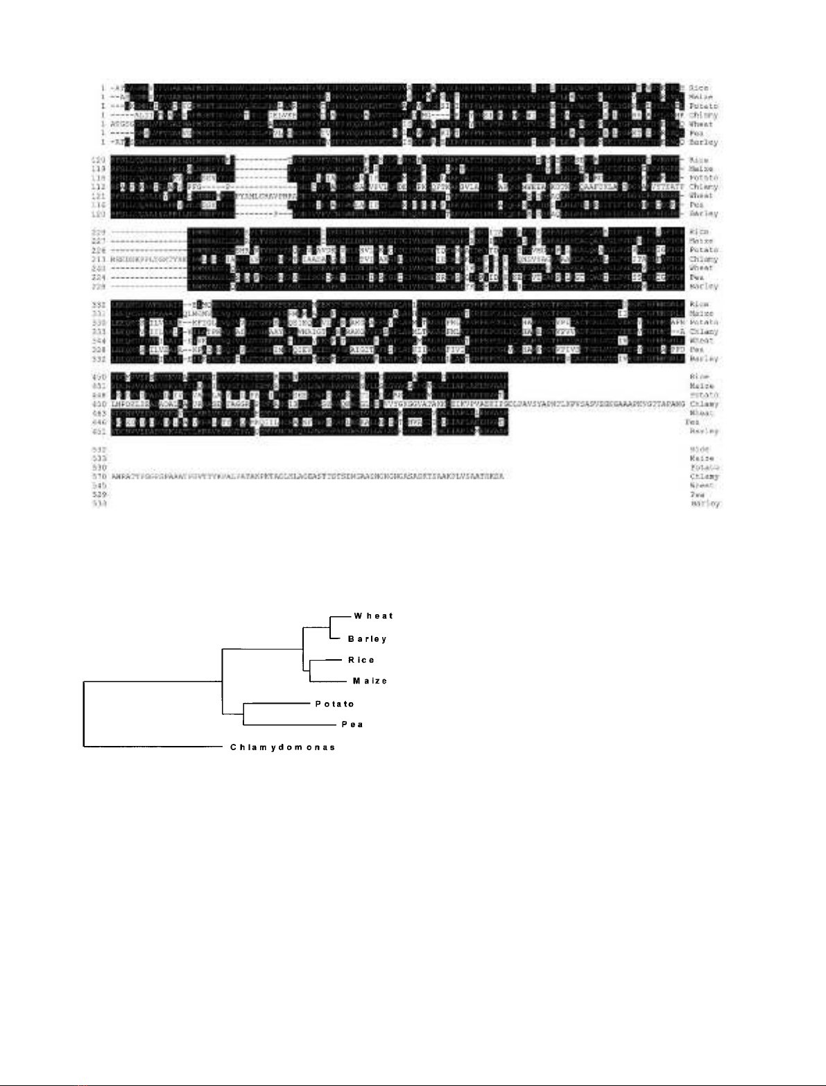
¼0.2) have enabled us to build the phylogenetic tree shown
in Fig. 2. It is clear from this analysis that divergence of
GBSSI sequences found by comparing several plant species
occurred at a very early stage during the evolution of
photosynthetic eukaryotes.
Characterization of the GBSSI gDNA sequences
The cDNA clone CD142 was used to select for correspond-
ing gDNAs from an indexed cosmid library [31]. A 6.5 kb
fragment in cosmid GB911 covering most of the GBSSI
coding sequences was subcloned in two overlapping parts of
3.0 and 4.5 kb and subjected to DNA sequencing thus
generating a 5856 bp gDNA sequence deposited in Gen-
Bank (accession number AF433156). Figure 3 displays the
length and position of the six introns within the GBSSI
sequence compared with those of rice and potato. The
number and position of the introns are unrelated to those
present in vascular plant genes and suggest an ancient
divergence of the GBSSI gene in green algae.
Establishing the nature of the STA2 locus
Two separate lines of evidence show that the cDNA and
gDNA clones correspond to the STA2 gene products. First,
a gDNA clone obtained in an indexed cosmid library [31]
complemented a sta2-1 mutation. Figure 4 shows the
various levels of phenotypic complementation obtained
with six independent transformants. GBSSI specific activ-
ities (calculated with respect to the quantity of Chlamydo-
monas starch involved in the assay) in the complemented
strains varied from 44 to 84% when compared with that of
the wild-type strain. It is clear that six strains (out of three
hundred) cotransformed with the GB911 gDNA restored
both amylose biosynthesis (at least partially) and the
FEBS 2002 In vitro synthesis of amylose (Eur. J. Biochem. 269) 3813
presence of the 69 kDa GBSSI protein (data not shown).
Restoration of amylose synthesis is likely to come as a
consequence of the random integration of the wild-type
STA2 gene in the nuclear genome of Chlamydomonas.
Nevertheless, depending on the integration site, expression
of this integrated wild-type copy of STA2 might vary
greatly. Indeed integrations in some genomic regions have
been reported to trigger silencing of the DNA introduced
[38–40]. These position effectscould therefore explain
variation in phenotype between transformants and only
partial restoration of amylose synthesis. It must be stressed
that in control experiments involving cotransformation with
randomly selected cosmids we never observed complemen-
tation of the sta2 mutations.
Second, the CD142 cDNA was used to find RFLPs in
strains disrupted for the STA2 gene (Fig. 5). We were able
to show that these differences cosegregated in 22 indepen-
dent meiotic recombinants in a cross involving strain IJ2
(sta2-29::ARG7 sta3-1) and an interfertile ecotype of
C. reinhardtii known as C. smithii (strain CS9). This latter
is wild-type regarding starch accumulation. Functional
complementation of sta2-1 mutation by the gDNA
sequence together with the demonstration of allele-specific
changes in this gDNA by particular STA2 mutations
demonstrates that the cloned gene defines STA2 and that
the latter encodes GBSSI.
Amylose in storage starch appears after a block
in amylopectin synthesis
Nitrogen starvation in Chlamydomonas offers a good
model with which to understand the basic physiology of
storage starch synthesis. During nitrogen starvation cellu-
lar components including thylakoid membranes are bro-
ken down and converted into both lipid droplets and
starch. We followed the kinetics of amylose synthesis over
a 5-day period of nitrogen starvation and measured the
amounts of starch, amylose, the k
max
of the starch
fractions, the degree of crystallinity and the X-ray
Fig. 2. Phylogenetic tree established from GBSSI proteins sequences
alignment as shown in Fig. 1.
Fig. 1. Peptide sequence comparison of Chlamydomonas GBSSI with those of other plant species. This analysis was done using mature proteins only.
Alignment was generated using the
CLUSTALW
method with PAM series residue weight matrix (gap penalty ¼10; gap-length penalty ¼0.2).
Residues matching the consensus GBSSI sequence derived from this comparison are shaded in black. Accession numbers for the different GBSSI
are as follows: wheat: P27736; Chlamydomonas: AF026420; maize: P04713; pea: X88789; rice: P19395; barley: X07931; potato: X58453.
3814 F. Wattebled et al. (Eur. J. Biochem. 269)FEBS 2002