A sucrose binding protein homologue from soybean exhibits
GTP-binding activity that functions independently of sucrose
transport activity
Carlos P. Pirovani
1
, Joci Neuby A. Mace
ˆdo
2
, Luı
´s Anto
ˆnio S Contim
2
, Fabiana S. V. Matrangolo
2
,
Marcelo E. Loureiro
1
and Elizabeth P. B. Fontes
2
Departments of
1
Biologia Vegetal and
2
Bioquı´mica e Biologia Molecular/BIOAGRO, Universidade Federal de Vic¸ osa, Brazil
The sucrose binding protein (SBP) has been implicated as an
important component of the sucrose uptake system in
plants. SBP-mediated sucrose transport displays unique ki-
netic features and the protein is not similar to other transport
proteins. Here, we report the characterization of a member
of the SBP family from soybean [Glycine max (L) Merrill]
designated S64 or SBP2. Subcellular fractionation and pre-
cipitation by GTP-agarose demonstrated that S64/SBP2 is a
membrane-associated protein that exhibits GTP binding
activity. Purified recombinant S64/SBP2 protein, expressed
as a histidine-tagged protein in Escherichia coli, exhibited
nucleotide-binding specificity to guanine nucleotides. The
GTP binding site was mapped to an imperfect Walker A
type-sequence, Ala279-Leu-Ala-Pro-Thr-Lys-Lys-Ser286,
by site-directed mutagenesis. Escherichia coli-produced wild-
type protein and a truncated version of the protein con-
taining the putative binding-sequence-bound GTP, although
not with the same efficiency. In contrast, replacement of
Thr283 and Lys284 residues to Leu and Glu residues pre-
vented GTP binding. The site directed mutant failed to bind
GTP but retained the ability to undergo oligomerization
and to promote growth of the susy7 yeast strain, deficient
in utilizing extracellular sucrose, on medium containing
sucrose as the sole carbon source. Our results indicate that
GTP binding and sucrose transport by SBP are separable
and function independently. The implications of our findings
with respect to the function and membrane topology of SBP
are discussed.
Keywords: sucrose transporter; soybean; yeast complemen-
tation assay; Glycine max.
In many higher plants, sucrose is the predominant form of
photoassimilate that is transported from mature leaves
(sourcetissues)tosinktissues,suchasseeds,stems,
reproductive organs and roots, via the vascular system [1].
Biochemical studies have demonstrated that sucrose uptake
kinetics in leaves is complex and consists of multiple
components; for example, in Vicia faba, two saturable
(high- and low-affinity) components and one linear,
low-affinity component have been described [2]. Our
understanding of sucrose translocation has advanced con-
siderably over the last decade with the molecular and
biochemical characterization of the sucrose transporter
(SUT) family of low- and high-affinity sucrose transporters
[1]. The SUT1 protein has been described as the proton-
motive-force-driven sucrose symporter that mediates
phloem loading and long-distance transport, the key
transport step in assimilate partitioning for many plants
[3–5]. SUT1 serves as a high-affinity transporter, whereas
SUT4, a second member of this sucrose transporter family,
corresponds to the low-affinity/high capacity saturable
component of sucrose uptake found in leaves [6]. A third
structurally related-member of the family has been identified
and designated SUT2 [7]. The SUT2 protein has been
proposed to act as a sugar sensor that controls sucrose
fluxes across the plasma membrane of sieve elements by
regulating expression, activity and turnover of SUT1 and
SUT4 [7]. This hypothesis was raised based on the lack of
transport activity of SUT2 and its colocalization with the
high and low-affinity sucrose transporter in sieve elements.
Nevertheless, direct evidence for a SUT2 sucrose sensor and
regulatory function has not been provided.
Earlier attempts to identify sucrose transporters resulted
in the identification of a sucrose binding protein from
soybean cotyledonary microsomal membrane fraction by
its capacity to bind to the sucrose analogue 6¢-deoxy-6¢-
(4-azido-2-hydroxy)-benzamido-sucrose [8]. Subsequent
progress in characterizing SBP led to the isolation of its
cDNA from an expression library prepared from cotyledon
mRNA [9]. Molecular characterization of the cDNA-
encoded product revealed that SBP was quite dissimilar
from the H
+
/sucrose symporter SUT. Despite the lack of
similarity between SBP and other known membrane
transport proteins, several lines of evidence have implicated
the SBP protein as the linear, low affinity component of
sucrose uptake system in plants. The SPB protein is
localized in the plasma membrane of cells that are actively
engaged in sucrose transport, such as mesophyll cells of
Correspondence to E. P. B. Fontes, DBB/BIOAGRO-Universidade
Federal de Vic¸ osa, Avenue. P.H. Rolfs s/n, 36571.000 Vic¸osa MG,
Brazil.
Fax: + 55 31 38992864, Tel.: + 55 31 38992949,
E-mail: bbfontes@ufv.br
Abbreviations: SUT, sucrose transporter; SBP, sucrose binding
protein; CaMV, cauliflower mosaic virus; rbcS, small subunit of
RUBISCO; ADH, alcohol dehydrogenase; DAF, days after flowering.
(Received 1 April 2002, revised 12 June 2002, accepted 2 July 2002)
Eur. J. Biochem. 269, 3998–4008 (2002) FEBS 2002 doi:10.1046/j.1432-1033.2002.03089.x

young sink leaves, the companion cells of mature phloem
and the cells of cotyledons undergoing differentiation [9,10].
In the cotyledon, expression of the SBP gene is temporally
regulated and accumulation of the protein is coordinated
with active sucrose uptake [9]. In spinach, a SBP homologue
was immunolocalized in the plasma membrane of sieve
elements in fully expanded leaves, shoots and roots [11,12]
and in V. faba developing seeds, SBP was colocalized with
the H
+
/sucrose symporter in the plasma membrane of
transfer cells [13]. A SBP homologue was also detected in
the microsomal fraction of young leaves from Nicotiana
tabacum [14]. Direct evidence implicating SBP in sucrose
transport has been obtained with complementation studies
using a secreted-invertase-deficient mutant yeast strain,
incapable of growth on medium containing sucrose as the
only carbon source [15,16]. Ectopic expression of the SBP
cDNA alone reverses the mutant yeast phenotype and SBP-
mediated specific sucrose uptake in yeast displays linear,
nonsaturable kinetics up to 30 m
M
external sucrose, being
relatively insensitive to pH gradient across the membrane
[15,17]. These biochemical features closely resemble the
kinetics properties of the previously characterized linear
component of sucrose uptake in higher plants [18–20].
Recently, we have conducted overexpression and antisense
repression studies in transgenic tobacco (Nicotiana tabacum
L. Cv Havana) to analyze the function of SBP in the long-
distance sucrose transport [14]. The antisense transgenic
plants developed symptoms consistent with inhibition of
sucrose translocation and displayed a reduction in plant
growth and development. Furthermore, both antisense
repression and overexpression of a SBP homologue in
transgenic lines altered carbohydrate partitioning in mature
leaves. These results indicated that SBP might represent an
important component of the sucrose translocation pathway
in plants.
More recently, we have addressed the role of SBP in
plant cell sucrose transport by performing radiolabeled
sucrose uptake experiments with transgenic tobacco cell
lines expressing the SBP sense or antisense gene [21]. In
this condition, the level of a SBP homologue correlated
with the efficiency of radiolabeled uptake by the trans-
genic tobacco cells. Furthermore, manipulation of SBP
levels altered sucrose-cleaving activities in a metabolic
compensatory manner. Enhanced accumulation of SBP
caused an increase in intracellular sucrose synthase activity
with a concomitant decline in cell-wall invertase activity.
This alteration in sucrose-cleaving activities is consistent
with a metabolic adjustment of the sense cell lines caused
by its high efficiency of direct sucrose uptake as
disaccharide. Although these studies clearly demonstrated
that SBP is involved in sucrose translocation-dependent
physiological processes, still unresolved is whether the
underlying mechanism involves SBP-mediated sucrose
transport or SBP-mediated regulation of alternative car-
bohydrate uptake systems.
Despite the functional characterization of SBP, potential
post-translational modifications that could regulate its
function have not been examined. In this investigation, we
describe the identification of an isoform of soybean SBP,
designated S64 or SBP2, and we show that the SBP
homologue is a membrane-associated GTP binding protein.
We have generated mutants that blocked its GTP binding
activity but not interfered in its oligomerization property
and S64/SBP-mediated sucrose transport in yeast. These
mutants should be valuable tools for determining the
physiological role of SBP as a G-protein in vivo.
EXPERIMENTAL PROCEDURES
Isolation of a SBP homologue cDNA from soybean
DNA manipulations were performed essentially as
described previously [22]. The S64 cDNA (GeneBank
accession number AF191299) was unintentionally isolated
from a soybean seed expression library using an antibody
raised against a partially purified microsomal membrane
fraction from immature soybean seeds [14]. The positive
clones resulted from this screening were designated by the
letter S from soybean seeds followed by 1 : 1000 of the
estimated M
r
of the encoded product. The identity of this
particular S64 clone was obtained by sequence comparison
analysis using the
BLAST
program [23]. The computer
program
CLUSTALW
was used for sequence alignment. The
S64 deduced protein shares 86% sequence identity with the
sucrose binding protein (GeneBank accession number
L06038) and is also referred to as SBP homologue or SBP2.
Construction of plasmids and antibody production
The S64/SBP homologue insert was released from the k
recombinant DNA with EcoRI digestion and subcloned
into the EcoRI site of pUC118 to obtain the clone
pUFVS64. The S64 protein was expressed as a fusion
protein using the pET-16b vector (Novagen), which
provides an N-terminal His tag. For this purpose, an EcoRI
site immediately adjacent to the stop codon was created by
PCR using the Pfu DNA polymerase, the forward primer
S64XHOF 5¢-AAGAAACTCGAGGTCGAAGA-3¢
(coordinates 103–121, XhoI site underlined) and the reverse
primer SEF97R 5¢-ATACATTCCCCGAATTCAGCCA
CCTCC-3¢(positions 1498–1524, EcoRI site underlined).
The amplified sequence, spanning the entire protein-coding
region and lacking the putative peptide signal coding
sequence and the 3¢untranslated sequences, was subcloned
into the EcoRI/SmaI-restricted pGEM7Zf(–) vector (Pro-
mega), and then moved as a XhoI insert into pET16b,
yielding pUFV120.
The construction was transformed into E. coli strain
BL21 (DE3) and the synthesis of the recombinant protein
was induced by isopropyl thio-b-
D
-galactoside (IPTG). The
induced protein was affinity-purified using Ni-chelating
Sepharose resin (Amersham Pharmacia Biotech.) and used
as an antigen to raise polyclonal antisera in rabbits, which
were immunized through subcutaneous injections during
2-week intervals. The specificity of the anti-S64 serum was
previously evaluated with protein extracts from transgenic
tobacco plants expressing the S64/SBP homologue cDNA
either in the sense or antisense orientation [14,21], in a yeast
expression system [14] and in a bacteria expression system
[14].
Truncated protein, mutagenesis and bacterial
overexpression
To produce a S64 truncated protein, an internal 916-bp
sequence of S64 cDNA was released from pUFVS64 with
FEBS 2002 GTP binding of sucrose binding protein (Eur. J. Biochem. 269) 3999

Sau3AI digestion and inserted into the BamHIsiteof
pET16b to create pUFV50. The inserted sequence spans
nucleotides 122–1041 of the cDNA and encodes the amino
acid residues from position 36–343.
The putative GTP-binding site was mutated using a
PCR-based mutagenesis strategy, in which overlapping
upstream and downstream sequences of the site were
individually amplified with sets of primers to create an
internal XbaI site within the putative site. The sets of
primers used were SPI97F 5¢-TCCTCACTGCAG
TCACCATGGCGACCA-3¢(coordinates 1–27, PstIsite
underlined) and SGTPXBAF 5¢- GGCCCCTCTAGA
GAAAAGCTC-3¢(coordinates 857–878, XbaIsite
underlined) for the S64 N-terminal encoding sequence as
well as SGTPXBAR 5¢-GCTTTTCTCTAGAGGGGCC
AACG-3¢(positions 853–876, XbaI site underlined)
and SEF97R 5¢-ATACATTCCCCGAATTCAGCCACC
TCC-3¢(positions 1498–1524, EcoRI site underlined) for the
adjacent C-terminal encoding sequence. The upstream-
amplified sequence was digested with PstIandXbaI,
whereas the downstream-amplified fragment was digested
with XbaIandEcoRI, and then they were inserted by triple
ligation into PstI–EcoRI sites of pUC118 to obtain
pUFV193. This restored the S64 coding region in which
an internal XbaI site was created and, as consequence, the
putative GTP-binding site Ala279-Leu-Ala-Pro-Thr-Lys-
Lys-Ser286 was mutated to Ala279-Leu-Ala-Pro-Leu-Glu-
Lys-Ser286. The mutations were confirmed by sequencing.
To transfer the mutated S64 sequence to pET16b, it was
amplified from pUFV193 with the sense primer S64XHOF
and the antisense primer SEF97R. The amplified sequence,
harboring the mutated protein-coding region and lacking
the putative peptide signal coding sequence and the
3¢untranslated sequences, was subcloned into the EcoRI/
SmaI-restricted pGEM7Zf(–) vector (Promega), and then
moved as a XhoI insert into pET16b to obtain pUFV232.
Constructions in pET16b were expressed in E. coli
strain BL21 (DE3) LysS following induction by IPTG.
N-Terminal His-tagged SBP fusion proteins were purified
according to manufacturer’s instructions (Novagen) for
soluble proteins. For oligomerization studies, after a first
round of purification, the His tag was removed from the
E. coli-produced proteins by treatment with catalytic
amounts of Factor Xa (10 lgÆmg
)1
of recombinant protein)
in 100 m
M
NaCl, 50 m
M
Tris/HCl, pH 8.0, 1 m
M
CaCl
2
at
37 C for 24 h.
Isolation of microsomal fraction
For microsomal membrane isolation, soybean cotyledons
were homogenized with 25 m
M
Tris/HCl, pH 7.0,
250 m
M
sucrose, 2.5 m
M
dithiothreitol, 10 m
M
MgSO
4
,
0.5% (w/v) gelatin and 0.5 m
M
phenylmethanesulfonyl
fluoride [8]. The homogenate was filtered and centrifuged
for 15 min at 13 000 gand 4 C. Microsomal prepara-
tions were isolated by centrifugation at 80 000 gfor
45 min [24].
Transient expression of S64/SBP homologue
in soybean suspension cells
The pUFVS64 clone was modified by site-directed muta-
genesis to create an EcoRI restriction site immediately
downstream of the stop codon, yielding pUFV32. A
plant expression cassette containing the S64/SBP homo-
logue gene was constructed by insertion of the S64
coding region that was released from pUFV32 with
EcoRI/BamHI digestion into pMON921 vector [25],
previously digested with BglII/EcoRI. The resulting
plasmid, pUFV52, harbors the S64 coding region in the
sense orientation placed between the CaMV 35S pro-
moter with a duplicated enhancer region and the 3¢end
of the pea E9 rbcS gene. A soybean cell culture line was
generated and established as described previously [26].
Transient expression of S64 was performed by electro-
poration (380 V, 975 lF) of 10 lg of expression cassette
DNA and 40 lg of sheared salmon sperm DNA into
0.8 mL of cultured soybean cells in electroporation buffer
(80 m
M
KCl, 5 m
M
CaCl
2
,10m
M
Mes, pH 6.7, 0.425
M
mannitol). Prior to electroporation, soybean suspension
cells at 4 days after passage were recovered by centrifu-
gation at 200 g, washed three times and concentrated
twice with electroporation buffer, incubated with plasmid
and carrier DNA at 37 C for 1 h and then at 0 Cfor
10 min. The electroporated cells were diluted into 10 mL
of MS medium [27], supplemented with complex vitamin
B5, 0.2 mgÆmL
)1
2,4-dichlorophenoxyacetic acid, 6%
(w/v) sucrose and 15 m
M
glutamine, pH 5.7. Total
protein was isolated from cells 48 h after transfection as
described [24], separated by SDS/PAGE and immuno-
blotted with anti-S64 serum.
Gel electrophoresis and immunoblotting analysis
SDS/PAGE was carried out as described previously [28]
and the proteins were transferred from 10% SDS/
polyacrylamide gels to nitrocellulose membrane by elec-
troblotting. The membrane was blocked with 3% (w/v)
BSA in NaCl/Tris/Tween [100 m
M
Tris/HCl, pH 8.0,
150 m
M
NaCl, 0.05% (v/v) Tween-20]. S64/SBP homo-
logue was detected using polyclonal anti-S64 serum at a
1 : 1000 dilution, followed by a goat anti-(rabbit IgG) Ig
conjugated to alkaline phosphatase (Sigma) at a 1 : 5000
dilution. Alkaline phosphatase activity was assayed using
5-bromo-4-chloro-3-indolyl phosphate (Life Technologies,
Inc.) and p-nitroblue tetrazolium (Life Technologies,
Inc.).
Binding of S64/SBP homologue to GTP-agarose
Whole cell protein extracts were obtained from transgenic
tobacco cell lines expressing a soybean S64 transgene [21]
and from soybean suspension cells transiently trans-
formed with a S64 expression cassette. Protein extracts
were prepared by homogenization of the cells with lysis
buffer [100 m
M
Tris/HCl, pH 7.5, 50 m
M
KCl, 1 m
M
EDTA, 1% (v/v) Triton X-100, 1 m
M
phenyl-
methanesulfonyl fluoride, 0.1 m
M
dithiothreitol, 5 m
M
MgCl
2
]ataratioof1mgofcellsper2lL of buffer
and then clarified by centrifugation at 20 000 gfor
20 min. The supernatant (2 mL) was incubated with
100 lL of 50% (v/v) GTP-agarose suspension in 50 m
M
Tris/HCl, pH 7.5, for 12 h under agitation at 4 C[29].
The agarose beads were pelleted by centrifugation,
washed extensively with cold 50 m
M
Tris/HCl, pH 7.5
and resuspended in 40 lL of SDS/PAGE sample buffer.
4000 C. P. Pirovani et al. (Eur. J. Biochem. 269)FEBS 2002

GTP bound proteins were fractionated by SDS/PAGE,
transferred to nitrocellulose and probed with anti-S64
serum, as described above.
Binding of S64/SBP homologue fusion protein
to nucleotide-agarose
The purified His-tagged S64 fusion protein (2 lg) was
incubated with either 50 lL of ATP-agarose, GTP-agarose
or Protein A-agarose suspension, previously equilibrated
with binding buffer [20 m
M
Tris/HCl pH 7.5, 100 m
M
NaCl, 0.01% (v/v) Triton X-100, 2 m
M
MgCl
2
]. After 1 h
at 4 C, the beads were washed three times with 500 mL of
cold binding buffer and eluted in SDS/PAGE sample
buffer at 100 C for 3 min. The eluted proteins were
separated by SDS/PAGE and stained with Coomassie Blue.
In competition assays, GTP, GDP, GTPcS (guanosine
5¢-O-3-thiotriphosphate), ATP, UTP or CTP were included
in the binding buffer at 2 m
M
andincubatedwiththe
recombinant protein (0.5 lg) for 30 min at 4 Cpriortothe
binding reaction to GTP-agarose.
GTP-binding assay
The GTP-binding assay was performed as described
previously [30]. Briefly, E. coli-expressed His fusion proteins
were affinity-purified and blotted onto nitrocellulose mem-
brane using BIO-DOTTM (Bio-Rad), according to the
manufacture’s instructions. Alternatively, affinity-purified
recombinant proteins were fractionated by SDS/PAGE and
transferred to nitrocellulose by electroblotting. The mem-
branes were washed twice with binding buffer [50 m
M
NaH
2
PO
4,
pH 7.5, 10 m
M
MgCl
2
,2m
M
dithiothreitol,
0.3% (v/v) Tween-20 and 4 l
M
ATP] and then incubated
with binding buffer supplemented with 1 lCÆmL
)1
(or
0.33 n
M
)[a-
32
P]GTP (3000 CiÆmmol
)1
; Amersham/Phar-
macia) for 2 h. After the incubation period, the membranes
were washed at least six times with binding buffer and
subjected to autoradiography at )80 C, using WOLF
L-PLUS 505504 LP intensifying screens (Sigma).
Yeast strain and plasmids
The generation of susy7 yeast strain has been described
previously [3]. It has the potato sucrose synthase gene stably
integrated into its genome but lacks an endogenous sucrose
transport system and invertase activity. Thus, susy7yeast
strain is incapable to grow on a medium containing sucrose
as the sole carbon source, unless a sucrose uptake system is
provided through ectopic expression. For complementation
assays in the mutant yeast strain, the intact S64 cDNA was
released from pUFVS64 with EcoRIandinsertedintothe
same site of the yeast expression vector 112AINE [3]. The
resulting plasmid, pUFV373, contains the S64 cDNA in the
right orientation placed between the ADH1 promoter and 3¢
end of ADH1 gene. A yeast expression cassette containing
the mutated S64 gene was constructed by insertion of the
GTP binding site mutated cDNA that was released from
pUFV193 with PstIandEcoRI digestion into the same
restriction sites of the 112AINE vector. The resulting
plasmid, pUFV375, harbors the mutated S64 coding region
in the sense orientation placed between the ADH1promoter
and the 3¢end of the ADH1 gene.
The susy7 yeast strain was transformed with either
pUFV373 or pUFV375 by electroporation [31], resulting in
susy7-S64 or susy7-MS64, respectively. To monitor growth
on sucrose medium, 200 lL of a 24-h-old liquid culture of
either susy7-S64 or susy7-MS64 growing in complete
medium supplemented with 2% (w/v) glucose were used
to inoculate 20 mL of complete medium with 2% (w/v)
sucrose as the only carbon source. Relative growth was
monitored by taking the D
600
during 24-h intervals, as
indicated in the figure legend. For each DNA construct, at
least three independent transformants were monitored.
RESULTS
Isolation of a second member of the
SBP
gene family
from soybean
Based on structural homology and functional analogy, we
have isolated a sucrose binding protein (SBP) homologue
cDNA from soybean. The predicted encoded protein was
first designated S64, has an estimated M
r
of 55 834 and pI
of 6.32. Sequence comparison analysis revealed that the
predicted encoded protein was quite similar to the sucrose
binding protein, first identified in soybean cotyledon (86%
sequence identity). It also showed a significant amino-acid
sequence similarity to heterologous SBP sequences from
other plant species (Fig. 1). Analysis of the deduced amino-
acid sequence allowed us to predict a signal peptide and its
processing site, which suggests that the protein be targeted
to the secretory pathway. In fact, the S64 protein was
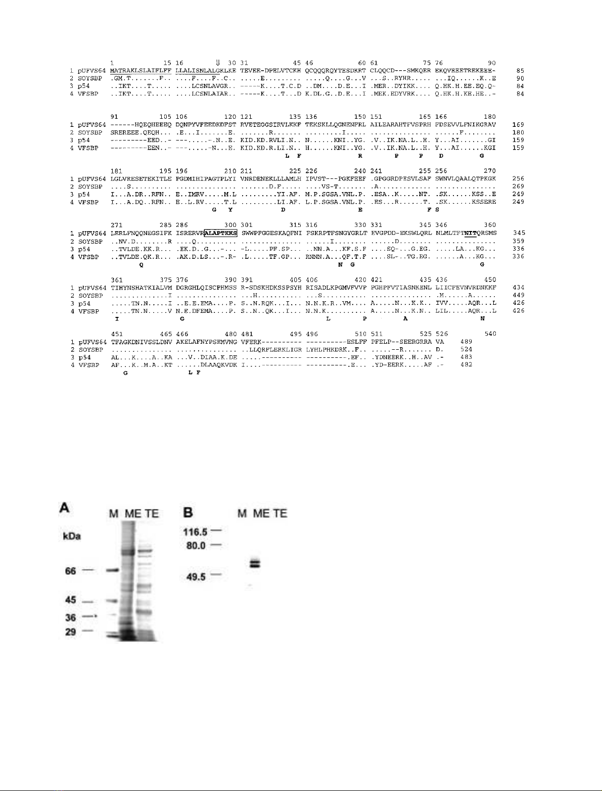
detected in microsomal fraction of soybean cotyledon
(Fig. 2).
In soybean cotyledon, the S64 antibody recognized two
cross-reacting polypeptides with slightly different electro-
phoretic mobility (Fig. 2B, lane ME). Because SBP has a
predicted M
r
of 60 522 and it is highly homologous to S64,
the reduced SDS/PAGE mobility polypeptide could repre-
sent SBP. Alternatively, the cross-reacting polypeptides
could be differentially processed forms of the same S64/SBP
homologue protein. The primary structure of the S64/SBP
homologue protein shows the presence of a consensus
sequence for nucleotide binding and a site for N-linked
glycosylation, as potential sites for post-translational mod-
ifications of the protein (Fig. 1). Furthermore, despite the
hydrophilic nature of SBP, solubilization and partitioning
studies of plasma membrane proteins have demonstrated
that 25% of SBPs are associated with a hydrophobic
portion of the plasma membrane [10]. This observation has
led to the suggestion that the putative leader peptide, which
corresponds to the only hydrophobic region of the protein,
is not quantitatively cleaved from the mature protein. The
presence of the leader peptide in a fraction of S64/SBP
homologue would explain the antibody cross-reactivity to
the higher molecular mass form. To examine these possi-
bilities, we transferred the S64 coding region to a plant
expression cassette and the recombinant protein was over-
synthesized in cultured soybean cells (Fig. 3, compare lanes
1 and 2). In the homologous system, the apparatus for
protein processing is expected to operate properly and with
similar specificity. As shown in Fig. 3, the S64/SBP
homologue protein was synthesized as single polypeptide
(lane 1) that comigrated with the faster migrating polypep-
tide detected in membrane fraction of soybean cotyledon
FEBS 2002 GTP binding of sucrose binding protein (Eur. J. Biochem. 269) 4001
(lane 3). This result indicates that the reduced SDS/PAGE
mobility polypeptide may represent SBP, whereas the faster
migrating form corresponds to S64/SBP homologue. Con-
sistent with this observation, the predicted M
r
of SBP
(60 552) is slightly higher than that of S64 (55 834). Thus,
SBP and S64 cDNAs may correspond to nonallelic SBP
genes from soybean. Investigation of the genomic complex-
ity of the SBP genes by Southern blot analysis revealed a
pattern of cross-hybridizing bands consistent with the
argument that SBP is encoded by a small gene family in
soybean (data not shown). In view of this observation, the
S64 protein may also be designated SBP2 (isoform 2),
whereas the previously identified SBP [9] would be SBP1
(isoform 1).
The S64/SBP2 protein is a membrane-associated
GTP-binding protein
The S64/SBP2 deduced protein contains a predicted
nucleotide-binding site (Fig. 1) that harbors classical
Walker-type consensus sequence for the P-loop, [Ala/
Gly]x(4)Gly-Lys[Ser/Thr] [32]. Despite the fact that the
Fig. 2. SDS/PAGE and immunoblotting of membrane fractions from
soybean cotyledons. (A) Whole cell protein extracts (TE) and
microsomal membranes (ME) were isolated from soybean seeds at
20 days after flowering (DAF), fractionated by SDS/PAGE and
stained with Coomassie Brilliant Blue. M corresponds to molecular
mass markers indicated on the left in kDa. (B) SDS/PAGE fraction-
ated protein was transferred to nitrocellulose membranes and probed
with an anti-S64 serum.
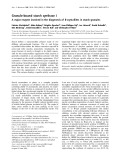
Fig. 1. Comparison of the amino acid sequence of S64 with SBP from soybean and other organisms. A multiple sequence alignment of the deduced
amino acid sequence of soybean S64 (pUFVS64, GeneBank accession number AF191299), soybean SBP (SOYSBP, GeneBank accession number
L06038), pea SBP homologue (p54, GeneBank accession number Y11207) and Vicia faba SBP homologue (VPSBP, GeneBank accession number
VFA292221) was obtained with the
CLUSTALW
program. The amino acid sequences are in the one-letter code and have been aligned by introducing
gaps (shown as dashes) to maximize identity. Dots represent identity to S64. The nucleotide-binding motif is boxed and the putative N-linked
glycosylation site is underlined. The open arrow indicates the putative signal peptide cleavage site of S64. Amino acid residues indicated below the
sequences correspond to highly conserved residues in equivalent positions of vicilin-like protein sequences that are important in maintaining their
three-dimensional structure.
4002 C. P. Pirovani et al. (Eur. J. Biochem. 269)FEBS 2002